Review
. 2022 Sep 1;71(9):1852-1861.
doi: 10.2337/dbi22-0004.

The Liver-α-Cell Axis in Health and in Disease

Affiliations
Review

The Liver-α-Cell Axis in Health and in Disease

Michael M Richter et al. Diabetes. .

Abstract

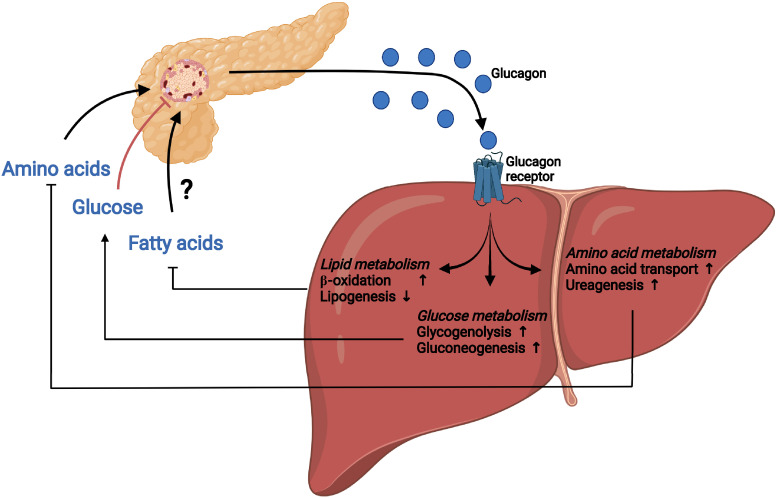
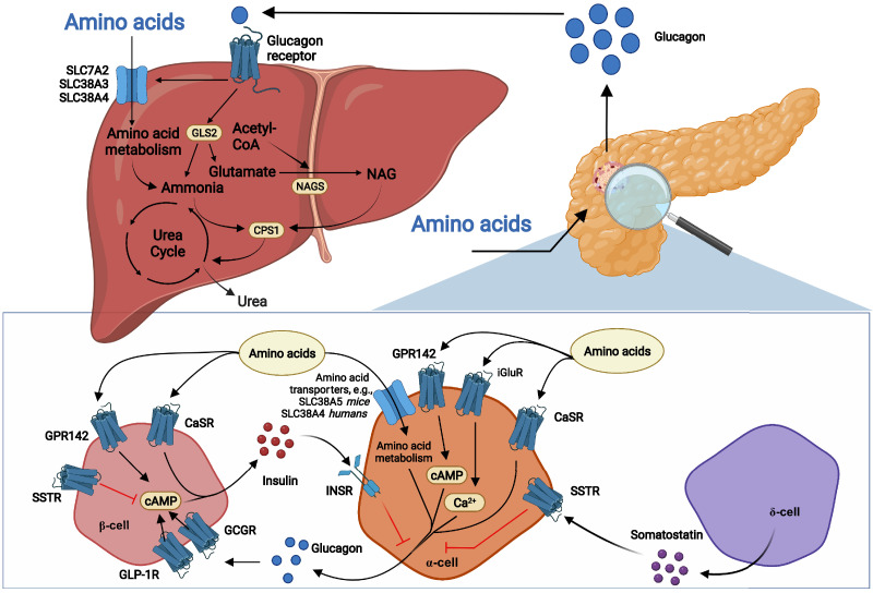
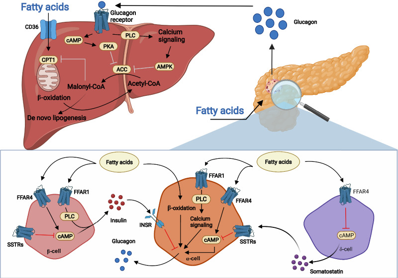
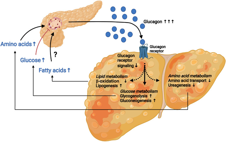
Glucagon and insulin are the main regulators of blood glucose. While the actions of insulin are extensively mapped, less is known about glucagon. Besides glucagon's role in glucose homeostasis, there are additional links between the pancreatic α-cells and the hepatocytes, often collectively referred to as the liver-α-cell axis, that may be of importance for health and disease. Thus, glucagon receptor antagonism (pharmacological or genetic), which disrupts the liver-α-cell axis, results not only in lower fasting glucose but also in reduced amino acid turnover and dyslipidemia. Here, we review the actions of glucagon on glucose homeostasis, amino acid catabolism, and lipid metabolism in the context of the liver-α-cell axis. The concept of glucagon resistance is also discussed, and we argue that the various elements of the liver-α-cell axis may be differentially affected in metabolic diseases such as diabetes, obesity, and nonalcoholic fatty liver disease (NAFLD). This conceptual rethinking of glucagon biology may explain why patients with type 2 diabetes have hyperglucagonemia and how NAFLD disrupts the liver-α-cell axis, compromising the normal glucagon-mediated enhancement of substrate-induced amino acid turnover and possibly fatty acid β-oxidation. In contrast to amino acid catabolism, glucagon-induced glucose production may not be affected by NAFLD, explaining the diabetogenic effect of NAFLD-associated hyperglucagonemia. Consideration of the liver-α-cell axis is essential to understanding the complex pathophysiology underlying diabetes and other metabolic diseases.


Figures

Figure 1
Figure 1
The liver–α-cell axis. The liver–α-cell axis constitutes a feedback loop in which circulating amino acids stimulate glucagon secretion from pancreatic α-cells and glucagon in turn controls hepatic amino acid uptake and metabolism, including increased amino acid transport and ureagenesis. The liver–α-cell axis also constitutes a feedback loop in which high levels of circulating glucose inhibit α-cell secretion of glucagon, resulting in decreased hepatic glucose metabolism and subsequent glucose production. In contrast, lower levels of circulating glucose result in increased α-cell secretion of glucagon and increased glucagon-mediated hepatic glucose metabolism, including increased glycogenolysis and gluconeogenesis, and glucose production. A final component of the liver–α-cell axis may be the regulation of hepatic lipid metabolism by increasing β-oxidation and decreasing lipogenesis; however, it is currently not established exactly how, and if, lipids regulate α-cell secretion.
Figure 2
Figure 2
Amino acids stimulate glucagon secretion, and glucagon in turn stimulates hepatic amino acid uptake and metabolism, including ureagenesis. Circulating amino acids stimulate glucagon secretion from pancreatic α-cells by mechanisms that may involve stimulation of ionotropic glutamate receptors (iGluR) and G protein–coupled receptors (GPRs), including CaSR and GPR142. Increased expression of amino acid transporters such as SLC38A5 (in mice) and SLC38A4 (in humans) and subsequent amino acid metabolism may also be involved. Amino acids may also stimulate insulin secretion from β-cells through GPR142 and CaSR, and insulin may inhibit glucagon release via the insulin receptor (INSR) on α-cells. Paracrine somatostatin inhibits cAMP generation and inhibits secretion via activation of somatostatin receptor (SSTR). Glucagon activates both the GLP-1R and the GCGR on β-cells and stimulates insulin secretion. Once secreted to the peripheral circulation, glucagon binds the GCGR and stimulates hepatic amino acid uptake (through amino acid transporters such as SLC7A2, SLC38A3, and SLC38A4) and metabolism. Glucagon receptor signaling activates glutaminase, yielding glutamate, a NAGS substrate, whereby NAG levels increase, activating CPS-1 and ureagenesis. Glutaminase activity also yields ammonia, a CPS-1 substrate, and thus activates CPS-1. GCGR activity also increases the expression and activity of urea cycle enzymes.
Figure 3
Figure 3
Model for stimulation of glucagon secretion by FAs and effects of glucagon signaling on hepatic lipid metabolism. Circulating FAs, primarily LCFAs, may stimulate glucagon secretion from pancreatic α-cells through various known and unknown mechanisms. This may involve FFAR1 and FFAR4 with subsequent increase in intracellular Ca2+ and cAMP. Likewise, β-oxidation in α-cells may result in the release of glucagon. Fatty acids may also inhibit somatostatin release from δ-cells through decreased intracellular cAMP via FFAR4 activation. The reduced somatostatin release results in decreased activation of several somatostatin receptors (SSTRs) on α-cells, which results in less inhibition of cAMP generation. FAs can also stimulate insulin secretion from β-cells through FFAR1 and FFAR4, and insulin may inhibit glucagon release through the insulin receptor (INSR) on α-cells. Once secreted, glucagon binds the hepatic GCGRs, stimulates β-oxidation, and inhibits de novo lipogenesis. GCGR signaling inhibits acetyl-CoA carboxylase (ACC) via protein kinase A (PKA) and AMP-activated protein kinase (AMPK), resulting in decreased malonyl-CoA formation. As a result, mitochondrial entry of FAs and β-oxidation are increased, while the availability of FAs for triglyceride synthesis and subsequent export is reduced. PLC, phospholipase C.
Figure 4
Figure 4
The liver–α-cell axis in disease. Under conditions of type 2 diabetes and fatty liver disease, the liver–α-cell feedback loop is affected. Impaired hepatic glucagon signaling (glucagon resistance) decreases amino acid uptake and metabolism, resulting in hyperaminoacidemia and increased stimulation of glucagon secretion from pancreatic α-cells. Likewise, reduced glucagon signaling decreases hepatic β-oxidation, increases lipogenesis, and elevates circulating free FA concentrations, which may contribute to increased α-cell secretion of glucagon. Lastly, the hepatic glucose production is not affected by the reduced glucagon signaling. This results in increased hepatic glucose metabolism and hyperglycemia by the increased circulating levels of glucagon secreted from pancreatic α-cells in response to increased levels of amino acids and possibly FAs.

Similar articles

Cited by

References

    1. Müller TD, Finan B, Clemmensen C, DiMarchi RD, Tschöp MH. The new biology and pharmacology of glucagon. Physiol Rev 2017;97:721–766 - PubMed
    1. Holst JJ, Holland W, Gromada J, et al. . Insulin and glucagon: partners for life. Endocrinology 2017;158:696–701 - PMC - PubMed
    1. Wewer Albrechtsen NJ, Kuhre RE, Pedersen J, Knop FK, Holst JJ. The biology of glucagon and the consequences of hyperglucagonemia. Biomarkers Med 2016;10:1141–1151 - PubMed
    1. Finan B, Capozzi ME, Campbell JE. Repositioning glucagon action in the physiology and pharmacology of diabetes. Diabetes 2020;69:532–541 - PMC - PubMed
    1. Scheen AJ, Paquot N, Lefèbvre PJ. Investigational glucagon receptor antagonists in phase I and II clinical trials for diabetes. Expert Opin Investig Drugs 2017;26:1373–1389 - PubMed

Publication types