

CD73 facilitates invadopodia formation and boosts malignancy of head and neck squamous cell carcinoma via the MAPK signaling pathway
- PMID: 35657703
- PMCID: PMC9357645
- DOI: 10.1111/cas.15452
CD73 facilitates invadopodia formation and boosts malignancy of head and neck squamous cell carcinoma via the MAPK signaling pathway
Abstract
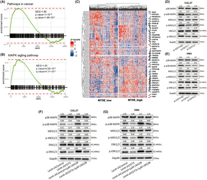
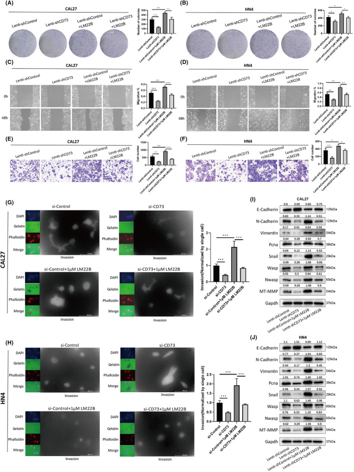
Elevated adenosine generated by CD73 (ecto-5'-nucleotidase; NT5E) could boost immunosuppressive responses and promote immune evasion in the tumor microenvironment. However, despite the immune response, CD73 could also promote tumor progression in a variety of cancers, and the nonimmunologic role and corresponding molecular mechanism of CD73 involved in head and neck squamous cell carcinoma (HNSCC) progression are not well characterized. Here, we demonstrated that CD73/NT5E is overexpressed in HNSCC tissues and predicts poor prognosis. Suppression of CD73 inhibited the proliferation, migration, and invasion of HNSCC cell lines (CAL27 and HN4) in vitro and in vivo. Gene set variation analysis (GSVA) and gene set enrichment analysis (GSEA) predicted that CD73 may be involved in invadopodia formation and MAPK signaling activation. As expected, knockdown of CD73 inhibited the MAPK signaling pathway, and the suppressive effect of CD73 knockdown on proliferation, migration, invasion, and invadopodia formation was reversed by a MAPK signaling activator. Our results suggest that CD73 could promote the proliferation, migration, invasion, and invadopodia formation of HNSCC via the MAPK signaling pathway and provide new mechanistic insights into the nonimmunological role of CD73 in HNSCC.
Keywords: CD73; EMT; HNSCC; MAPK signaling pathway; invadopodia.
© 2022 The Authors. Cancer Science published by John Wiley & Sons Australia, Ltd on behalf of Japanese Cancer Association.
Figures


Similar articles
-
5'-Ectonucleotidase CD73/NT5E supports EGFR-mediated invasion of HPV-negative head and neck carcinoma cells.J Biomed Sci. 2023 Aug 24;30(1):72. doi: 10.1186/s12929-023-00968-6. J Biomed Sci. 2023. PMID: 37620936 Free PMC article.
-
NT5E upregulation in head and neck squamous cell carcinoma: A novel biomarker on cancer-associated fibroblasts for predicting immunosuppressive tumor microenvironment.Front Immunol. 2022 Aug 26;13:975847. doi: 10.3389/fimmu.2022.975847. eCollection 2022. Front Immunol. 2022. PMID: 36091055 Free PMC article.
-
CD73 is associated with poor prognosis in HNSCC.Oncotarget. 2016 Sep 20;7(38):61690-61702. doi: 10.18632/oncotarget.11435. Oncotarget. 2016. PMID: 27557512 Free PMC article.
-
Adenosinergic Signalling in Cervical Cancer Microenvironment.Expert Rev Mol Med. 2025 Jan 7;27:e5. doi: 10.1017/erm.2024.30. Expert Rev Mol Med. 2025. PMID: 39762204 Free PMC article. Review.
-
The roles of CD73 in cancer.Biomed Res Int. 2014;2014:460654. doi: 10.1155/2014/460654. Epub 2014 Jul 14. Biomed Res Int. 2014. PMID: 25126561 Free PMC article. Review.
Cited by
-
Immune Escape Strategies in Head and Neck Cancer: Evade, Resist, Inhibit, Recruit.Cancers (Basel). 2024 Jan 11;16(2):312. doi: 10.3390/cancers16020312. Cancers (Basel). 2024. PMID: 38254801 Free PMC article. Review.
-
Effects of abnormal expression of CD73 on malignant phenotype of nasopharyngeal carcinoma.J Mol Histol. 2023 Dec;54(6):633-644. doi: 10.1007/s10735-023-10165-2. Epub 2023 Oct 24. J Mol Histol. 2023. PMID: 37874500
-
CD73: a new immune checkpoint for leukemia treatment.Front Immunol. 2025 Mar 6;16:1486868. doi: 10.3389/fimmu.2025.1486868. eCollection 2025. Front Immunol. 2025. PMID: 40114928 Free PMC article. Review.
-
A novel tertiary lymphoid structure-associated signature accurately predicts patient prognosis and facilitates the selection of personalized treatment strategies for HNSCC.Front Immunol. 2025 Mar 13;16:1551844. doi: 10.3389/fimmu.2025.1551844. eCollection 2025. Front Immunol. 2025. PMID: 40181975 Free PMC article.
-
Integrated multi-omics analysis identifies CD73 as a prognostic biomarker and immunotherapy response predictor in head and neck squamous cell carcinoma.Front Immunol. 2022 Nov 16;13:969034. doi: 10.3389/fimmu.2022.969034. eCollection 2022. Front Immunol. 2022. PMID: 36466881 Free PMC article.
References
-
- Chow LQM. Head and neck cancer. N Engl J Med. 2020;382:60‐72. - PubMed
-
- Leemans CR, Snijders PJF, Brakenhoff RH. The molecular landscape of head and neck cancer. Nat Rev Cancer. 2018;18:269‐282. - PubMed
-
- Budach V, Tinhofer I. Novel prognostic clinical factors and biomarkers for outcome prediction in head and neck cancer: a systematic review. Lancet Oncol. 2019;20:e313‐e326. - PubMed
-
- Rischin D, Ferris RL, Le Q‐T. Overview of advances in head and neck cancer. J Clin Oncol. 2015;33:3225‐3226. - PubMed
-
- Kaidar‐Person O, Gil Z, Billan S. Precision medicine in head and neck cancer. Drug Resist Updat. 2018;40:13‐16. - PubMed
MeSH terms
Substances
Grants and funding
- ZDB202001/Key Project of Health Commission of Jiangsu Province
- LGY2019088/LIUGEYI Project
- 81672678/National Natural Science Foundation of China
- JSKLOD-KF-2105/Open Project from Jiangsu Key Laboratory of Oral Diseases, Nanjing Medical University
- PAPD, 2018-87/Priority Academic Program Development of Jiangsu Higher Education Institutions
LinkOut - more resources
Full Text Sources
Medical
Research Materials
Miscellaneous
