. 2022 Jun 3;17(6):e0268391.
doi: 10.1371/journal.pone.0268391. eCollection 2022.

Targeting mitochondrial DNA polymerase gamma for selective inhibition of MLH1 deficient colon cancer growth

Affiliations

Targeting mitochondrial DNA polymerase gamma for selective inhibition of MLH1 deficient colon cancer growth

Berna Somuncu et al. PLoS One. .

Abstract

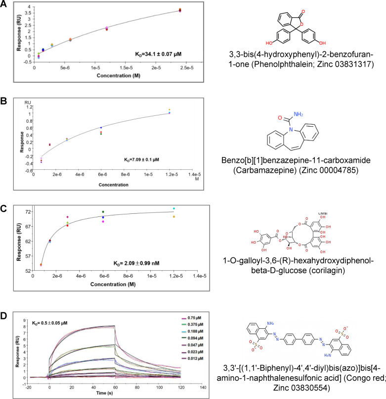
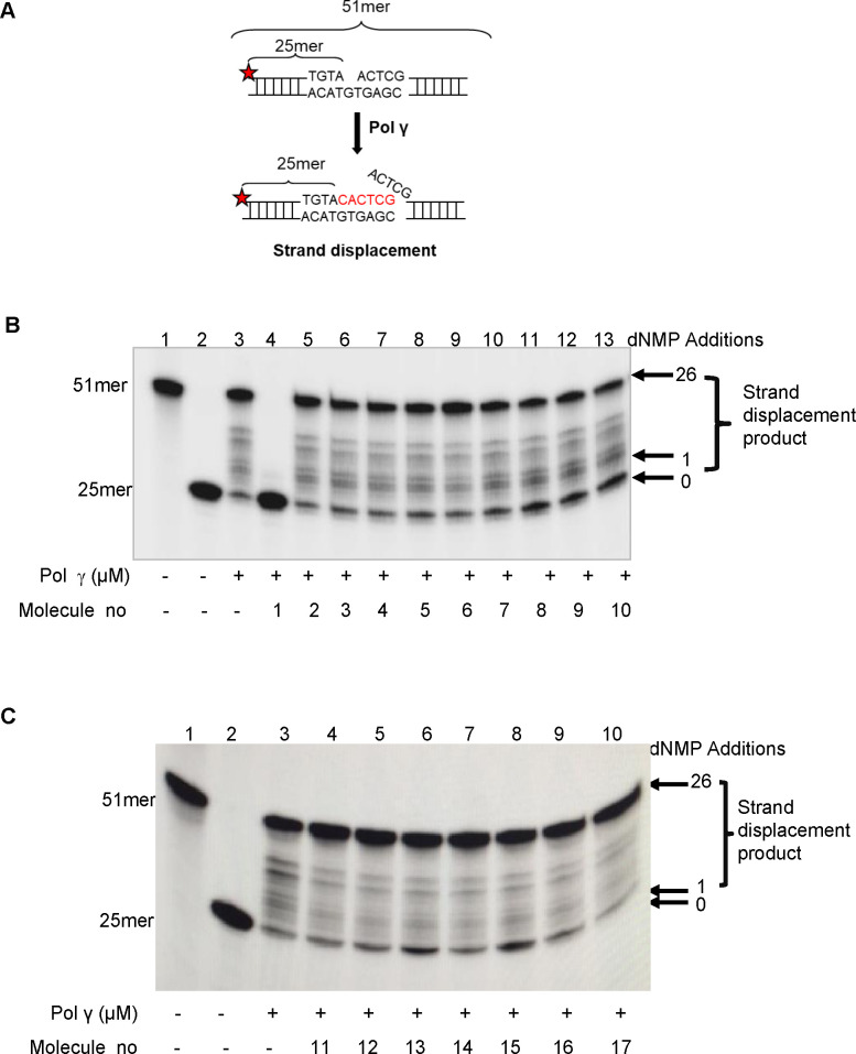
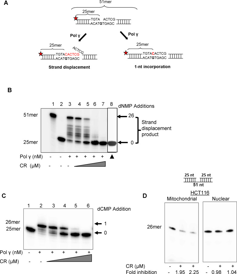
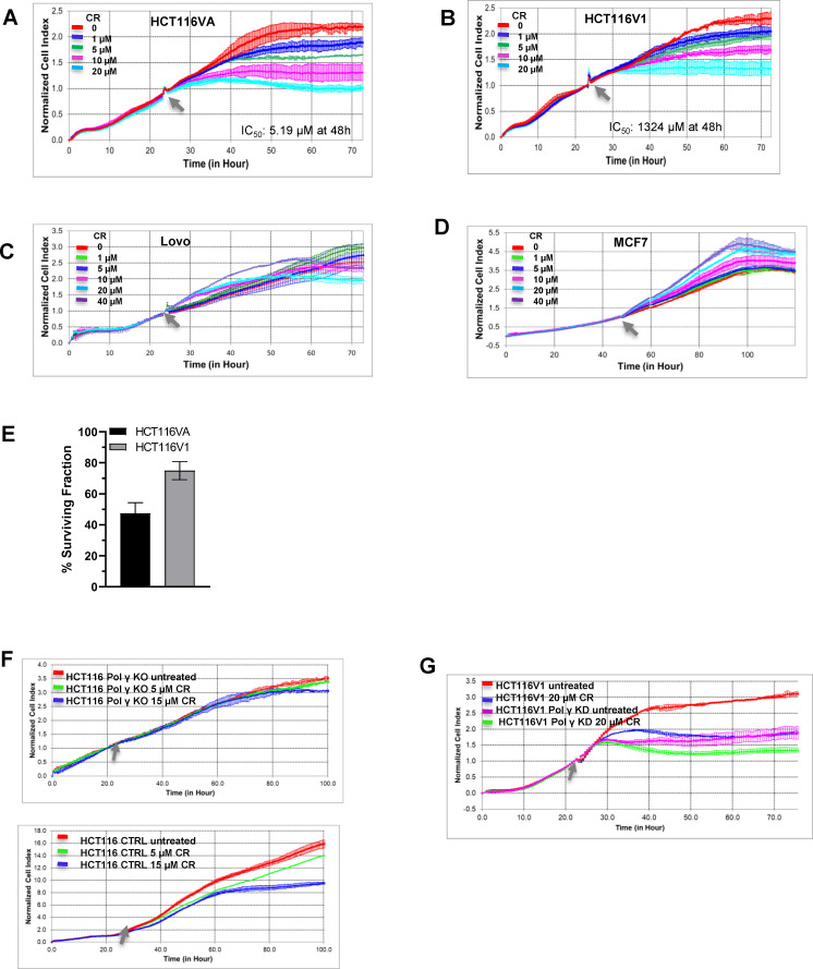
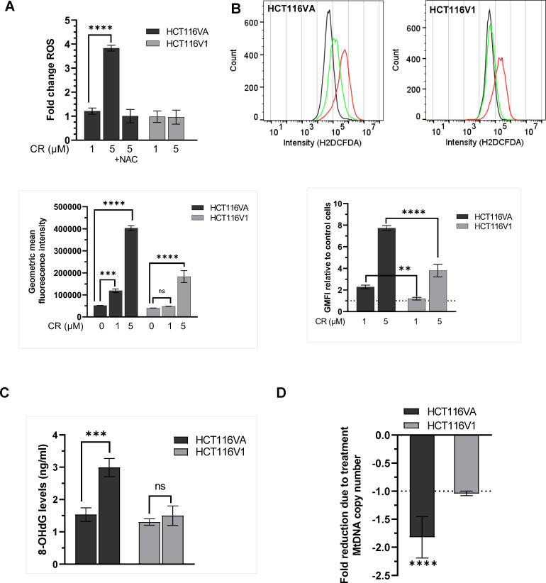
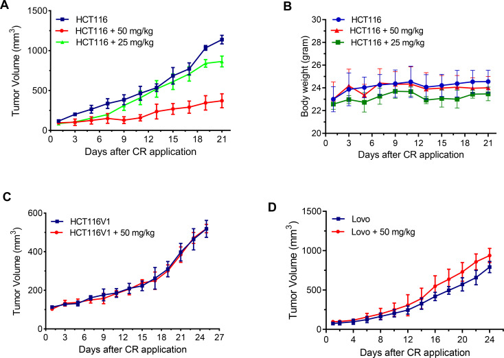
Synthetic lethality in DNA repair pathways is an important strategy for the selective treatment of cancer cells without harming healthy cells and developing cancer-specific drugs. The synthetic lethal interaction between the mismatch repair (MMR) protein, MutL homolog 1 (MLH1), and the mitochondrial base excision repair protein, DNA polymerase γ (Pol γ) was used in this study for the selective treatment of MLH1 deficient cancers. Germline mutations in the MLH1 gene and aberrant MLH1 promoter methylation result in an increased risk of developing many cancers, including nonpolyposis colorectal and endometrial cancers. Because the inhibition of Pol γ in MLH1 deficient cancer cells provides the synthetic lethal selectivity, we conducted a comprehensive small molecule screening from various databases and chemical drug library molecules for novel Pol γ inhibitors that selectively kill MLH1 deficient cancer cells. We characterized these Pol γ inhibitor molecules in vitro and in vivo, and identified 3,3'-[(1,1'-Biphenyl)-4',4'-diyl)bis(azo)]bis[4-amino-1-naphthalenesulfonic acid] (congo red; CR; Zinc 03830554) as a high-affinity binder to the Pol γ protein and potent inhibitor of the Pol γ strand displacement and one-nucleotide incorporation DNA synthesis activities in vitro and in vivo. CR reduced the cell proliferation of MLH1 deficient HCT116 human colon cancer cells and suppressed HCT116 xenograft tumor growth whereas it did not affect the MLH1 proficient cell proliferation and xenograft tumor growth. CR caused mitochondrial dysfunction and cell death by inhibiting Pol γ activity and oxidative mtDNA damage repair, increasing the production of reactive oxygen species and oxidative mtDNA damage in MLH1 deficient cells. This study suggests that the Pol γ inhibitor, CR may be further evaluated for the MLH1 deficient cancers' therapy.


Conflict of interest statement

MM, BuE and BaE are co-inventors on a patent application (WO2020005171). The competing interest does not alter our adherence to PLOS ONE policies on sharing data and materials.

Figures

Fig 1
Fig 1. Screening of small molecules for direct binding to Pol γ by SPR technology.
(A-D) SPR binding kinetics of computationally predicted small molecules to Pol γ. Sensorgrams show the interaction between compounds and Pol γ at different concentrations and KD values. All data are presented as the means ± standard deviation (STD) of three independent experiments. The chemical structures of small molecules are given.
Fig 2
Fig 2. Screening the small molecules on the strand displacement DNA synthesis activity of Pol γ.
(A) Schematic of the 32P-labeled (red star) 51 bp DNA substrate containing 1-nt gap at position 26 is shown before and after Pol γ strand displacement activity. Reactions contained the substrate and Pol γ alone (40 nM; lane 3) or together with 20 μM of the small molecules. (B) Molecule no 1–10: CR, corilagin, carbamazepine, phenolphthalein, acemetacin, indomethacin, amikacin hydrate, chenodiol, econazole nitrate, and ketoconazole, respectively. (C) Molecule no 11–17: Nicardipine hydrochloride, miconazole, nialamide, nystatin, misoprostol, pimethixene maleate, and rifampicin, respectively. Lane 1, 51mer oligodeoxynucleotide; lane 2, substrate alone.
Fig 3
Fig 3. CR inhibits the DNA synthesis activity of purified Pol γ protein and that of mitochondrial cell extracts.
(A) Schematic of the 32P-labeled (red star) 51 bp DNA substrate containing 1-nt gap at position 26 is shown before and after Pol γ strand displacement and 1-nt incorporation activities (B) Strand displacement reactions contained Pol γ alone (40 nM; lane 3) or together with increasing concentrations of CR (lanes 4–7; 0,5, 1.25, 2.5, and 5 μM). Lane 1, 51mer oligodeoxynucleotide; lane 2, substrate alone. ▲, 40 nM heat-denatured Pol γ protein (lane 8). (C) 1-nt gap incorporation reactions contained Pol γ alone (40 nM; lane 2) or together with increasing concentrations of CR (0,5, 1.25, 2.5, and 5 μM; lanes 3–6, respectively). Lane 1, substrate alone. (D) Single nucleotide DNA synthesis activity in mitochondrial and nuclear extracts treated with/without CR. Treated samples were run in duplicate.
Fig 4
Fig 4. The effects of Pol γ inhibitor molecules on MLH1 deficient and proficient cancer cell proliferation.
Real-time dynamic monitoring of the cytotoxic effects of CR on (A) HCT116VA (MLH1 deficient) (B) HCT116V1 (MLH1 proficient) (C) Lovo (MLH1 proficient) (D) MCF7 (MLH1 proficient) cells using the xCELLigence system. Cell growth was continuously monitored every 30 min. Cell index was normalized to the time point of CR administration. Normalized cell index was plotted as the mean value from triplicates; error bars represent the standard deviation of the mean. The Grey arrow indicates the time of CR administration. The xCELLigence RTCA software was used to determine IC50 values at 48h post-treatment time point. (E) Effect of 5 μM CR (IC50 = 5.19 μM) on colony formation in HCT116VA and HCT116V1 cells. Data are expressed as the mean ± standard deviation of three independent experiments. (F) Upper panel, Real-time dynamic monitoring of the cytotoxic effects of CR on CRISPR/Cas9 Pol γ knockout HCT116 cells and lower panel HCT116 control cells (empty vector control). (G) Real-time dynamic monitoring of the cytotoxic effects of CR on shRNA Pol γ knockdown HCT116V1 cells and HCT116V1 control cells.
Fig 5
Fig 5. CR increases ROS and decreases the mtDNA copy number in MLH1 deficient cells.
(A) ROS measured using the H2DCFDA assay in HCT116VA and HCT116V1 cells treated with CR for 48h and normalized to the corresponding viable cells. Data are shown as mean fold change to untreated control ± STD. (B) Flow cytometry measurement of ROS levels by H2DCFDA fluorescence assay. Plots of the fluorescence intensities of the DCF dye in cells exposed to 1 μM CR (green color), 5 μM CR (red color), or untreated cells (black color) for 1 h. Data are given as the ratio of geometric mean fluorescence intensity (GMFI) and GMFI of treated relative to untreated cells (control). (C) The quantification of 8-OHdG levels in HCT116VA and HCT116V1 cells upon treatment of CR. (D) MtDNA copy number. The fold change in mtDNA copy number due to treatment was calculated by 2-ΔΔCt method. Each experiment was performed in triplicate. **p = 0.002; ***p = 0.0003; ****p< 0.0001; ns, not significant.
Fig 6
Fig 6. CR suppresses the growth of HCT116 xenograft tumors.
(A) Growth curves of HCT116 xenograft tumors (n = 6, tumor volume mm3) treated with CR and 0.02% DMSO (n = 6, control). p < 0.01, 50 mg/kg compared with control group; p˃0.05, 25 mg/kg compared with control group. (B) The corresponding body weight changes during the treatment. p˃0.05, compared with a control group. (C) Growth curves of HCT116V1 xenograft tumors (n = 3, tumor volume mm3) treated with CR and 0.02% DMSO (n = 3, control). p˃0.05, compared with a control group. (D) Growth curves of Lovo xenograft tumors (n = 5, tumor volume mm3) treated with CR and 0.02% DMSO (n = 5, control). p˃0.05, compared with a control group.

References

    1. Klinakis A, Karagiannis D, Rampias T. Targeting DNA repair in cancer: current state and novel approaches. Cell Mol Life Sci. 2020;77(4):677–703. Epub 2019/10/16. doi: 10.1007/s00018-019-03299-8 . - DOI - PMC - PubMed
    1. Nickoloff JA, Jones D, Lee SH, Williamson EA, Hromas R. Drugging the Cancers Addicted to DNA Repair. J Natl Cancer Inst. 2017;109(11). Epub 2017/05/19. doi: 10.1093/jnci/djx059 ; PubMed Central PMCID: PMC5436301. - DOI - PMC - PubMed
    1. Martin SA, McCabe N, Mullarkey M, Cummins R, Burgess DJ, Nakabeppu Y, et al.. DNA polymerases as potential therapeutic targets for cancers deficient in the DNA mismatch repair proteins MSH2 or MLH1. Cancer Cell. 2010;17(3):235–48. Epub 2010/03/17. doi: 10.1016/j.ccr.2009.12.046 ; PubMed Central PMCID: PMC2845806. - DOI - PMC - PubMed
    1. de Souza-Pinto NC, Mason PA, Hashiguchi K, Weissman L, Tian J, Guay D, et al.. Novel DNA mismatch-repair activity involving YB-1 in human mitochondria. DNA Repair (Amst). 2009;8(6):704–19. Epub 2009/03/11. doi: 10.1016/j.dnarep.2009.01.021 ; PubMed Central PMCID: PMC2693314. - DOI - PMC - PubMed
    1. Alencar RR, Batalha C, Freire TS, de Souza-Pinto NC. Enzymology of mitochondrial DNA repair. Enzymes. 2019;45:257–87. Epub 2019/10/20. doi: 10.1016/bs.enz.2019.06.002 . - DOI - PubMed

Publication types

MeSH terms