. 2022 Jun 7;119(23):e2200794119.
doi: 10.1073/pnas.2200794119. Epub 2022 Jun 3.

Atomic view of the HIV-1 matrix lattice; implications on virus assembly and envelope incorporation

Affiliations

Atomic view of the HIV-1 matrix lattice; implications on virus assembly and envelope incorporation

Alexandra B Samal et al. Proc Natl Acad Sci U S A. .

Abstract

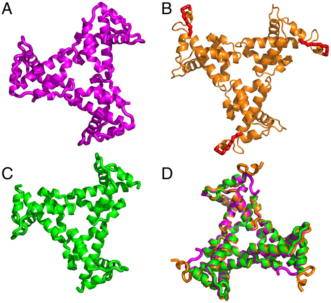
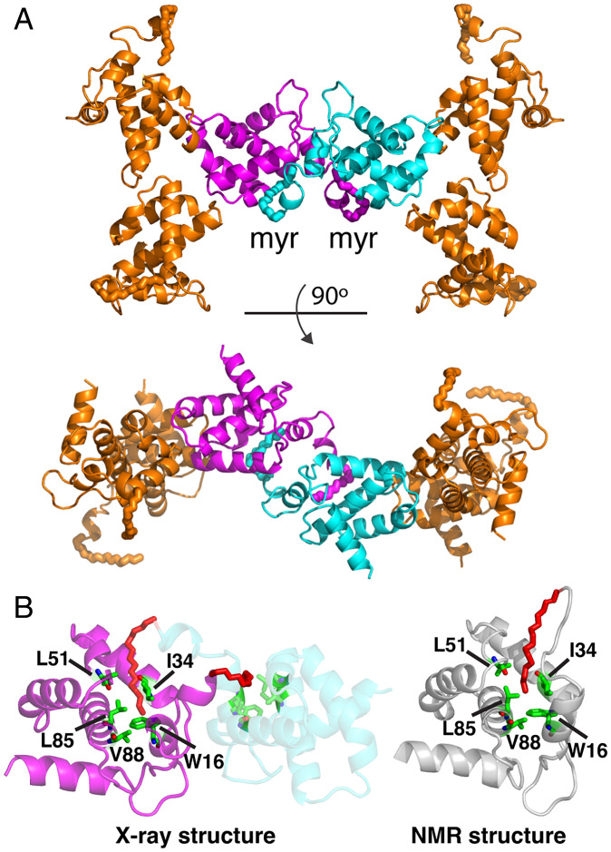
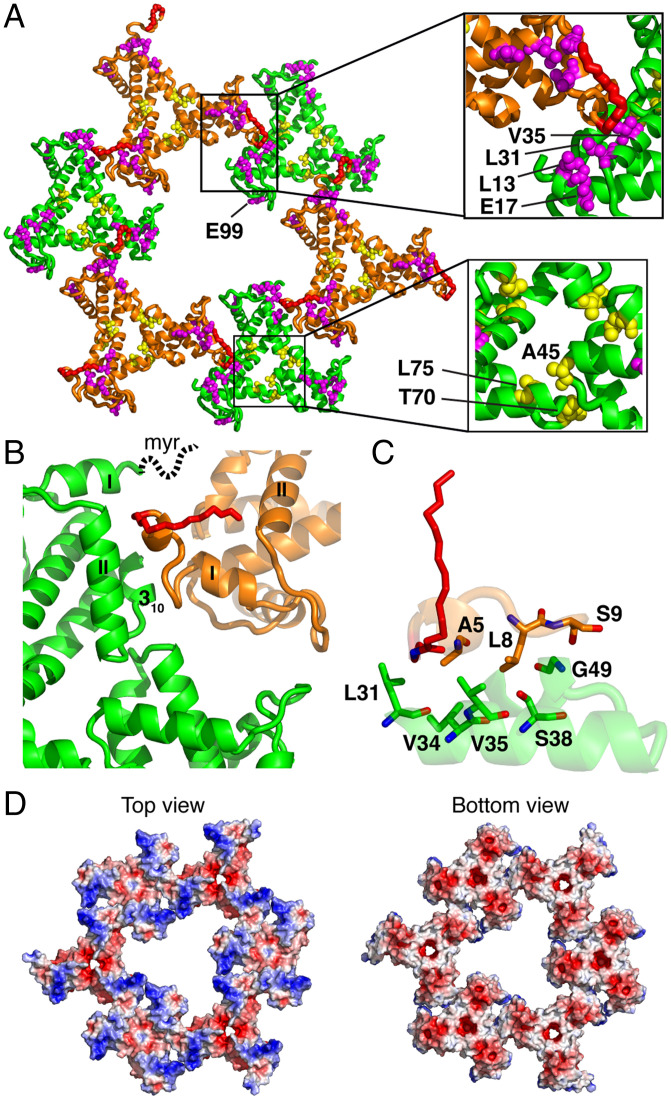
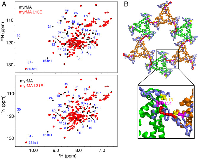
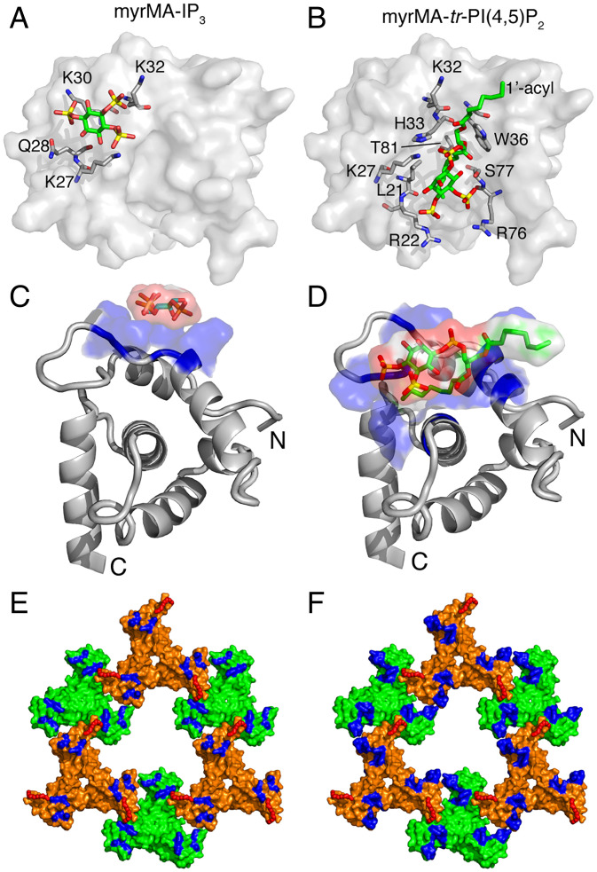
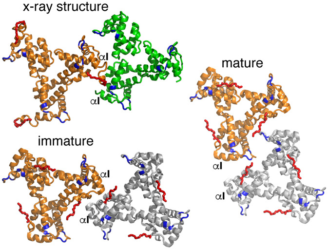
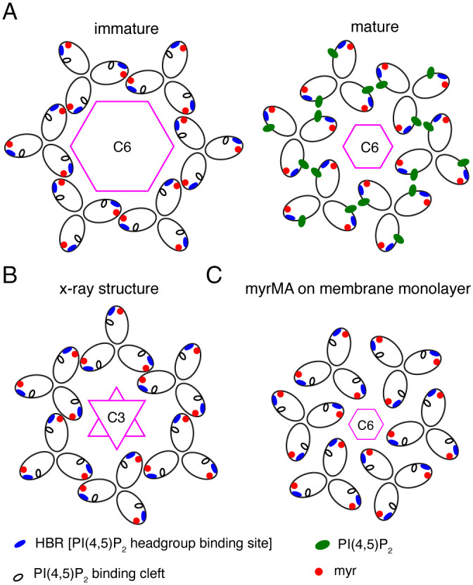
During the late phase of HIV type 1 (HIV-1) infection cycle, the virally encoded Gag polyproteins are targeted to the inner leaflet of the plasma membrane (PM) for assembly, formation of immature particles, and virus release. Gag binding to the PM is mediated by interactions of the N-terminally myristoylated matrix (myrMA) domain with phosphatidylinositol 4,5-bisphosphate. Formation of a myrMA lattice on the PM is an obligatory step for the assembly of immature HIV-1 particles and envelope (Env) incorporation. Atomic details of the myrMA lattice and how it mediates Env incorporation are lacking. Herein, we present the X-ray structure of myrMA at 2.15 Å. The myrMA lattice is arranged as a hexamer of trimers with a central hole, thought to accommodate the C-terminal tail of Env to promote incorporation into virions. The trimer–trimer interactions in the lattice are mediated by the N-terminal loop of one myrMA molecule and α-helices I–II, as well as the 310 helix of a myrMA molecule from an adjacent trimer. We provide evidence that substitution of MA residues Leu13 and Leu31, previously shown to have adverse effects on Env incorporation, induced a conformational change in myrMA, which may destabilize the trimer–trimer interactions within the lattice. We also show that PI(4,5)P2 is capable of binding to alternating sites on MA, consistent with an MA–membrane binding mechanism during assembly of the immature particle and upon maturation. Altogether, these findings advance our understanding of a key mechanism in HIV-1 particle assembly.

Keywords: Gag; HIV-1; envelope; matrix; retrovirus.


Conflict of interest statement

The authors declare no competing interest.

Figures

Fig. 1.
Fig. 1.
X-ray structure of HIV-1 myrMA112. Three independent monomeric chains (a, b, and c) of MA112 were found in the asymmetric unit of the crystal. Trimeric assemblies are shown of chain a (A), chain b (B), and chain c (C). These assembled units are generated by imposing crystallographic symmetry. In B, the complete N terminus of myrMA112 is shown with attached myr group (red) and linkage to the N-terminal glycine shown in stick model. For chain b, well-defined electron density was observed for the entire N terminus (SI Appendix, Fig. S1). For chains a and c, electron density for the myr group as well as residues 2–5 and 2–9, respectively, was not observed presumably due to flexibility of the N terminus. (D) Overlay of the three trimers showing that the structures of the MA molecules and the trimer arrangements are nearly identical.
Fig. 2.
Fig. 2.
Myristoyl swap. (A) Cartoon representation of myrMA112 chain b showing the dimeric interface formed by two trimers through a myr swapping mode (cyan and magenta). The two trimers are aligned in an antiparallel mode. (B) Cartoon representation showing two myrMA112 molecules (cyan and magenta) in which the myr group (red) is sequestered in a preexisting hydrophobic cavity of an adjacent molecule. For comparison, the NMR structure of the monomeric myrMA protein (gray; PDB ID code 2H3I) is shown with the myr group buried in an identical hydrophobic cavity formed by the side chains of residues Trp16, Ile34, Leu51, Leu85, and Val88.
Fig. 3.
Fig. 3.
Hexamer of trimer lattice formation. (A) Hexamer of trimer is formed by trimers b and c. In this lattice, the myr group (red) is extruded and projecting out-of-plane. All six trimers are in a parallel mode with a threefold rotation axis. Residues implicated in Env incorporation (Glu17, Leu13, Leu31, and Val35; magenta) are located in the trimer–trimer interface. Glu99, which was also shown to impact Env incorporation, is located in the interior of the central hole of the hexamer lattice. Other residues that have been shown to impact Env incorporation by stabilizing the myrMA trimer are shown as yellow spheres. (B) Trimer–trimer interactions are mediated by the N-terminal residues of one molecule (orange) and α-helices I–II and 310 helix of an adjacent molecule (green). The myr group in chain c, which is not observed due to its dynamic nature, is drawn as a dotted line. (C) A close-up view of the residues involved in the trimer–trimer interface. (D) Electrostatic surface potential maps of the myrMA112 lattice. The blue (+5 kT/e) (e, electron charge; k, Boltzmann constant; T, temperature) and red (−5 kT/e) colors indicate positively and negatively charged electric potentials, respectively.
Fig. 4.
Fig. 4.
Structural analysis of myrMA mutants that impair Env incorporation. (A) Overlay of 2D 1H–15N HSQC spectra for WT (black) and mutant L13E (Top) and L31E (Bottom) myrMA proteins collected at 120 μM (red). Substantial CSPs or severe broadening of signals corresponding to residues 2 to 40 are observed in the NMR spectra of myrMA L13E and L31E mutants when compared with the spectrum of the WT myrMA protein. (B) CSPs induced by the L13E and L31 mutants are mapped to the N-terminal loop and α-helices I–II of myrMA (slate). These CSPs are suggestive of a conformational change in the packing of α-helices, which also altered the position of the myr group (red) and ultimately lattice formation.
Fig. 5.
Fig. 5.
Alternate PI(4,5)P2–binding mechanism. (A and C) Surface and cartoon representation, respectively, of the HIV-1 myrMA structure (PDB ID code 2H3I) highlighting basic residues that exhibited substantial chemical-shift changes upon binding of IP3. The IP3 molecule was modeled on the myrMA structure using PyMOL. (B and D) Surface and cartoon representation, respectively, of the HIV-1 myrMA structure bound to tr-P(4,5)P2 (PDB ID code 2H3V). Residues involved in tr-P(4,5)P2 binding are shown in sticks. Structures in A and B, and those in C and D, are viewed in identical orientations. The myr group is not shown in AD. (E and F) Surface representation of the myrMA112 hexamer of trimer lattice highlighting residues that exhibited CSPs upon binding of IP3 and tr-PI(4,5)P2, respectively. The myr groups are shown as red sticks in E and F.
Fig. 6.
Fig. 6.
Comparison of trimer–trimer contacts in the lattice. Cartoon illustration of the HIV-1 myrMA trimer–trimer units obtained by the X-ray structure and those reconstructed from the cryotomography data of the immature (PDB ID code 7OVQ) and mature (PDB ID code 7OVR) particles. The orange trimers are in an identical orientation. As shown, the trimer–trimer relationship and interface in the X-ray structure is relatively similar to that of the immature particle. The HBR and myr groups are shown in blue and red, respectively.
Fig. 7.
Fig. 7.
Comparison of myrMA lattices. (A) Illustration of the myrMA lattice for the immature and mature particles based on the cryotomography data (36). The trimer–trimer interactions are mediated by the N-terminal domain in the vicinity of the myr group, while the PI(4,5)P2 binding pocket is empty. In the mature myrMA lattice, PI(4,5)P2 is bound to the cleft and myrMA trimer–trimer interactions are formed by the HBR and PI(4,5)P2. (B) Schematic illustration of the myrMA lattice based on the X-ray structure of myrMA112. In this lattice, myrMA–myrMA interaction at the trimer–trimer interface is mediated by the N-terminal residues. Of note, myrMA–myrMA interaction at the trimer–trimer interface places the myr groups (red) in juxtaposition. The HBR and PI(4,5)P2 binding cleft are also shown. (C) A schematic representation of the myrMA lattice based on the assembly of the protein on a membrane monolayer containing PI(4,5)P2 (23).

References

    1. Freed E. O., HIV-1 assembly, release and maturation. Nat. Rev. Microbiol. 13, 484–496 (2015). - PMC - PubMed
    1. Murphy R. E., Saad J. S., The interplay between HIV-1 Gag binding to the plasma membrane and Env incorporation. Viruses 12, 548 (2020). - PMC - PubMed
    1. Mücksch F., Laketa V., Müller B., Schultz C., Kräusslich H. G., Synchronized HIV assembly by tunable PIP2 changes reveals PIP2 requirement for stable Gag anchoring. eLife 6, e25287 (2017). - PMC - PubMed
    1. Hendrix J., et al. , Live-cell observation of cytosolic HIV-1 assembly onset reveals RNA-interacting Gag oligomers. J. Cell Biol. 210, 629–646 (2015). - PMC - PubMed
    1. Gousset K., et al. , Real-time visualization of HIV-1 GAG trafficking in infected macrophages. PLoS Pathog. 4, e1000015 (2008). - PMC - PubMed

Publication types

MeSH terms

Substances