. 2022 Jun 3;13(1):222.
doi: 10.1186/s13287-022-02901-4.

Clearance of senescent cells with ABT-263 improves biological functions of synovial mesenchymal stem cells from osteoarthritis patients

Affiliations

Clearance of senescent cells with ABT-263 improves biological functions of synovial mesenchymal stem cells from osteoarthritis patients

Yugo Miura et al. Stem Cell Res Ther. .

Abstract

Background: Osteoarthritis (OA) is an age-related joint disease characterized by progressive cartilage loss. Synovial mesenchymal stem cells (MSCs) are anticipated as a cell source for OA treatment; however, synovial MSC preparations isolated from OA patients contain many senescent cells that inhibit cartilage regeneration through their senescence-associated secretory phenotype (SASP) and poor chondrogenic capacity. The aim of this study was to improve the biological function of OA synovial MSCs by removing senescent cells using the senolytic drug ABT-263.

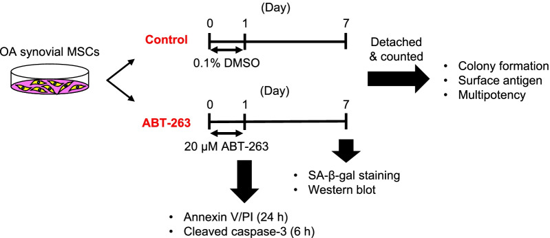
Methods: We pretreated synovial MSCs derived from 5 OA patients with ABT-263 for 24 h and then evaluated senescence-associated beta-galactosidase (SA-β-gal) activity, B cell lymphoma 2 (BCL-2) activity, apoptosis, surface antigen expression, colony formation ability, and multipotency.

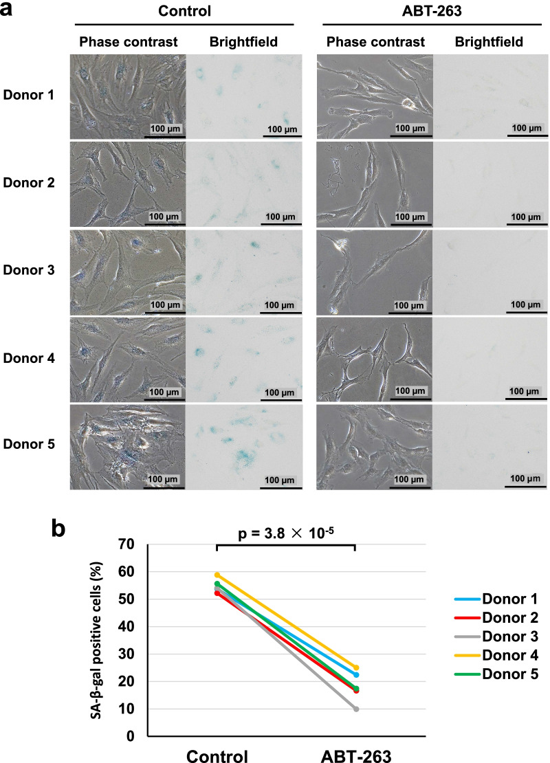
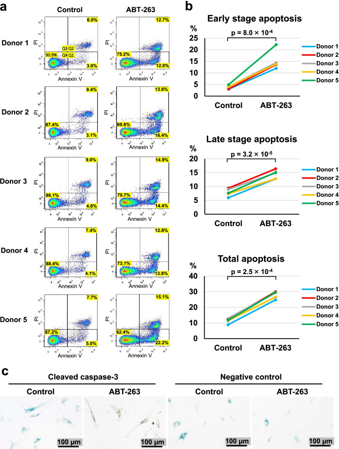
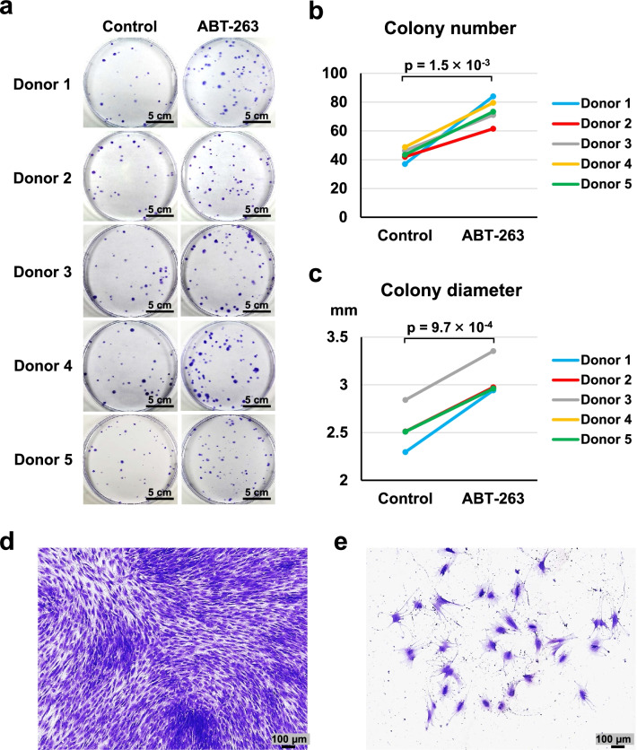
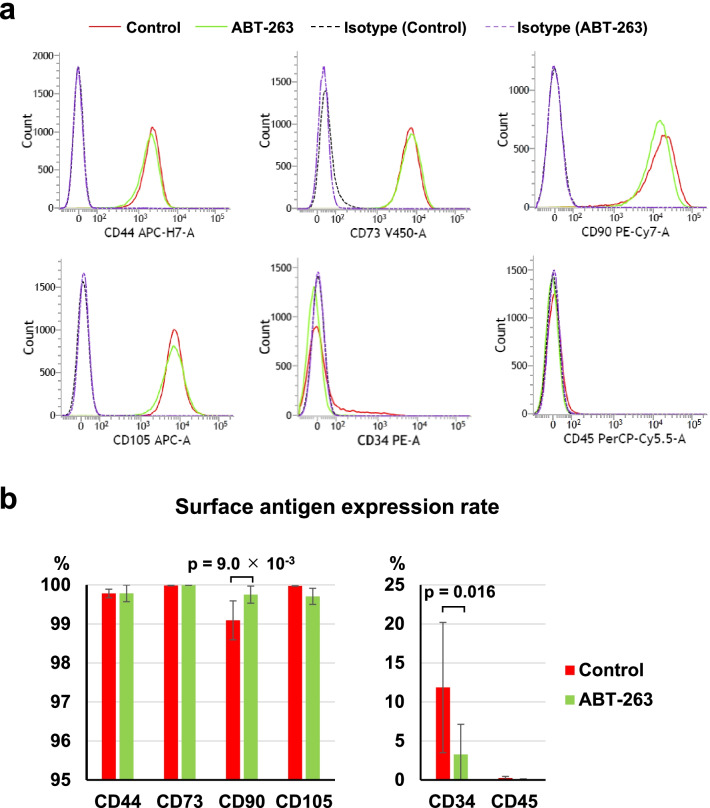
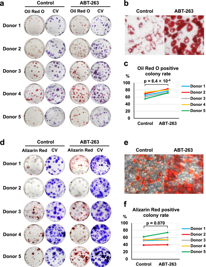
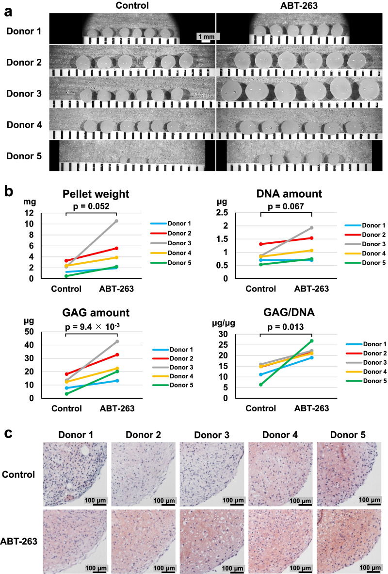
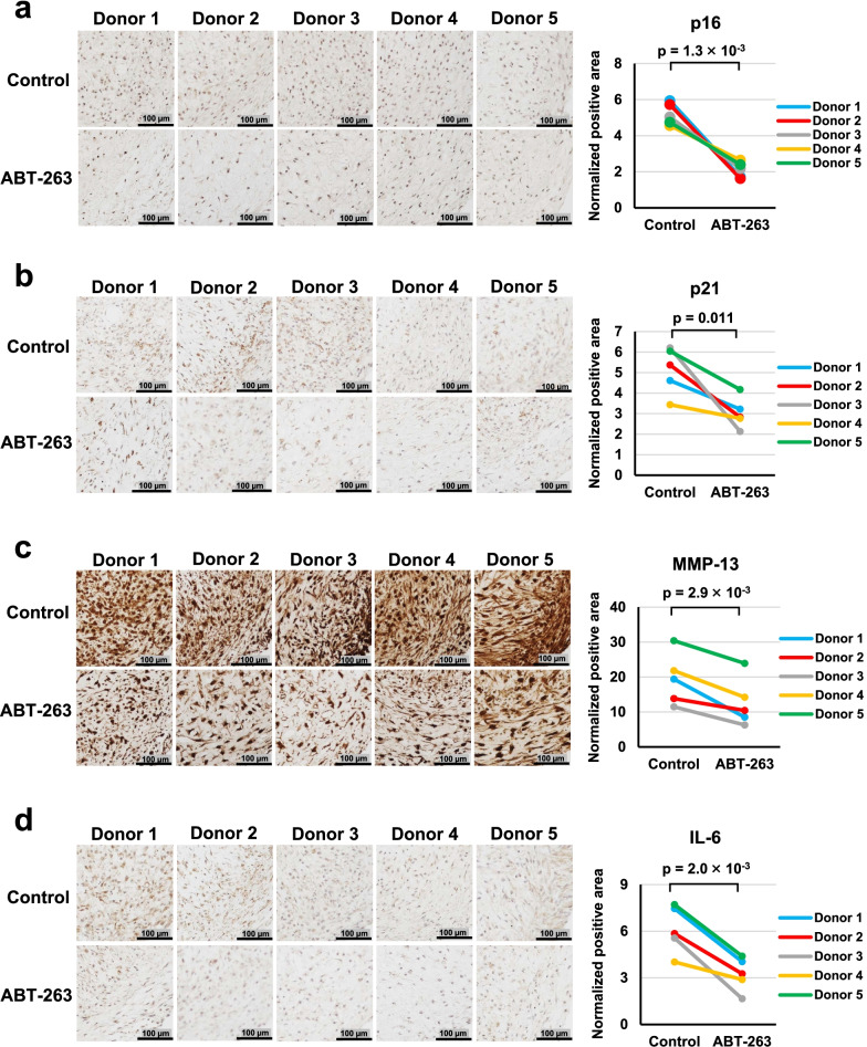
Results: The ABT-263 pretreatment significantly decreased the percentage of SA-β-gal-positive cells and BCL-2 expression and induced early- and late-stage apoptosis. Cleaved caspase-3 was expressed in SA-β-gal-positive cells. The pretreated MSCs formed greater numbers of colonies with larger diameters. The expression rate of CD34 was decreased in the pretreated cells. Differentiation assays revealed that ABT-263 pretreatment enhanced the adipogenic and chondrogenic capabilities of OA synovial MSCs. In chondrogenesis, the pretreated cells produced greater amounts of glycosaminoglycan and type II collagen and showed lower expression of senescence markers (p16 and p21) and SASP factors (MMP-13 and IL-6) and smaller amounts of type I collagen.

Conclusion: Pretreatment of synovial MSCs from OA patients with ABT-263 can improve the function of the cells by selectively eliminating senescent cells. These findings indicate that ABT-263 could hold promise for the development of effective cell-based OA therapy.

Keywords: ABT-263; Mesenchymal stem cells; Osteoarthritis; Senescence; Senolytic drug; Synovium.


Conflict of interest statement

The authors declare that they have no competing interests.

Figures

Fig. 1
Fig. 1
Scheme of the experiments. Synovial mesenchymal stem cells (MSCs) from osteoarthritis (OA) patients were treated with 0.1% dimethyl sulfoxide (DMSO) or 20 µM ABT-263 in DMSO for 24 h. After the treatment, the cells were expanded for another 6 days and used for the experiments. Annexin V/propidium iodide (PI) staining and cleaved caspase-3 immunostaining were performed immediately after the treatment for 24 and 6 h, respectively
Fig. 2
Fig. 2
Senescence-associated beta-galactosidase (SA-β-gal) staining. a Phase contrast and brightfield images. b The ratio of SA-β-gal-positive cells in control and ABT-263 group
Fig. 3
Fig. 3
Apoptosis assay. a Flow cytometry analysis of annexin V/propidium iodide (PI). Dot plots show the percentages of viable (Q4), early apoptotic (Q1), and late apoptotic (Q2) cells. b The ratio of early, late, and total apoptotic cells in the control and ABT-263 groups. c Co-staining of senescence-associated beta-galactosidase (blue) and cleaved caspase-3 (brown) after the treatment with ABT-263
Fig. 4
Fig. 4
Colony-forming ability. a Representative images of colonies stained with crystal violet (4 replicate dishes per donor). b Colony numbers per dish in the control and ABT-263 groups. c Diameter of colonies in the control and ABT-263 groups. d Representative image of colony-forming cells. e Representative image of flattened and enlarged cells that did not form colonies
Fig. 5
Fig. 5
Surface antigen expression. a Representative flow cytometry histograms. Red solid lines indicate the control group, and green solid lines indicate the ABT-263 group. Dotted lines show the isotype control. b The expression rate of each surface antigen. Values are shown as mean ± SD from 5 donors
Fig. 6
Fig. 6
Adipogenic and osteogenic differentiation. a Representative images of Oil Red O and crystal violet (CV) staining (3 replicate dishes per donor). b Reperesentative microscopic images of Oil Red O staining. c The percentage of Oil Red O-stained colonies in the control and ABT-263 groups. d Representative images of Alizarin Red and CV staining (3 replicate dishes per donor). e Reperesentative microscopic images of Alizarin Red staining. f The percentage of Alizarin Red-stained colonies in the control and ABT-263 groups
Fig. 7
Fig. 7
Chondrogenic differentiation. a Gross appearance of chondrogenic pellets (5 or 6 replicate pellets per donor). b Pellet wet weight, DNA quantification, glycosaminoglycan (GAG) quantification, and GAG/DNA ratio (3 or 4 replicate pellets per donor). c Representative images of Safranin O staining of chondrogenic pellets
Fig. 8
Fig. 8
Immunohistochemistry of senescence markers in chondrogenic pellets. Representative images and positive ratios of a p16, b p21, c MMP-13, and d IL-6 immunostaining in the control and ABT-263 groups

References

    1. Jeon OH, David N, Campisi J, Elisseeff JH. Senescent cells and osteoarthritis: a painful connection. J Clin Invest. 2018;128(4):1229–1237. doi: 10.1172/JCI95147. - DOI - PMC - PubMed
    1. Sellam J, Berenbaum F. The role of synovitis in pathophysiology and clinical symptoms of osteoarthritis. Nat Rev Rheumatol. 2010;6(11):625–635. doi: 10.1038/nrrheum.2010.159. - DOI - PubMed
    1. Taruc-Uy RL, Lynch SA. Diagnosis and treatment of osteoarthritis. Prim Care. 2013;40(4):821–836. doi: 10.1016/j.pop.2013.08.003. - DOI - PubMed
    1. Bianco P, Robey PG, Simmons PJ. Mesenchymal stem cells: revisiting history, concepts, and assays. Cell Stem Cell. 2008;2(4):313–319. doi: 10.1016/j.stem.2008.03.002. - DOI - PMC - PubMed
    1. Barry F. MSC therapy for osteoarthritis: an unfinished story. J Orthop Res. 2019;37(6):1229–1235. doi: 10.1002/jor.24343. - DOI - PubMed

Publication types