A clinical trial of super-stable homogeneous lipiodol-nanoICG formulation-guided precise fluorescent laparoscopic hepatocellular carcinoma resection
- PMID: 35658966
- PMCID: PMC9164554
- DOI: 10.1186/s12951-022-01467-w
A clinical trial of super-stable homogeneous lipiodol-nanoICG formulation-guided precise fluorescent laparoscopic hepatocellular carcinoma resection
Abstract
Background: Applying traditional fluorescence navigation technologies in hepatocellular carcinoma is severely restricted by high false-positive rates, variable tumor differentiation, and unstable fluorescence performance.
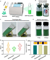
Results: In this study, a green, economical and safe nanomedicine formulation technology was developed to construct carrier-free indocyanine green nanoparticles (nanoICG) with a small uniform size and better fluorescent properties without any molecular structure changes compared to the ICG molecule. Subsequently, nanoICG dispersed into lipiodol via a super-stable homogeneous intermixed formulation technology (SHIFT&nanoICG) for transhepatic arterial embolization combined with fluorescent laparoscopic hepatectomy to eliminate the existing shortcomings. A 52-year-old liver cancer patient was recruited for the clinical trial of SHIFT&nanoICG. We demonstrate that SHIFT&nanoICG could accurately identify and mark the lesion with excellent stability, embolism, optical imaging performance, and higher tumor-to-normal tissue ratio, especially in the detection of the microsatellite lesions (0.4 × 0.3 cm), which could not be detected by preoperative imaging, to realize a complete resection of hepatocellular carcinoma under fluorescence laparoscopy in a shorter period (within 2 h) and with less intraoperative blood loss (50 mL).
Conclusions: This simple and effective strategy integrates the diagnosis and treatment of hepatocellular carcinoma, and thus, it has great potential in various clinical applications.
Keywords: Fluorescent laparoscope; Hepatectomy; Indocyanine green nanoparticles; Theranostics; Translational medicine.
© 2022. The Author(s).
Conflict of interest statement
The authors declare that they have no competing interests.
Figures


References
-
- Zhou M, Wang H, Zeng X, Yin P, Zhu J, Chen W, Li X, Wang L, Wang L, Liu Y, Liu J, Zhang M, Qi J, Yu S, Afshin A, Gakidou E, Glenn S, Krish VS, Miller-Petrie MK, Mountjoy-Venning WC, Mullany EC, Redford SB, Liu H, Naghavi M, Hay SI, Wang L, Murray CJL, Liang X. Mortality, morbidity, and risk factors in China and its provinces, 1990–2017: A systematic analysis for the global burden of disease study 2017. The Lancet. 2019;394(10204):1145–1158. doi: 10.1016/S0140-6736(19)30427-1. - DOI - PMC - PubMed
-
- Urano Y, Sakabe M, Kosaka N, Ogawa M, Mitsunaga M, Asanuma D, Kamiya M, Young MR, Nagano T, Choyke PL, Kobayashi H. Rapid cancer detection by topically spraying a γ-glutamyltranspeptidase-activated fluorescent probe. Sci Transl Med. 2011;3(110):110ra119. doi: 10.1126/scitranslmed.3002823. - DOI - PMC - PubMed
MeSH terms
Substances
Grants and funding
- HYX20003/Nuclear Medicine and Molecular Imaging Key Laboratory of Sichuan province open project
- 2020S02/Chengdu Gaoxin Medical Association 2020 Annual Cancer Intervention Special Research Fund
- 81925019, 81422023, 82003147, 81603015, 81871404, 82003147 and U1705281/National Natural Science Foundation of China
- 2017YFA0205201/Major State Basic Research Development Program of China
- 20720190088, 20720200019/Fundamental Research Funds for the Central Universities
LinkOut - more resources
Full Text Sources
Medical

