. 2022 Jun 3;19(1):129.
doi: 10.1186/s12974-022-02495-x.

DKK3 ameliorates neuropathic pain via inhibiting ASK-1/JNK/p-38-mediated microglia polarization and neuroinflammation

Affiliations

DKK3 ameliorates neuropathic pain via inhibiting ASK-1/JNK/p-38-mediated microglia polarization and neuroinflammation

Long-Qing Zhang et al. J Neuroinflammation. .

Abstract

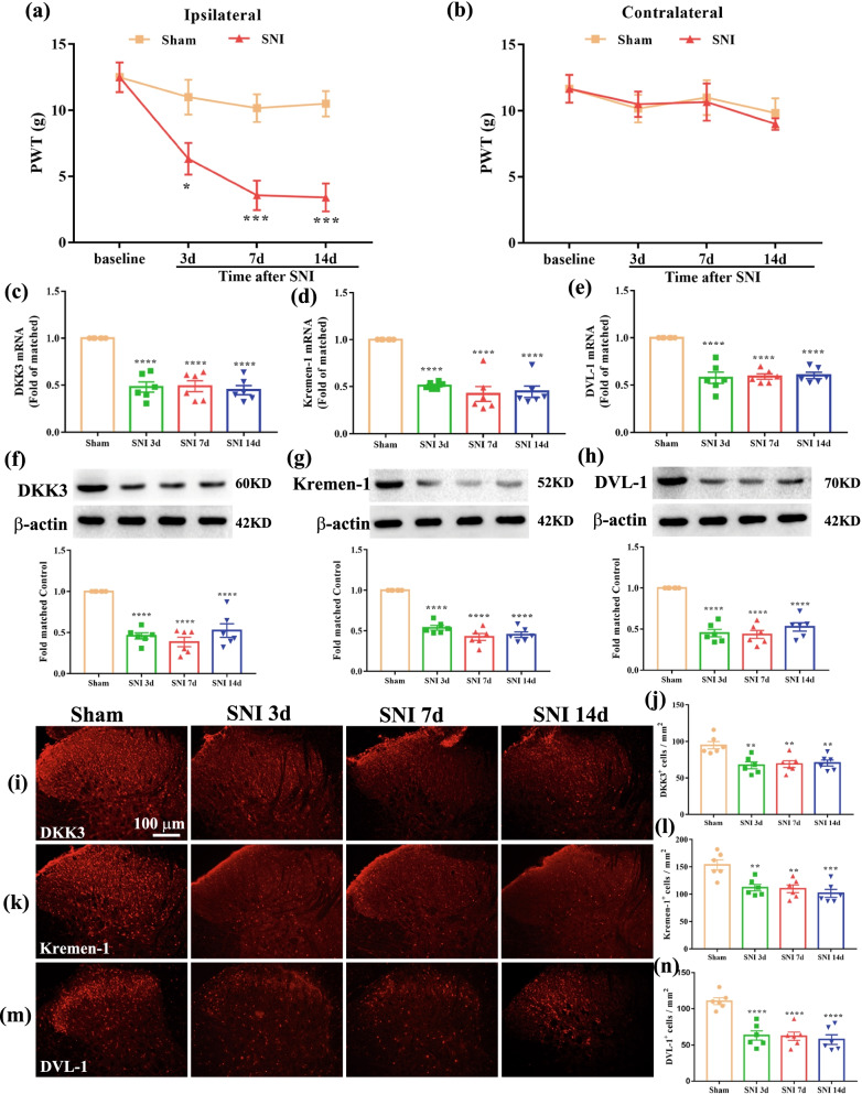
Background: Neuropathic pain is a common and severely disabling state that affects millions of people worldwide. Microglial activation in the spinal cord plays a critical role in the pathogenesis of neuropathic pain. However, the mechanisms underlying spinal microglial activation during neuropathic pain remain incompletely understood. Here, we investigated the role of Dickkopf (DKK) 3 and its interplay with microglial activation in the spinal cord in neuropathic pain.

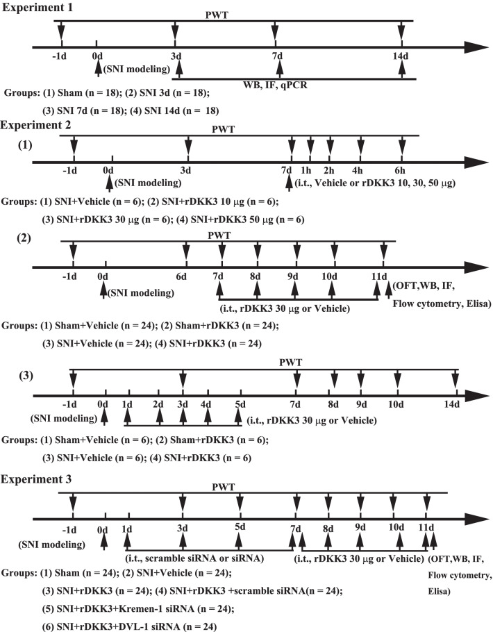
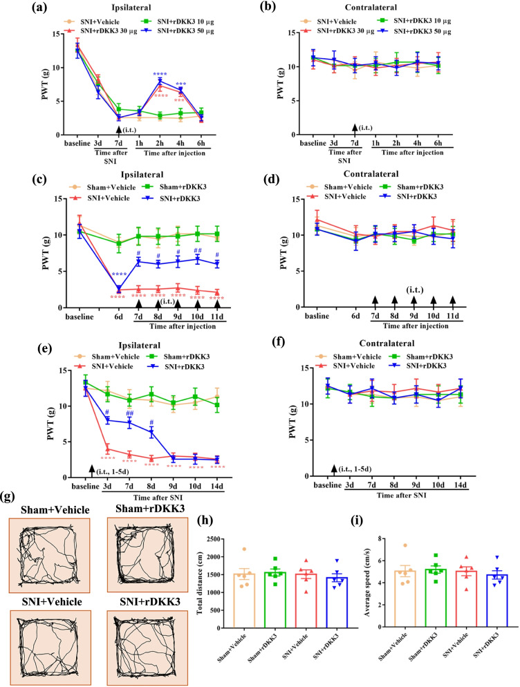
Methods: In this study, we investigated the effects of intrathecal injection of recombinant DKK3 (rDKK3) on mechanical allodynia and microglial activation in the spinal cord after spared nerve injury (SNI) in rats by western blot (WB), immunofluorescence (IF), quantitative polymerase chain reaction (qPCR), and enzyme-linked immunosorbent assay (ELISA).

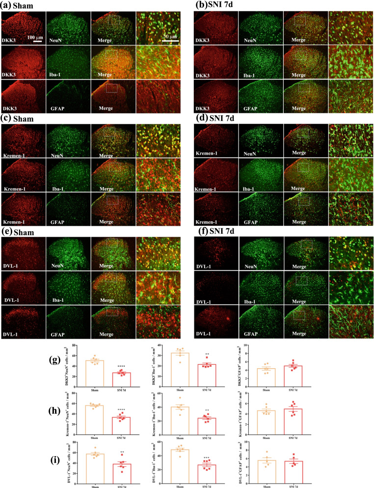
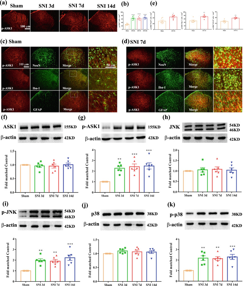
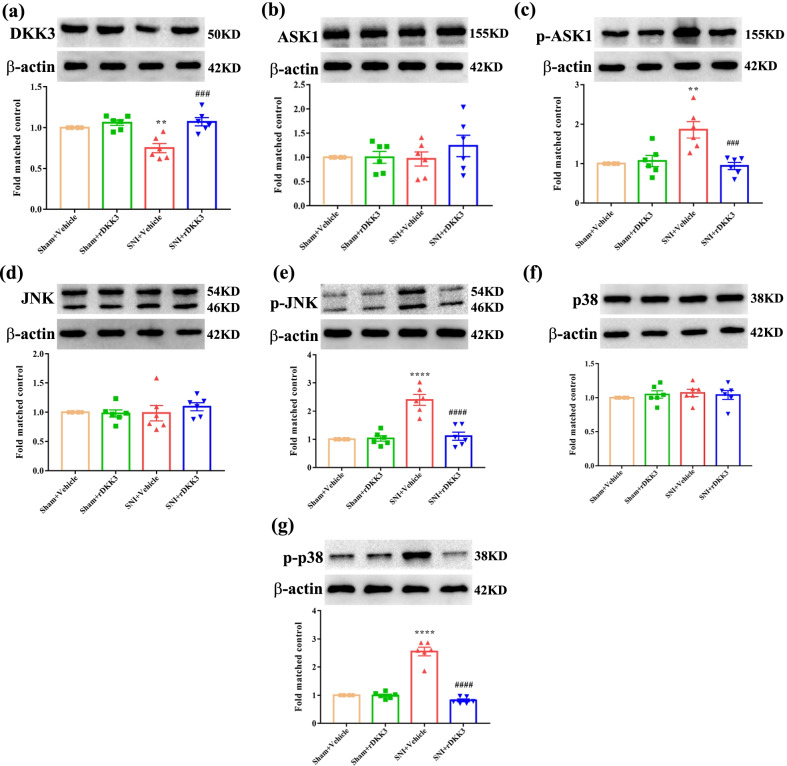
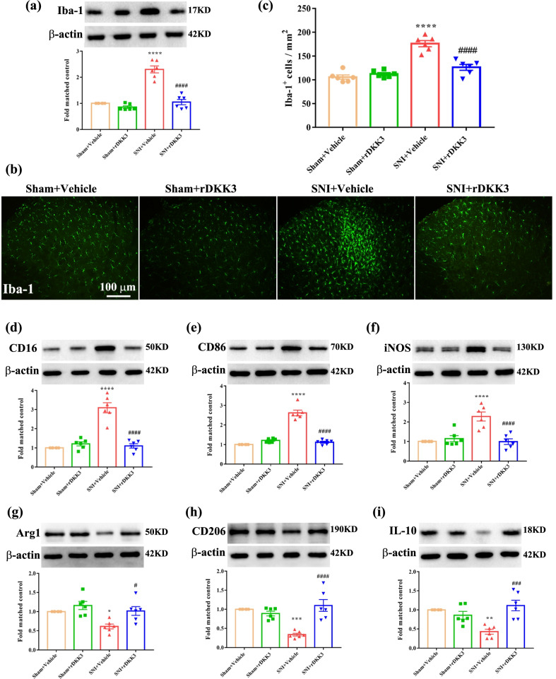
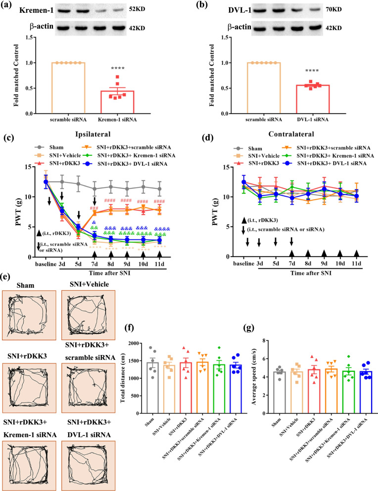
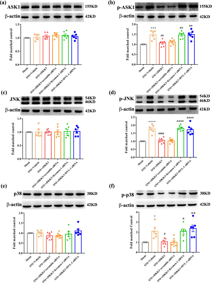
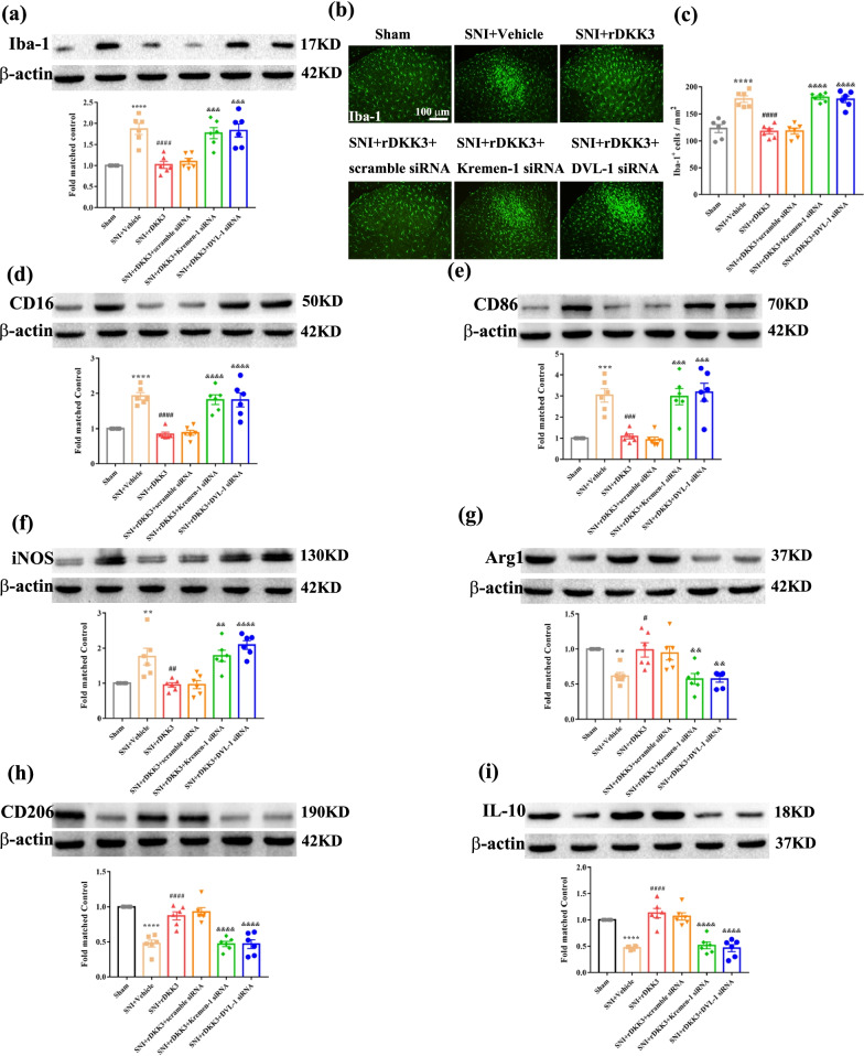
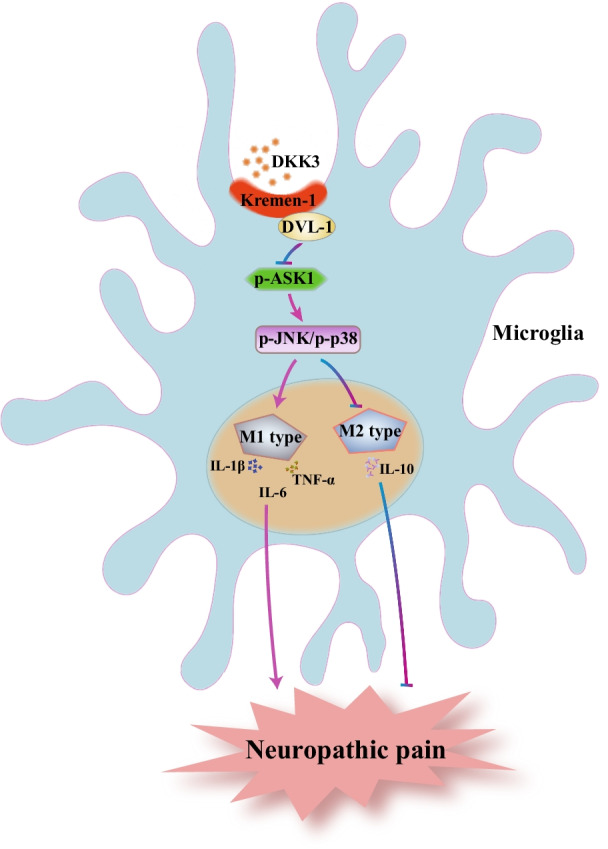
Results: We found that SNI induced a significant decrease in the levels of DKK3, Kremen-1 and Dishevelled-1 (DVL-1) and up-regulated the expression of phosphorylated apoptosis signal-regulating kinase 1 (p-ASK1), phosphorylated c-JUN N-terminal kinase (p-JNK), phosphorylated p38 (p-p38) in the spinal cord. Moreover, our results showed that exogenous intrathecal administration of rDKK3 inhibited expression of p-ASK1, p-JNK, p-p38, promoted the transformation of microglia from M1 type to M2 type, and decreased the production of pro-inflammatory cytokines compared to the rats of SNI + Vehicle. However, these effects were reversed by intrathecal administration of Kremen-1 siRNA or Dishevelled-1 (DVL-1) siRNA.

Conclusions: These results suggest that DKK3 ameliorates neuropathic pain via inhibiting ASK-1/JNK/p-38-mediated microglia polarization and neuroinflammation, at least partly, by the Kremen-1 and DVL-1 pathways.

Keywords: DKK3; Microglial polarization; Neuroinflammation; Neuropathic pain.


Conflict of interest statement

The authors declare that they have no competing interests.

Figures

Fig. 1
Fig. 1
Experimental designs and animal groups. Experiment 1: Changes in mechanical allodynia; the expression of DKK3, Kremen-1, DVL-1, and ASK-1/JNK/p38 MAPK pathway after SNI in rats. Experiment 2: The effects of exogenous rDKK3 administration on mechanical allodynia, microglia polarization and neuroinflammation induced by neuropathic pain. Experiment 3: Kremen-1 siRNA or DVL-1 siRNA abolished the effects of rDKK3 on mechanical allodynia, microglia polarization and neuroinflammation caused by neuropathic pain
Fig. 2
Fig. 2
Expression of DKK3, Kremen-1, DVL-1 in the spinal cord following SNI. a, b Mechanical allodynia evaluated by the paw withdraw threshold (PWT) at baseline and 3, 7 and 14 days after surgery. There is no significant difference regarding the PWT among sham and SNI group at baseline. However, the ipsilateral PWT in SNI rats was markedly decreased from day 3 to day 14 (*p < 0.05, ***p < 0.001 compared with sham group, n = 6 in each group). In contrast, the contralateral PWT in sham and SNI rats had no significant change during the observation period. ce qPCR result showed that the mRNA levels of DKK3, Kremen-1, and DVL-1 in SNI rats was markedly decreased from day 3 to day 14 (****p < 0.0001 compared with sham group, n = 6 in each group). fh Western blot results showed that the protein level of DKK3, Kremen-1, and DVL-1 in the spinal cord of rats with SNI was markedly decreased from day 3 to day 14 (****p < 0.0001 compared with sham group, n = 6 in each group). in Immunofluorescence result indicated that the expression levels of DKK3, Kremen-1, and DVL-1 in SNI rats spinal dorsal horn was markedly decreased from day 3 to day 14 (**p < 0.01, ***p < 0.001, ****p < 0.0001 compared with sham group, n = 6 in each group)
Fig. 3
Fig. 3
Double-immunofluorescence of DKK3, Kremen-1, DVL-1, and NeuN, Iba1, GFAP in the spinal cord of sham and neuropathic pain rats. a Double-immunofluorescence of DKK3 and NeuN, Iba1, GFAP in the spinal cord of sham rats. b Double-immunofluorescence of DKK3 and NeuN, Iba1, GFAP in the spinal cord of SNI rats. c Double-immunofluorescence of Kremen-1 and NeuN, Iba1, GFAP in the spinal cord of sham rats. d Double-immunofluorescence of Kremen-1 and NeuN, Iba1, GFAP in the spinal cord of SNI rats. e Double-immunofluorescence of DVL-1 and NeuN, Iba1, GFAP in the spinal cord of sham rats. f Double-immunofluorescence of DVL-1 and NeuN, Iba1, GFAP in the spinal cord of SNI rats. g Histogram showed that DKK3 co-localization with NeuN, or Iba-1 was decreased in SNI rats (**p < 0.01, ****p < 0.0001 compared with sham group, n = 6 in each group). h Histogram showed that Kremen-1 co-localization with NeuN, or Iba-1 was decreased in SNI rats (**p < 0.01, ****p < 0.0001 compared with sham group, n = 6 in each group). i Histogram showed that DVL-1 co-localization with NeuN, or Iba-1 was decreased in SNI rats (**p < 0.01, ***p < 0.001 compared with sham group, n = 6 in each group)
Fig. 4
Fig. 4
Expression of ASK1/p-ASK1, JNK/p-JNK and p38/p-p38 in the spinal cord following SNI. a, b Histogram showed the expression level of p-ASK1 in SNI rats spinal dorsal horn was elevated from 3 to 14 days following nerve injury (****p < 0.0001 compared with sham group, n = 6 in each group). c Double-immunofluorescence of p-ASK1 and NeuN, Iba-1, GFAP in the spinal cord of sham rats. d Double-immunofluorescence of p-ASK1 and NeuN, Iba-1, GFAP in the spinal cord of SNI rats. e Histogram showed that p-ASK1 co-localization with NeuN, or Iba-1, or GFAP was increased in SNI rats (*p < 0.05, **p < 0.01 compared with sham group, n = 6 in each group). fg Western blot result showed that there is no significant difference regarding the protein levels of ASK1 among sham and SNI group, while the protein levels of p-ASK1 in SNI rats was markedly increased from day 3 to day 14 (**p < 0.01, ***p < 0.001 compared with sham group, n = 6 in each group). h, i Western blot result showed that there is no significant difference regarding the protein levels of JNK among sham and SNI group, while the protein levels of p-JNK in SNI rats was markedly increased from day 3 to day 14 (**p < 0.01, ***p < 0.001 compared with sham group, n = 6 in each group). j, k Western blot result showed that there is no significant difference regarding the protein levels of p38 among sham and SNI group, while the protein levels of p-p38 in SNI rats was markedly increased from day 3 to day 14 (**p < 0.01, ***p < 0.001 compared with sham group, n = 6 in each group)
Fig. 5
Fig. 5
Analgesic effect of rDKK3 on mechanical allodynia in neuropathic pain rats. a, b A single dose of rDKK3 (30 and 50 μg) markedly increased the ipsilateral PWT in SNI rats, beginning at 2 h and lasted for at least 4 h, and didn’t affect the contralateral PWT (***p < 0.001, ****p < 0.0001 compared with SNI + Vehicle group, n = 6 in each group). c, d Repetitive injections of rDKK3 (30 μg, i.t.) considerably reversed established mechanical allodynia in the ipsilateral side of SNI rats, and didn’t affect the contralateral PWT (****p < 0.0001 compared with Sham + Vehicle group, #p < 0.05, ##p < 0.01 compared with SNI + Vehicle group, n = 6 in each group). e, f The ipsilateral PWT was significantly increased from day 3 to day 8 in rDKK3-treated SNI rats compared with vehicle-treated SNI rats (****p < 0.0001 compared with Sham + Vehicle group, #p < 0.05, ##p < 0.01 compared with SNI + Vehicle group, n = 6 in each group). And the contralateral PWT in sham and SNI rats treated with Vehicle or rDKK3 had no significant change during the observation period (p > 0.05). g Horizontal movement traces in the OFT of sham and SNI rats treated with Vehicle or rDKK3. h, i The OFT result showed that there was no significant difference in the total distance and average speed between the Sham + Vehicle group, Sham + rDKK3 group, SNI + Vehicle group, and SNI + rDKK3 group (p > 0.05)
Fig. 6
Fig. 6
rDKK3 inhibited the activation of ASK1/JNK/p-38 signaling pathway in rats with neuropathic pain. a rDKK3 could increase the reduced protein expression level of DKK3 in neuropathic pain rats (**p < 0.01 compared with Sham + Vehicle group, ###p < 0.001 compared with SNI + Vehicle group, n = 6 in each group). b, c Western blot results indicated that the protein level of ASK1 in sham and SNI rats treated with Vehicle or rDKK3 had no significant change, while rDKK3 down-regulated the elevated protein expression level of p-ASK1 induced by neuropathic pain in the spinal cord (**p < 0.01 compared with Sham + Vehicle group, ###p < 0.001 compared with SNI + Vehicle group, n = 6 in each group). d, e Western blot results indicated that the protein level of JNK in sham and SNI rats applied with Vehicle or rDKK3 had no significant change, while rDKK3 down-regulated the increased protein expression level of p-JNK caused by neuropathic pain in the spinal cord (****p < 0.0001 compared with Sham + Vehicle group, ####p < 0.0001 compared with SNI + Vehicle group, n = 6 in each group). f, g Western blot result indicated that the protein level of p38 in sham and SNI rats administrated with Vehicle or rDKK3 had no significant change, while rDKK3 alleviated the increased protein expression level of p-p38 in the spinal cord of rats with neuropathic pain (****p < 0.0001 compared with Sham + Vehicle group, ####p < 0.0001 compared with SNI + Vehicle group, n = 6 in each group)
Fig. 7
Fig. 7
rDKK3 promoted the switch of microglia from M1 type to M2 type in rats with neuropathic pain. ac Western blot and immunofluorescence results showed that rDKK3 ameliorated the up-regulated protein expression level of Iba-1 in the spinal cord and suppressed the activated microglia caused by neuropathic pain (****p < 0.0001 compared with Sham + Vehicle group, ####p < 0.0001 compared with SNI + Vehicle group, n = 6 in each group). df Western blot results indicated that treatment with rDKK3 attenuated the increased protein expression level of CD16, CD86 and iNOS in the spinal cord induced by neuropathic pain (****p < 0.0001 compared with Sham + Vehicle group, ####p < 0.0001 compared with SNI + Vehicle group, n = 6 in each group). gi Western blot result indicated that application with rDKK3 up-regulated the decreased protein expression level of Arg1, CD206 and IL-10 in the spinal cord caused by neuropathic pain (*p < 0.05, **p < 0.01, ***p < 0.001 compared with Sham + Vehicle group, #p < 0.05, ###p < 0.001, ####p < 0.0001 compared with SNI + Vehicle group, n = 6 in each group)
Fig. 8
Fig. 8
The improved mechanical allodynia was reversed by intrathecal injection with Kremen-1 siRNA or DVL-1 siRNA. a Western blot results showed that Kremen-1 siRNA effective down-regulated the protein level of Kremen-1 (****p < 0.0001 compared with scramble siRNA group, n = 6 in each group). b Western blot results showed that DVL-1 siRNA successful down-regulated the protein level of DVL-1 (****p < 0.0001 compared with scramble siRNA group, n = 6 in each group). c, d rDKK3 alleviated the decreased ipsilateral PWT induced by neuropathic pain, while application with Kremen-1 siRNA or DVL-1 siRNA both could abolish the analgesic effect of rDKK3 (****p < 0.0001 compared with Sham group, ###p < 0.001, ####p < 0.001 compared with SNI + Vehicle group, &p < 0.05, &&p < 0.01, &&&p < 0.001 &&&&p < 0.0001 compared with SNI + rDKK3 group, n = 6 in each group). In contrast, the contralateral PWT in the six groups had no significant change. (e) Horizontal movement traces in the OFT of the Sham group, SNI + Vehicle group, SNI + rDKK3 group, SNI + rDKK3 + scramble siRNA group, SNI + rDKK3 + Kremen-1 siRNA group, and SNI + rDKK3 + DVL-1 siRNA group. f, g The OFT result showed that there was no significant difference in the total distance and average speed between the Sham group, SNI + Vehicle group, SNI + rDKK3 group, SNI + rDKK3 + scramble siRNA group, SNI + rDKK3 + Kremen-1 siRNA group, and SNI + rDKK3 + DVL-1 siRNA group (p > 0.05)
Fig. 9
Fig. 9
rDKK3 inhibited ASK1/JNK/p38 MAPK signaling pathway via Kremen-1 and DVL-1. a, b The expression level of ASK1 in the six groups had no significant change. rDKK3 reduced the increased p-ASK1 in neuropathic pain rats, while Kremen-1 siRNA or DVL-1 siRNA both reversed the down-regulated p-ASK1 caused by rDKK3 in neuropathic pain rats (***p < 0.001 compared with Sham group, ##p < 0.01 compared with SNI + Vehicle group, &&p < 0.01 compared with SNI + rDKK3 group, n = 6 in each group). c, d The expression level of JNK in the six groups had no significant change. rDKK3 reduced the increased p-JNK in rats with SNI, while Kremen-1 siRNA or DVL-1 siRNA both up-regulated the decreased p-JNK caused by rDKK3 in rats with neuropathic pain (****p < 0.0001 compared with Sham group, ####p < 0.0001 compared with SNI + Vehicle group, &&&&p < 0.0001 compared with SNI + rDKK3 group, n = 6 in each group). e, f The expression level of p38 in the six groups had no significant change. rDKK3 dampened the increased p-p38 in neuropathic pain rats, while Kremen-1 siRNA or DVL-1 siRNA both elevated the decreased p-p38 caused by rDKK3 in neuropathic pain rats (*p < 0.05 compared with Sham group, #p < 0.05 compared with SNI + Vehicle gro up, &p < 0.05, &&p < 0.01 compared with SNI + rDKK3 group, n = 6 in each group)
Fig. 10
Fig. 10
rDKK3 promoted the switch of microglia from M1 type to M2 type in neuropathic pain rats via Kremen-1 and DVL-1. ac Western blot and immunofluorescence results showed that rDKK3 ameliorated the up-regulated protein expression level of Iba-1 in the spinal cord and suppressed the activated microglia caused by neuropathic pain, while treatment with Kremen-1 siRNA or DVL-1 siRNA abolished the effect of rDKK3 on microglia activation (****p < 0.0001 compared with Sham group, ####p < 0.0001 compared with SNI + Vehicle group, &&&&p < 0.0001 compared with SNI + rDKK3 group, n = 6 in each group). df Western blot results indicated that treatment with rDKK3 attenuated the increased protein expression level of CD16, CD86 and iNOS in the spinal cord induced by neuropathic pain, while Kremen-1 siRNA or DVL-1 siRNA abolished the effect of rDKK3 on M1 type biomarkers (**p < 0.01, ***p < 0.001, ****p < 0.0001 compared with Sham group, ##p < 0.01, ###p < 0.001, ####p < 0.0001 compared with SNI + Vehicle group, &&p < 0.01, &&&p < 0.001, &&&&p < 0.0001 compared with SNI + rDKK3 group, n = 6 in each group). gi Western blot results indicated that application with rDKK3 up-regulated the decreased protein expression level of Arg1, CD206 and IL-10 in the spinal cord caused by neuropathic pain, while administration with Kremen-1 siRNA or DVL-1 siRNA attenuated the effect of rDKK3 on M2 type biomarkers (**p < 0.01, ****p < 0.0001 compared with Sham group, #p < 0.05, ####p < 0.0001 compared with SNI + Vehicle group, &&p < 0.01, &&&&p < 0.0001 compared with SNI + rDKK3 group, n = 6 in each group)
Fig. 11
Fig. 11
Schematic illustration of the role DKK3 in neuropathic pain

References

    1. Basbaum AI, Bautista DM, Scherrer G, Julius D. Cellular and molecular mechanisms of pain. Cell. 2009;139:267–284. doi: 10.1016/j.cell.2009.09.028. - DOI - PMC - PubMed
    1. Kuner R. Central mechanisms of pathological pain. Nat Med. 2010;16:1258–1266. doi: 10.1038/nm.2231. - DOI - PubMed
    1. Grace PM, Hutchinson MR, Maier SF, Watkins LR. Pathological pain and the neuroimmune interface. Nat Rev Immunol. 2014;14:217–231. doi: 10.1038/nri3621. - DOI - PMC - PubMed
    1. Inoue K, Tsuda M. Microglia in neuropathic pain: cellular and molecular mechanisms and therapeutic potential. Nat Rev Neurosci. 2018;19:138–152. doi: 10.1038/nrn.2018.2. - DOI - PubMed
    1. Campbell JN, Meyer RA. Mechanisms of neuropathic pain. Neuron. 2006;52:77–92. doi: 10.1016/j.neuron.2006.09.021. - DOI - PMC - PubMed

MeSH terms

Substances