. 2022 Nov 17;33(8):1119-1131.
doi: 10.1080/09537104.2022.2078488. Epub 2022 Jun 5.

Apolipoprotein A-I, elevated in trauma patients, inhibits platelet activation and decreases clot strength

Affiliations

Apolipoprotein A-I, elevated in trauma patients, inhibits platelet activation and decreases clot strength

Wilbert L Jones et al. Platelets. .

Abstract

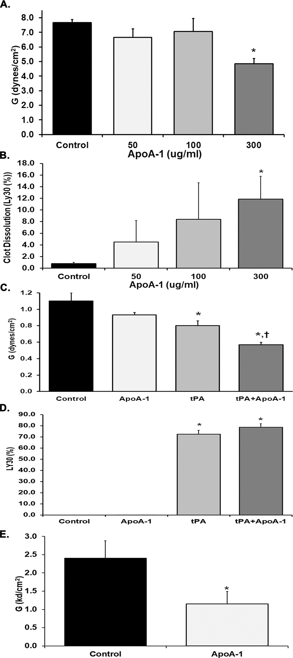
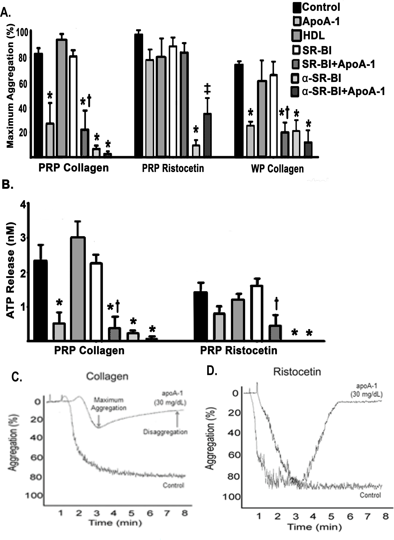
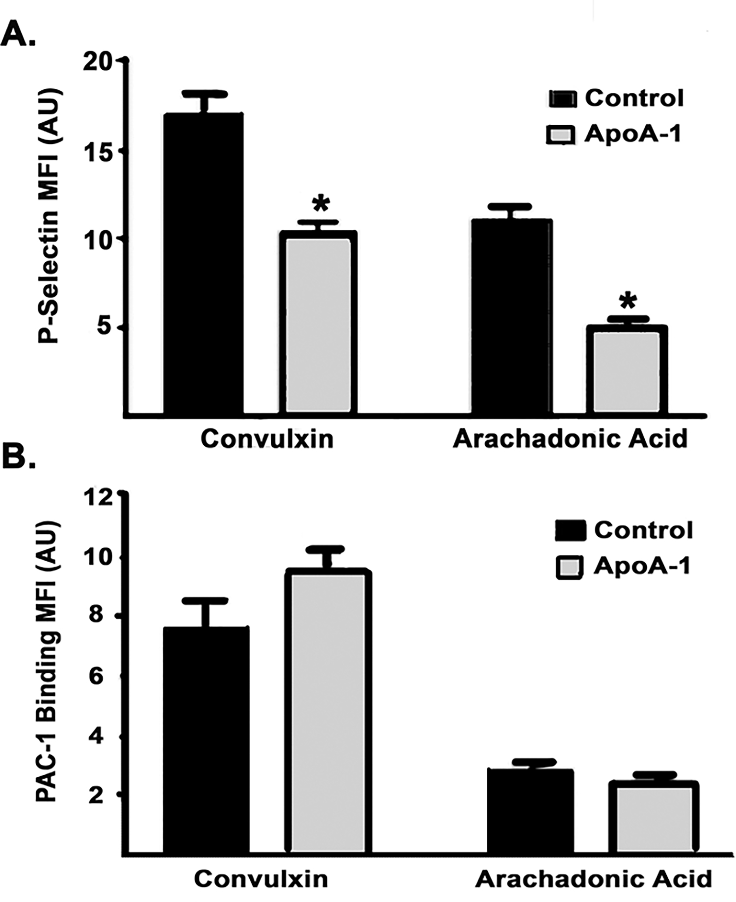
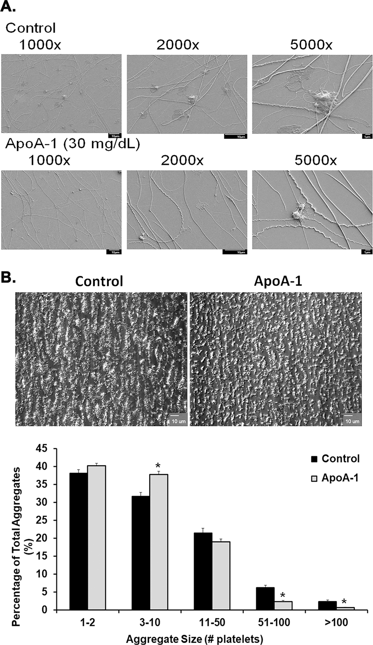
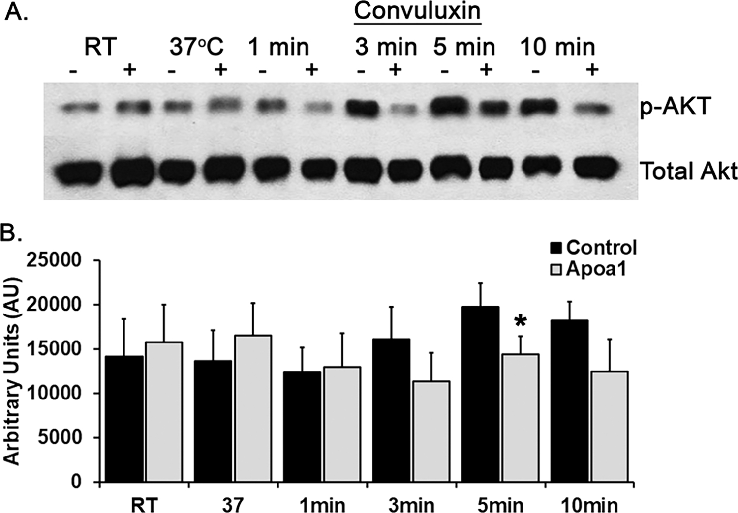
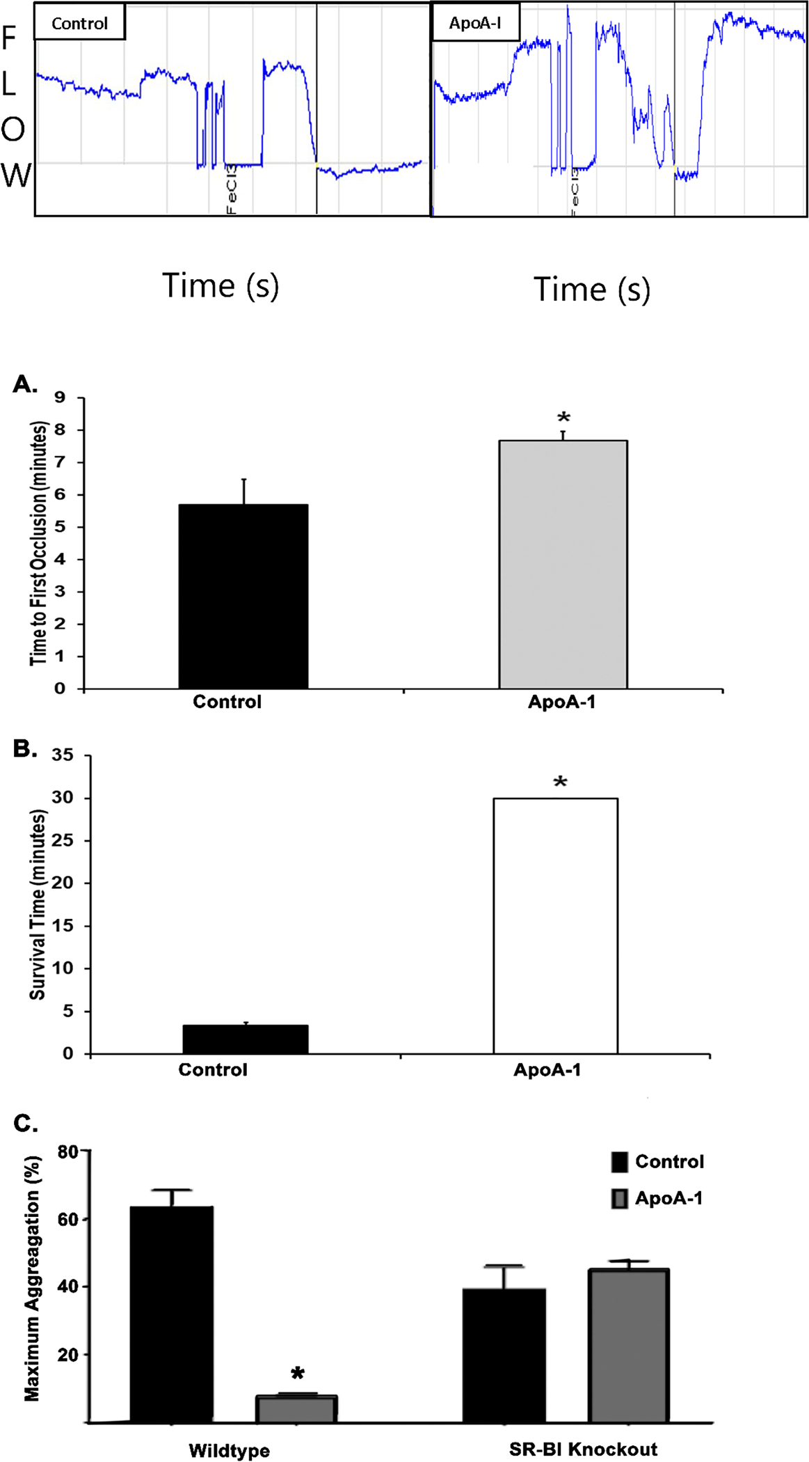
Apolipoprotein A-I (ApoA-I) is elevated in the plasma of a subgroup of trauma patients with systemic hyperfibrinolysis. We hypothesize that apoA-I inhibits platelet activation and clot formation. The effects of apoA-I on human platelet activation and clot formation were assessed by whole blood thrombelastography (TEG), platelet aggregometry, P-selectin surface expression, microfluidic adhesion, and Akt phosphorylation. Mouse models of carotid artery thrombosis and pulmonary embolism were used to assess the effects of apoA-I in vivo. The ApoA-1 receptor was investigated with transgenic mice knockouts (KO) for the scavenger receptor class B member 1 (SR-BI). Compared to controls, exogenous human apoA-I inhibited arachidonic acid and collagen-mediated human and mouse platelet aggregation, decreased P-selectin surface expression and Akt activation, resulting in diminished clot strength and increased clot lysis by TEG. ApoA-I also decreased platelet aggregate size formed on a collagen surface under flow. In vivo, apoA-I delayed vessel occlusion in an arterial thrombosis model and conferred a survival advantage in a pulmonary embolism model. SR-BI KO mice significantly reduced apoA-I inhibition of platelet aggregation versus wild-type platelets. Exogenous human apoA-I inhibits platelet activation, decreases clot strength and stability, and protects mice from arterial and venous thrombosis via the SR-BI receptor.

Keywords: Hyperfibrinolysis; SR-B1 receptor; microfluidics; platelet inhibition; thrombelastography.


Conflict of interest statement

Disclosure Statement

No authors have relevant conflicts of interest to declare.

Figures

Figure 1:
Figure 1:. Clot Strength and Stability.
Evaluation of thrombus strength and stability in 3.2% citrate-treated human whole blood (A-D) or heparinized (17 U/mL) human whole blood (E), demonstrating median +/− interquartile range. A. Clot strength, G (Dynes/cm2), as determined by Kaolin-TEG after pre-treatment with vehicle control (black bar) or increasing concentrations of apoA-I (white, gray; and charcoal gray bars). Pretreatment with 300 mg/ml of ApoA-1 significantly decreased clot strength vs. NS-treated controls (*=p<0.01). B. Clot dissolution after 30 minutes, Ly30 (%), as determined by Kaolin-TEG following pre-treatment with vehicle control (black) or increasing concentrations of apoA-I ((white, gray; and charcoal gray bars). Preincubation with apoA-I at 300 mg/ml significantly increased Ly30 versus controls (*=p<0.01). C. Clot strength (Dynes/cm2) was evaluated by the TEG citrated functional fibrinogen (CFF) assay in samples pre-treated with vehicle control (black), 300 μg/ml apoA-I (white), 100 ng/mL tPA (gray), or combination of tPA and apoA-I (charcoal gray). Preincubation with tPA inhibited formation of the functional fibrinogen clot vs. controls and apoA-I (*=p<0.01) and pre-treatment with the combination of apoA-I and tPA caused significant decrease in the functional fibrinogen clots versus both NS-treated controls and pre-incubation with apoA-I (*=p<0.01 vs. NS; †=P<.01 vs. apoA-I). D. Clot dissolution (%) was evaluated by the TEG CFF assay in samples pre-treated with vehicle control (black, no detectable value), 300 μg/ml apoA-I (clear, no detectable value), 100 ng/mL tPA (gray), or combination of tPA and apoA-I (charcoal gray) Both tPA and tPA+ apoA-I treatment induced significant amounts of CFF clot lysis as compared to NS-apoA-I treated samples (*=p<0.01). E. Clot strength (Dynes/cm2) was evaluated by thrombin-independent platelet mapping studies using factor XIIIa, reptilase, and arachidonic acid as agonists in vehicle controls (black) compared to samples treated with apoA-I (white). ApoA-I inhibited the clot strength in the platelet mapping assay (*=P<0.01). Statistical significance was determined by Mann Whitney; *=p<0.01.
Figure 2.
Figure 2.. Platelet aggregation and granule release are inhibited by ApoA-I.
A. The maximum light transmission aggregometry (percent) of PRP or WP stimulated with collagen (1 μg/mL) or ristocetin (1.25 mg/mL) after pre-treatment with vehicle control (NS, n=5), apoA-I (n=5), HDL (n=5), SR-BI±apoA-I (n=5), and anti-SR-BI (α-SR-BI) antibody±apoA-I (n=3) expressed as the mean±SEM. ApoA-I, SR-BI + apoA-I, α-SR-BI and α-SR-BI+ apoA-I pretreatments inhibited PRP platelet aggregation to collagen as compared to NS-treated controls (*=p<0.05). Pretreatment of platelets from PRP with α-SR-BI inhibited aggregation to ristocetin vs. controls (*=p<0.05). This inhibition by α-SR-BI inhibition was partially ameliorated by the addition of apoA-I and was significantly increased (‡=p<0.05 vs. α-SR-BI). Preincubation with ApoA-1, SR-BI+apoA-I, α-SR-BI, and α-SR-BI+apoA-I of washed platelets (right) stimulated with collagen showed a significant inhibition vs. controls (*=p<0.05). SB-RI+apoA-I pre-incubation inhibited collagen-induced platelet aggregation compared to SR-BI preincubation alone (†=p<0.05). B. ATP release is shown as the mean±SEM (nM) from PRP samples stimulated with collagen (1μg/mL, left) or ristocetin (1.25 mg/mL, right) after pre-treatment with control (saline, n=5, black bars), apoA-I [300 μg/ml] (n=5, light gray bars), HDL (n=5, darker gray bars), SR-BI +/− apoA-I (n=5, white and darkest gray bars). In response to collagen apoA-I inhibited ATP release as compared to controls (*=p<0.05), SR-BI +apoA-I together inhibited platelet ATP release vs. SAR-BI treated platelets alone (†=p<0.05), and anti-SR-BI antibody-treated platelets released significantly less ATP than control platelets. Ristocetin activation demonstrated that only platelet treated with SR-BI+ ApoA-1 released less ATP than SR-BI-treated platelets alone (†=p<0.05) and antibodies to SR-BI + ApoA-1 released less ATP than controls (*=p<0.05). Statistical significance was determined by Kruskal-Wallis one-way ANOVA with Dunn’s multiple corrections. C. and D. Representative light-transmission aggregation tracings demonstrate reaction after addition of collagen or ristocetin, respectively, of apoA-I treated human samples compared to vehicle treated controls. PRP, platelet rich plasma; WP, washed platelets.
Figure 3.
Figure 3.. ApoA-I inhibits human the surface expression P-selectin surface expression but not that of αiibβiii-integrin in washed human platelets.
A. P-selectin surface expression quantified by Mean fluorescence intensity (MFI)±SEM of anti-P-selectin antibody (arbitrary units, AU) on platelets stimulated with convulxin or AA after pre-treatment with NS (black bar) or apoA-I (n=4, charcoal gray bar). ApoA-I pretreatment inhibited the convulxin- or AA-induced increase in P-selectin surface expression (*=p<0.05). B. αiibβiii-integrin (PAC-1) surface expression (MFI) expressed as the mean±SEM on platelets stimulated with convulxin or AA after pre-treatment with normal saline (black) or apoA-I (charcoal gray) apoA-I preincubation did not alter PAC-1 surface expression to either convulxin or AA.
Figure 4.
Figure 4.. ApoA-I inhibits platelet spreading on fibrillary collagen, and limits the formation of large platelet aggregates on collagen under physiologic flow conditions.
Panel A: representative scanning electron microscopy of NS- (top row of micrographs) or apoA-I-pretreated (bottom row of micrographs) platelets forming aggregates on fibrillar collagen at different magnifications: 1,000X, 2,000X, and 5,000X. ApoA-I visually reduced the size of the human platelet aggregates on collagen fibrils versus the NS-treated controls. Panel B: two representative bright-field microscopic images of platelet aggregates: NS (vehicle)-treated controls on the left and apoA-I [300 μg/ml]-treated platelets on the right (100X). The ApoA-1 pretreatment diminished the size of the aggregates vs. the NS-treated controls. Panel C: Percentage of the total aggregates represented by the different size groups of various platelets per aggregate. Controls (black bars) and apoA-I-pre-treated samples (light gray bars) are divided into groups of various aggregate sizes along the x-axis, and corresponding proportions of the surface area covered by aggregates of each size (percent) shown on the y-axis. ApoA-I caused an increased in platelet aggregates of 3–10 and decreased the large aggregates 51–100 and >100 platelets on collagen versus NS-pretreated controls (*=p<0.05).
Figure 5.
Figure 5.. ApoA-I inhibits Akt phosphorylation.
A representative western blot comparing phosphorylated Akt from NS-pre-treated human platelet controls (−) to platelets pre-treated with ApoA-1 (+) for 5 min at 37°C and activated with convulxin [10 ng/mL] over a time course of 1–10 min at 37°C. Apolipoprotein A-I pretreatment decreased the amounts of Akt phosphorylation over the entire time course as seen both visually (upper panel) and by densitometry (lower panel) with statistical significance between total and phosphorylated Akt present at 5 min (*=p<0.05, n=3).
Figure 6.
Figure 6.. ApoA-I inhibits thrombosis in mice and SR_BI is necessary for apoA-I inhibition.
A. Time (minutes) to vessel occlusion in a 6% ferric chloride-induced carotid artery injury model with vehicle control-treated WT mice (black bar, n=5) compared to those treated with apoA-I (gray bar). ApoA-I preincubation significantly prolonged the time to vessel occlusion versus NS-pretreated control mice (n=5; *=p<0.05). B. Survival time (minutes) following pulmonary embolism induced by intravenous injection of collagen and epinephrine in mice treated with vehicle control (black, n=5) or apoA-I (red, n=5). All the NS-treated controls died via a pulmonary embolus within 5 minutes following injury. ApoA-I pre-treated mice survived and did not develop pulmonary emboli during the observation period. Statistical significance determined by Mann-Whitney test; *=p<0.05. C. Maximal aggregation (%) was compared between experimental groups with no difference seen in SR-BI KO mice treated with apoA-I (n=3) or vehicle (normal saline, n=3), but significant decrease in aggregation seen in WT mice treated with apoA-I (n=4) compared to controls treated with vehicle only (n=7, *=p<0.05).

References

    1. Chang R, Cardenas JC, Wade CE, Holcomb JB. Advances in the understanding of trauma-induced coagulopathy. Blood. 2016;128(8):1043–1049. - PMC - PubMed
    1. Gonzalez E, Moore EE, Moore HB, Chapman MP, Silliman CC, Banerjee A. Trauma-Induced Coagulopathy: An Institution’s 35 Year Perspective on Practice and Research. Scand J Surg. 2014;103(2):89–103. - PMC - PubMed
    1. Moore EE, Moore HB, Kornblith LZ, et al. Trauma-induced coagulopathy. Nat Rev Dis Primers. 2021;7(1):30. - PMC - PubMed
    1. Neal MD, Moore HB, Moore EE, et al. Clinical assessment of trauma-induced coagulopathy and its contribution to postinjury mortality: A TACTIC proposal. J Trauma Acute Care Surg. 2015;79(3):490–492. - PMC - PubMed
    1. Sumislawski JJ, Christie SA, Kornblith LZ, et al. Discrepancies between conventional and viscoelastic assays in identifying trauma-induced coagulopathy. Am J Surg. 2019;217(6):1037–1041. - PMC - PubMed