Review
. 2022 Jun 3;15(1):77.
doi: 10.1186/s13045-022-01292-6.

Hypoxia-inducible factors: master regulators of hypoxic tumor immune escape

Affiliations
Review

Hypoxia-inducible factors: master regulators of hypoxic tumor immune escape

Qinghua Wu et al. J Hematol Oncol. .

Abstract

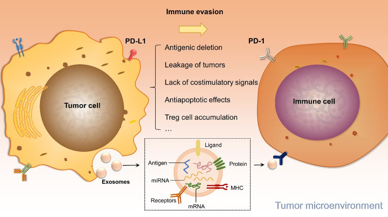
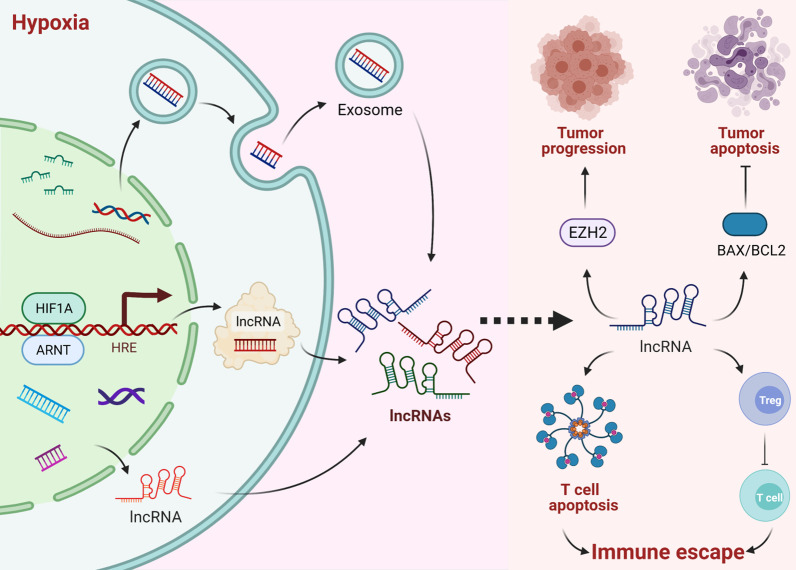
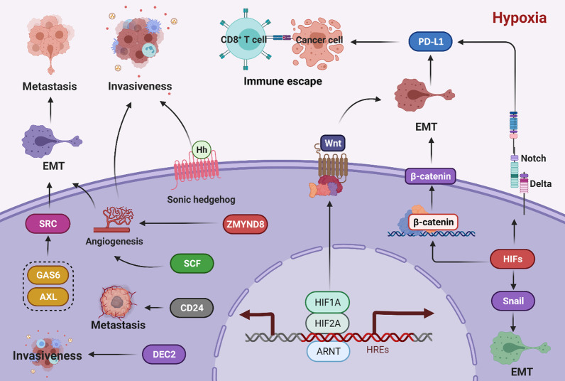
Hypoxia, a common feature of the tumor microenvironment in various types of cancers, weakens cytotoxic T cell function and causes recruitment of regulatory T cells, thereby reducing tumoral immunogenicity. Studies have demonstrated that hypoxia and hypoxia-inducible factors (HIFs) 1 and 2 alpha (HIF1A and HIF2A) are involved in tumor immune escape. Under hypoxia, activation of HIF1A induces a series of signaling events, including through programmed death receptor-1/programmed death ligand-1. Moreover, hypoxia triggers shedding of complex class I chain-associated molecules through nitric oxide signaling impairment to disrupt immune surveillance by natural killer cells. The HIF-1-galactose-3-O-sulfotransferase 1-sulfatide axis enhances tumor immune escape via increased tumor cell-platelet binding. HIF2A upregulates stem cell factor expression to recruit tumor-infiltrating mast cells and increase levels of cytokines interleukin-10 and transforming growth factor-β, resulting in an immunosuppressive tumor microenvironment. Additionally, HIF1A upregulates expression of tumor-associated long noncoding RNAs and suppresses immune cell function, enabling tumor immune escape. Overall, elucidating the underlying mechanisms by which HIFs promote evasion of tumor immune surveillance will allow for targeting HIF in tumor treatment. This review discusses the current knowledge of how hypoxia and HIFs facilitate tumor immune escape, with evidence to date implicating HIF1A as a molecular target in such immune escape. This review provides further insight into the mechanism of tumor immune escape, and strategies for tumor immunotherapy are suggested.

Keywords: Hypoxia; Hypoxia-inducible factors; Immunotherapy; Personalized medicine; Tumor disease.


Conflict of interest statement

The authors declare that they have no competing interests.

Figures

Fig. 1
Fig. 1
The protein structure of HIF1A and HIF2A. HIF1A and HIF2A contain basic helixloop-helix (bHLH) and Par-Arnt-SIM (PAS) transcription factors. And they also have N/C-terminal transactivation domain (N/C-TAD), inhibitory domain (ID), and oxygen-dependent degradation domain (ODD). Their DNA has a high degree of similarity. NLS: nuclear localization signal, pVHL: von Hippel-Lindau protein
Fig. 2
Fig. 2
Proposed mechanism underlying tumor immune escape. Tumor immune escape is related to antigenic deletion, tumor cell leakage, lack of costimulatory signals on the tumor cell surface, and the antiapoptotic effects of tumor cells. Tumor cells bind to PD-1 on tumor-infiltrating lymphocytes via PD-L1 that mediates immune escape in the tumor microenvironment. Tumor cells also inhibit immune cells by releasing exosomes rich in surface PD-L1 to escape from immune system recognition
Fig. 3
Fig. 3
HIF1A regulates tumor immune escape under hypoxic conditions. Under normoxic conditions, HIF1A is hydrolyzed by prolyl-4-hydroxylase (PHD). Von Hippel-Lindau (VHL) then recognizes and binds to HIF1A, leading to rapid proteasomal degradation of HIF1A, rendering tumor cells susceptible to immune surveillance. Under hypoxic conditions, HIF1A is protected from degradation and translocates to the nucleus, where it dimerizes with hydrocarbon receptor nuclear translocator (ARNT) to form HIF1 and binds to target gene hypoxic response elements (HREs). HIF1 induces cytotoxic T lymphocyte (CTL) apoptosis by upregulating PD-L1 expression, which enhances tumor cell resistance to lysis. Moreover, HIF1A causes MIC shedding by impairing NO signaling, allowing escape from NK cell immune surveillance. The HIF1-GAL3ST1-sulfatide signaling axis promotes immune escape by increasing tumor cell-platelet binding. HIF1 also regulates CD47 expression to promote escape from phagocytosis and promotes tumor angiogenesis through upregulation of vascular endothelial growth factor (VEGF), which facilitates immune escape by enabling metastasis
Fig. 4
Fig. 4
Effects of lncRNAs on hypoxic tumor immune escape. HIF1A induces lncRNA overexpression, inhibiting cyclin-dependent kinase inhibitor 1A (CDKN1A) expression by recruiting enhancer of zeste 2 polycomb repressive complex 2 subunit (EZH2) and causing tumor progression. Under hypoxia, lncRNAs inhibit tumor cell apoptosis by regulating the BAX/BCL2 ratio. Hypoxia induces production of lncRNA-containing exosomes in tumor cells and promotes proliferation, migration, and invasion. Notably, lncRNAs also contribute to immune escape by increasing Tregs levels, inhibiting CTLs, and modulating the sensitivity of tumor-infiltrating T cells to apoptosis
Fig. 5
Fig. 5
The role of HIFs in cancer invasiveness and metastasis. HIF1A regulates the sonic hedgehog signaling pathway to promote the invasive abilities of cancer cells. Under hypoxic conditions, HIF1A upregulates differentiated embryonic chondrocyte gene 2 (DEC2) at the transcriptional level, which in turn promotes HIF1A activation, ultimately contributing to cancer cell metabolic reprogramming, angiogenesis, and invasiveness. HIF2A directly induces transcription of the stem cell factor (SCF) gene via the hypoxia response element in the SCF promoter and upregulates SCF expression, thereby promoting angiogenesis and metastasis. Zinc finger MYND-type containing 8 (ZMYND8) is regarded as a direct target gene of HIF1A and HIF2A. The ZMYND8/HIF axis increases breast tumor angiogenesis and decreases cancer cell death to promote cancer metastasis. Protein kinase growth arrest-specific 6 (GAS6)/AXL is activated by HIF1A and HIF2A. The SRC proto-oncogene nonreceptor tyrosine kinase is a direct target of GAS6/AXL signaling; it activates MET proto-oncogenes, thereby regulating the epithelial-mesenchymal transition (EMT) and tumor metastasis. Overexpression of HIF1A promotes production of CD24, which leads to tumor growth and metastasis, and HIFs regulate EMT to promote cancer metastasis. Hypoxia-induced HIFs promote EMT by enhancing snail, β-catenin, Wnt, and Notch signaling, thereby inducing cancer cell survival, metastasis, and immune escape. EMT promotes expression of PD-L1, facilitating tumor cells escape and recognition and attack by immune cells

References

    1. Galon J, Bruni D. Approaches to treat immune hot, altered and cold tumours with combination immunotherapies. Nat Rev Drug Discov. 2019;18(3):197–218. doi: 10.1038/s41573-018-0007-y. - DOI - PubMed
    1. Lane AN, Higashi RM, Fan TW. Metabolic reprogramming in tumors: contributions of the tumor microenvironment. Genes Dis. 2020;7(2):185–198. doi: 10.1016/j.gendis.2019.10.007. - DOI - PMC - PubMed
    1. Binnewies M, Roberts EW, Kersten K, Chan V, Fearon DF, Merad M, et al. Understanding the tumor immune microenvironment (TIME) for effective therapy. Nat Med. 2018;24(5):541–550. doi: 10.1038/s41591-018-0014-x. - DOI - PMC - PubMed
    1. Bose S, Panda AK, Mukherjee S, Sa G. Curcumin and tumor immune-editing: resurrecting the immune system. Cell Div. 2015;10:1–13. doi: 10.1186/s13008-015-0012-z. - DOI - PMC - PubMed
    1. Lei X, Lei Y, Li JK, Du WX, Li RG, Yang J, et al. Immune cells within the tumor microenvironment: biological functions and roles in cancer immunotherapy. Cancer Lett. 2020;470:126–133. doi: 10.1016/j.canlet.2019.11.009. - DOI - PubMed

Publication types

Substances