. 2022 Jun 6;9(1):1.
doi: 10.1186/s44156-022-00001-w.

Mid-term follow-up and outcomes of patients with prosthetic heart valves: a single-centre experience

Affiliations

Mid-term follow-up and outcomes of patients with prosthetic heart valves: a single-centre experience

Sadie Bennett et al. Echo Res Pract. .

Abstract

Background: Patients with prosthetic heart valves (PHV) require long-term follow-up, usually within a physiologist led heart valve surveillance clinic. These clinics are well established providing safe and effective patient care. The disruption of the COVID-19 pandemic on services has increased wait times thus we undertook a service evaluation to better understand the patients currently within the service and PHV related complications.

Methods: A clinical service evaluation of the heart valve surveillance clinic was undertaken to assess patient demographics, rates of complications and patient outcomes in patients who had undergone a PHV intervention at our institute between 2010 and 2020.

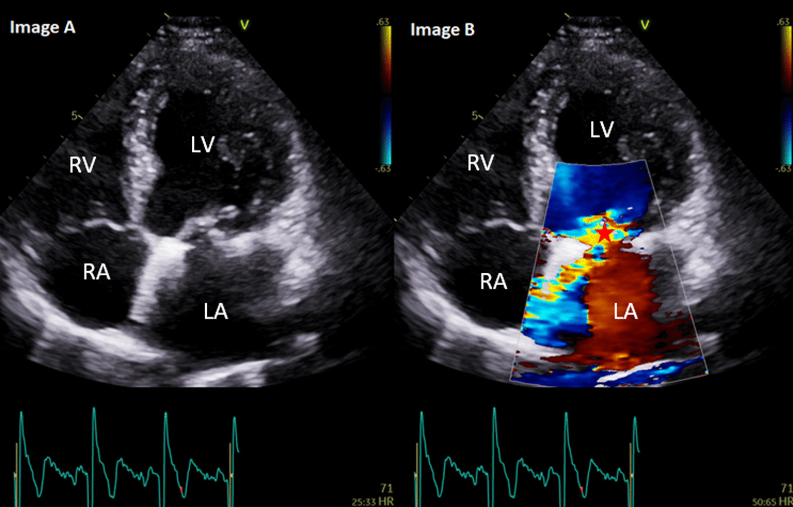
Results: A total of 294 patients (mean age at time of PHV intervention: 71 ± 12 years, 68.7% male) were included in this service evaluation. Follow-up was 5.9 ± 2.7 years (range: 10 years). 37.1% underwent baseline transthoracic echo (TTE) assessment and 83% underwent annual TTE follow-up. Significant valve related complications were reported in 20 (6.8%) patients. Complications included a change in patient functional status secondary to significant PHV regurgitation (0.3%) or stenosis (0.3%), PHV thrombosis (0.3%) or infective endocarditis (3.7%). Significant valve related complications resulted in ten hospital admission (3.4%), two re-do interventions (0.6%), and four deaths (1.3%).

Conclusions: This service evaluation highlights the large number of patients requiring ongoing surveillance. Only a small proportion of patients develop significant PHV related complications resulting in a low incidence of re-do interventions and deaths.

Keywords: Echocardiography; Patient outcomes; Prosthetic heart valves.


Conflict of interest statement

The authors declare that they have no competing interests.

Figures

Fig. 1
Fig. 1
Images A and B: A 68-year-old male with mitral valve repair + 34 mm annuloplasty ring for severe mitral regurgitation secondary to P2 mitral valve prolapse in 2016. Follow-up in 2019 demonstrated a new finding of moderate, eccentric and anteriorly directed jet of mitral regurgitation (*) secondary to a leaflet co-aptation defect. The left ventricle was mildly dilated by indexed volumes with normal left ventricular systolic function (biplane ejection fraction: 61%). The patient was asymptomatic without any reduction in exercise tolerance. The patient remains on 12 month follow-up. LV: left ventricle, RV: right ventricle, LA: left atrium, RA: right atrium
Fig. 2
Fig. 2
Images C and D: A 54-year-old female with a mechanical mitral valve replacement in 2012 for severe mitral stenosis secondary to rheumatic fever. In April 2020, the patient presented acutely with fever and night sweats. Blood cultures were positive for Staphylococcus aureus. Transthoracic echocardiography identified a stable in-situ mechanical mitral valve with good occluder mobility. There was turbulent forward flow (* in image C) and significantly elevated transvalvular mean gradient of 15 mmHg (documented as 3.3 mmHg on transthoracic echocardiography 13 months prior). There was a linear mobile mass (* in image D) on the left ventricular size of the mechanical valve replacement which was not visible on previous imaging. There was a high suspicion of infective endocarditis which was confirmed on a subsequent transesophageal echocardiography. The patient was commenced on antibiotic therapy, re-do mitral valve replacement was undertaken 16 weeks later, after which the patient made a good and uneventful recovery. At last follow-up, there was a stable in-situ mechanical mitral valve replacement, mean gradient: 3.4 mmHg, normal left ventricular size and systolic function, biplane ejection fraction: 59%. The patient was asymptomatic without any reduction in exercise tolerance. The patient remains on 12 monthly follow-up. LV: left ventricle, LA: left atrium, Ao: aorta

References

    1. Baumgartner H, Falk V, Bax JJ, et al. 2017 ESC/EACTS Guidelines for the management of valvular disease. Eur Heart J. 2017;38:2739–2791. doi: 10.1093/eurheartj/ehx391. - DOI - PubMed
    1. Chambers J, Garbi M, Briffa N, Sharma V, Steeds RP. Indications for echocardiography of replacement heart valves: a joint statement from the British Heart Valve Society and British Society of Echocardiography. Echo Res Prac. 2019;6:G9–G15. doi: 10.1530/ERP-18-0079. - DOI - PMC - PubMed
    1. Chambers JB, Parkin D, Rimington H, Subbiah S, Campbell D, Demetrescu C, Hayes A, Rajani R. Specalist valve clinic in a cardiac centre: 10 year experience. Open Heart. 2020;7(1):e001262. doi: 10.1136/openhrt-2020-001262. - DOI - PMC - PubMed
    1. Turpie D, Maycock M, Crawford C, Aitken K, Macdonald M, Farman C, Thompson MLP, Smith J, Cross SJ, Leslie SJ. Establishing an aortic stenosis surveillance clinic. Br J Cardiol. 2010;17:286–289.
    1. Edmunds LH, Clark RE, Cohn LH, Grunkemeier GL, Miller DC. Weisel RD guidelines for reporting morbidity and mortality after cardiac valvular operations. Ann Thorac Surg. 1996;63(3):932–935. doi: 10.1016/S0003-4975(96)00531-0. - DOI - PubMed

LinkOut - more resources