

A scoping review of methods used in musculoskeletal soft tissue and nerve shear wave elastography studies
- PMID: 35659822
- PMCID: PMC9394639
- DOI: 10.1016/j.clinph.2022.04.013
A scoping review of methods used in musculoskeletal soft tissue and nerve shear wave elastography studies
Abstract
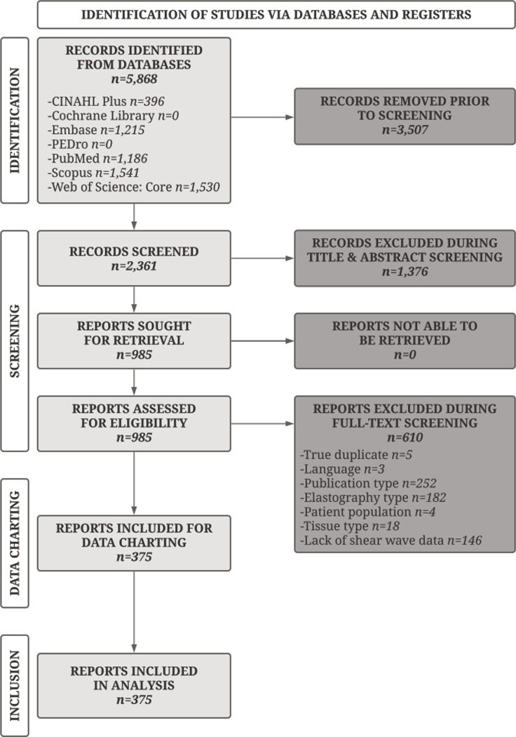
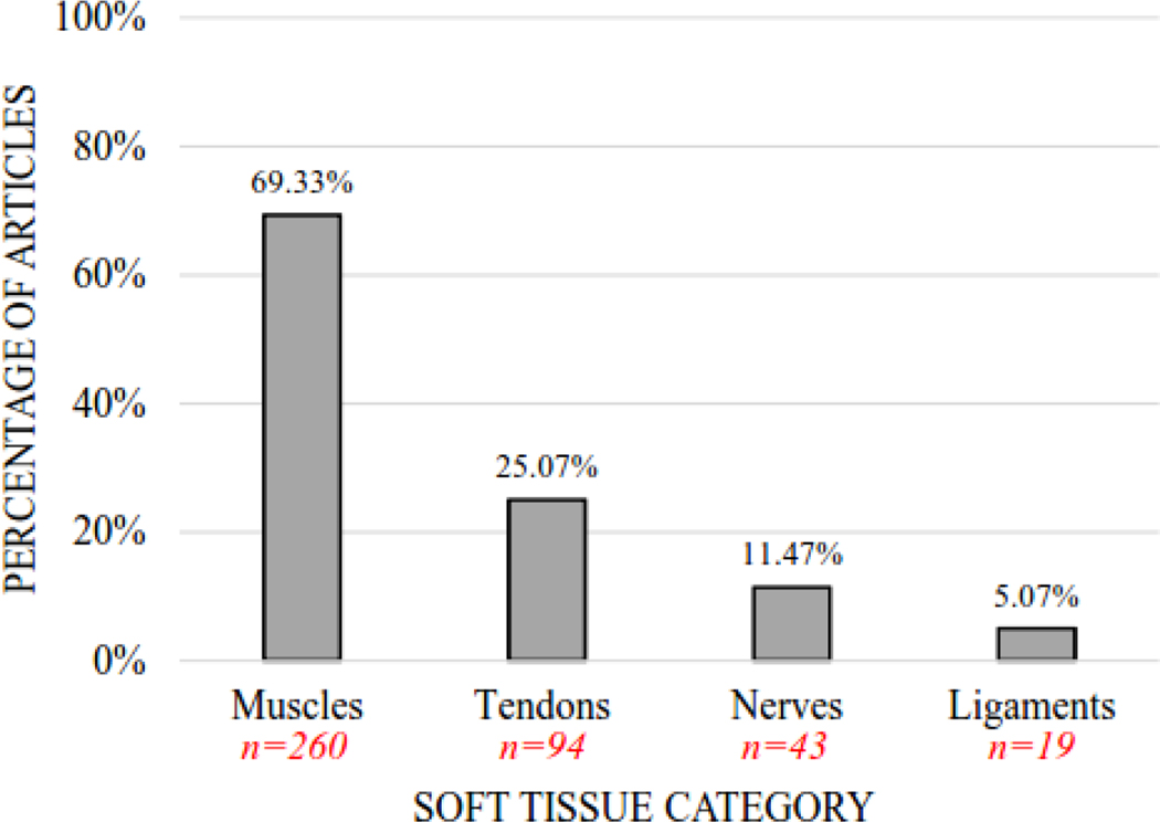
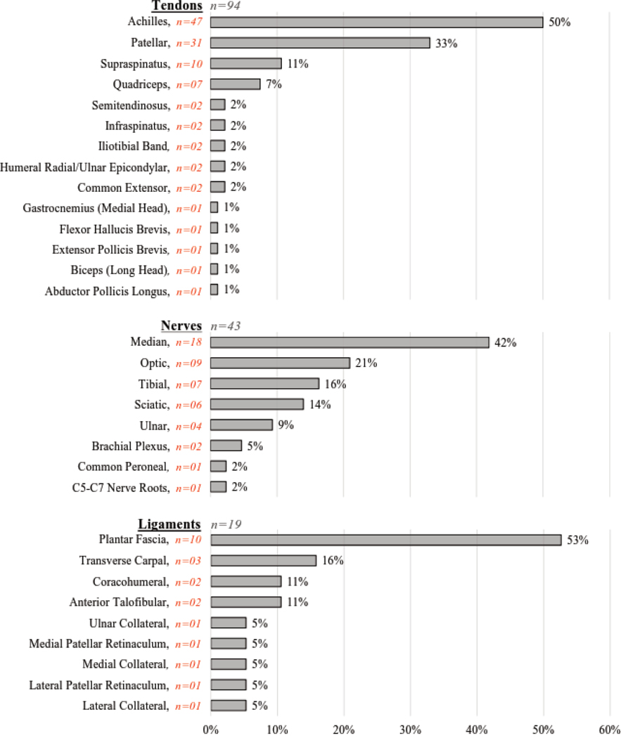
This scoping review of shear wave elastography (SWE) articles in musculoskeletal soft tissue and nerve research demonstrates methodological heterogeneity resulting from a lack of standardized data collection and reporting requirements. Seven literature databases were searched for original articles published in English from 2004-2020 that examine human skeletal muscles, tendons, and nerves in vivo. Although 5,868 records were initially identified, only 375 reports met inclusion criteria. Of the 375 articles, 260 examined 89 unique muscles, 94 examined 14 unique tendons, and 43 examined 8 unique nerves. Cohorts were often small (n = 11-20) and young (mean = 20-29 years), and participants were typically tested in the prone position. Regarding equipment, a variety of ultrasound systems (n = 11), ultrasound models (n = 18), and transducers (n = 19) were identified. Only 11% of articles contained information on the use of electromyography to confirm absence of muscle activity, and only 8% reported measurement depth. Since musculoskeletal soft tissue and nerve stiffness can vary significantly based on data collection methods, it is essential to standardize SWE collection and reporting procedures. This will allow SWE to serve as a valid and reproducible tool for assessing tissue pathology, disease progression, and response to intervention within a variety of musculoskeletal and nerve-related disorders.
Keywords: Elastic modulus; Elastogram; Musculoskeletal ultrasound; Neuromuscular ultrasound; Shear wave elastography; Sonoelastography.
Copyright © 2022 International Federation of Clinical Neurophysiology. All rights reserved.
Conflict of interest statement
Declaration of Competing Interest The authors declare that they have no known competing financial interests or personal relationships that could have appeared to influence the work reported in this paper.
Figures
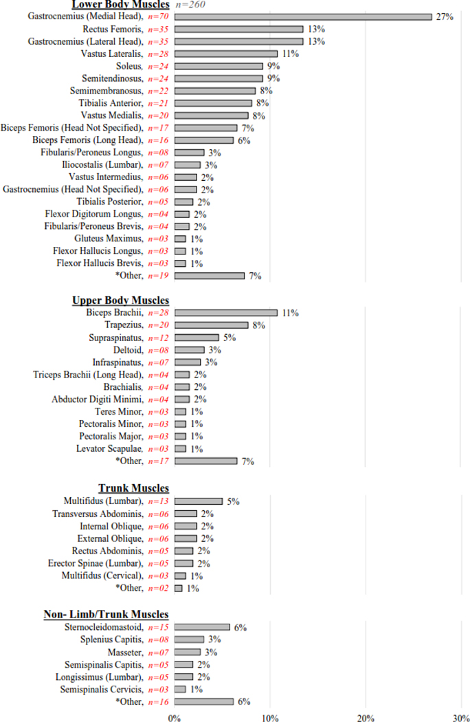
Lower Body Muscles: Adductor Magnus, Biceps Femoris (Short Head), Extensor Digitorum Longus, Flexor Digitorum Brevis, Gluteus Medius, Gracilis, Iliacus, Iliopsoas, Psoas Major, Quadricep (Unspecified), Quadriceps Femoris, Sartorius, Tensor Fasciae Latae, Triceps Surae
Upper Body Muscles: Abductor Hallucis, Brachioradialis, Extensor Carpi Radialis Brevis, Extensor Carpi Ulnaris, Flexor Carpi Radialis, Flexor Digitorum Profundus, Flexor Digitorum Superficialis, Forearm Flexor, Hypothenar, Latissimus Dorsi, Pronator Quadratus, Teres Major, Thenar, Triceps Brachii (Head Not Specified)
Trunk Muscles: Multifidus (Sacral), Serratus Anterior
Non-Limb/Trunk Muscles: Anterior Scalene, Diaphragm, Digastric (Anterior Belly), Geniohyoid, Lateral Rectus, Levator Ani, Medial Rectus, Musculus Rectus Inferior, Musculus Rectus Medialis, Musculus Rectus Superior, Musculus Rectus Temporalis, Spinalis Capitis, Urethral Rhabdosphincter
Comment in
-
Ultrasonic shear-wave elastography: A novel method for assessing musculoskeletal soft tissue and nerves.Clin Neurophysiol. 2022 Aug;140:163-164. doi: 10.1016/j.clinph.2022.05.006. Epub 2022 May 18. Clin Neurophysiol. 2022. PMID: 35618565 No abstract available.
References
Publication types
MeSH terms
Grants and funding
LinkOut - more resources
Full Text Sources
Miscellaneous
