

Reticular Pseudodrusen: The Third Macular Risk Feature for Progression to Late Age-Related Macular Degeneration: Age-Related Eye Disease Study 2 Report 30
- PMID: 35660417
- PMCID: PMC9509418
- DOI: 10.1016/j.ophtha.2022.05.021
Reticular Pseudodrusen: The Third Macular Risk Feature for Progression to Late Age-Related Macular Degeneration: Age-Related Eye Disease Study 2 Report 30
Abstract
Purpose: To analyze reticular pseudodrusen (RPD) as an independent risk factor for progression to late age-related macular degeneration (AMD), alongside traditional macular risk factors (soft drusen and pigmentary abnormalities) considered simultaneously.
Design: Post hoc analysis of 2 clinical trial cohorts: Age-Related Eye Disease Study (AREDS) and AREDS2.
Participants: Eyes with no late AMD at baseline in AREDS (6959 eyes, 3780 participants) and AREDS2 (3355 eyes, 2056 participants).
Methods: Color fundus photographs (CFPs) from annual visits were graded for soft drusen, pigmentary abnormalities, and late AMD. Presence of RPD was from grading of fundus autofluorescence images (AREDS2) and deep learning grading of CFPs (AREDS). Proportional hazards regression analyses were performed, considering AREDS AMD severity scales (modified simplified severity scale [person] and 9-step scale [eye]) and RPD presence simultaneously.
Main outcome measures: Progression to late AMD, geographic atrophy (GA), and neovascular AMD.
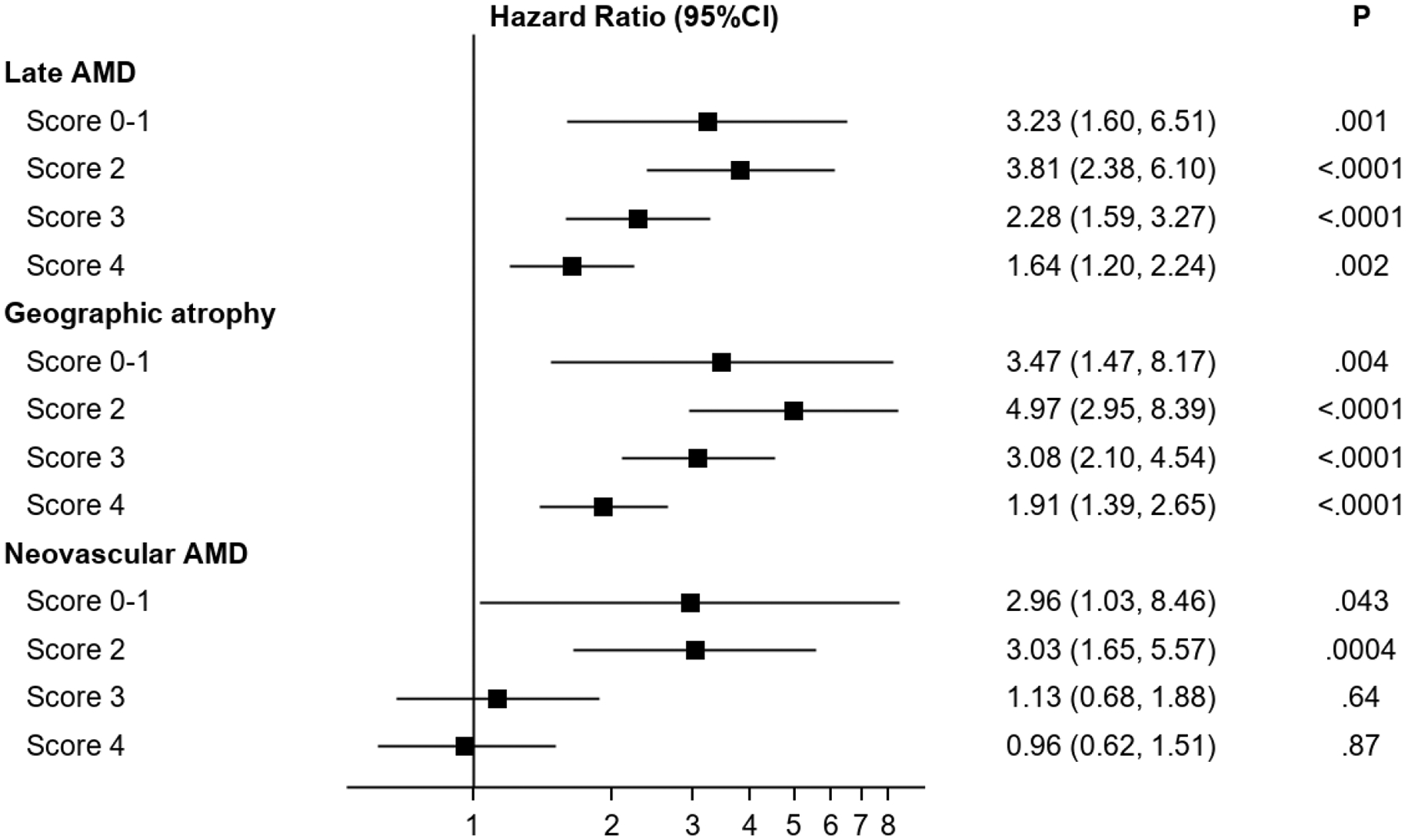
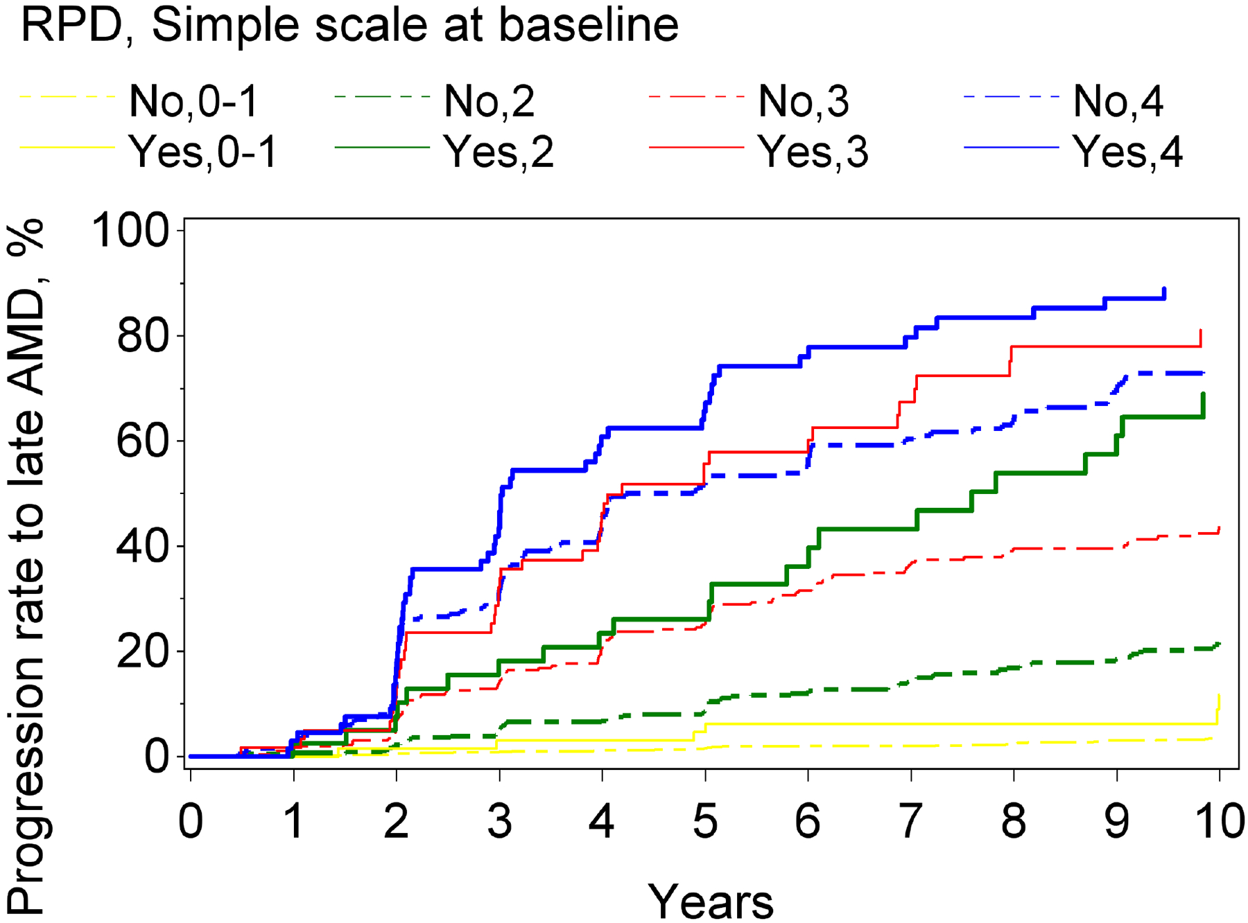
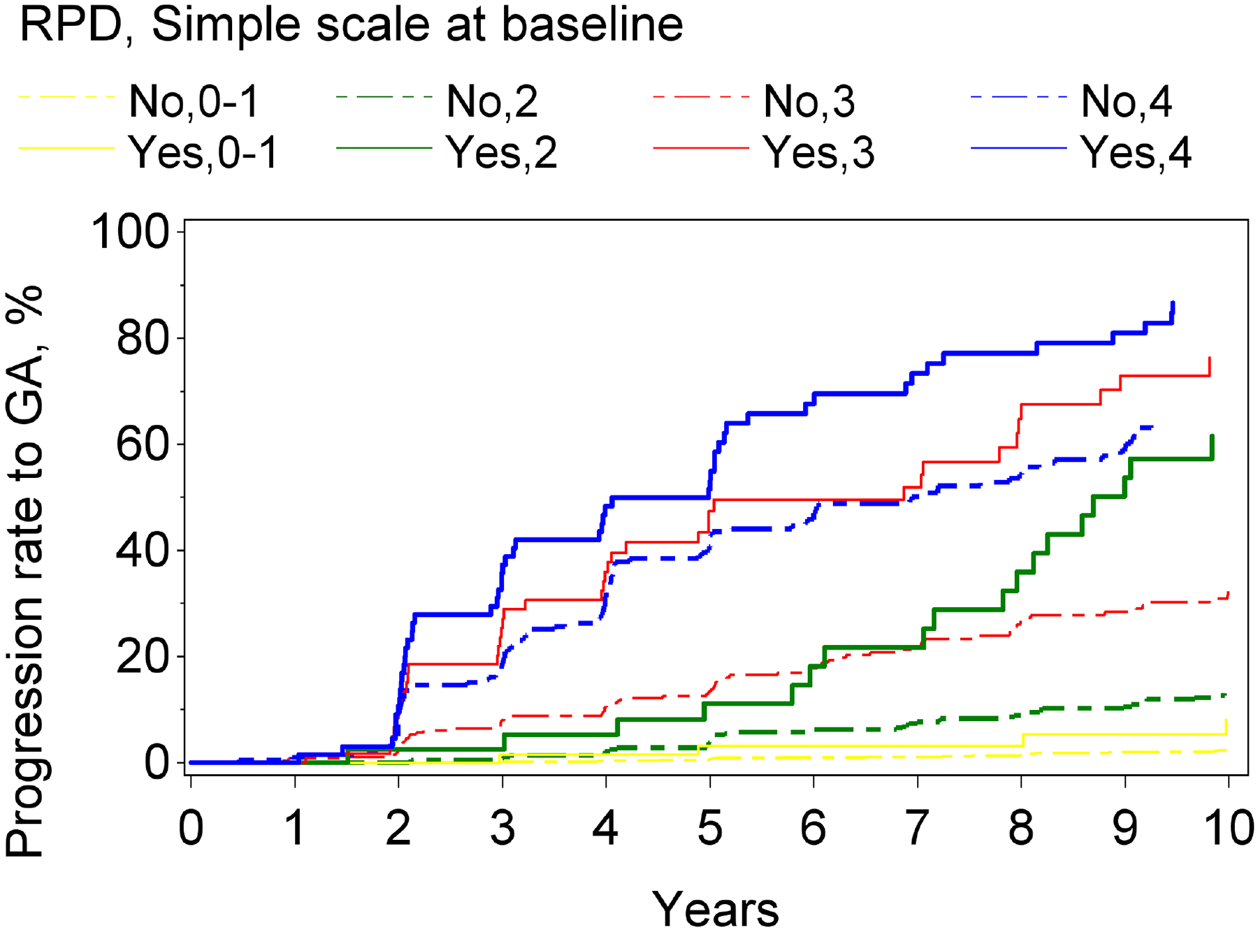
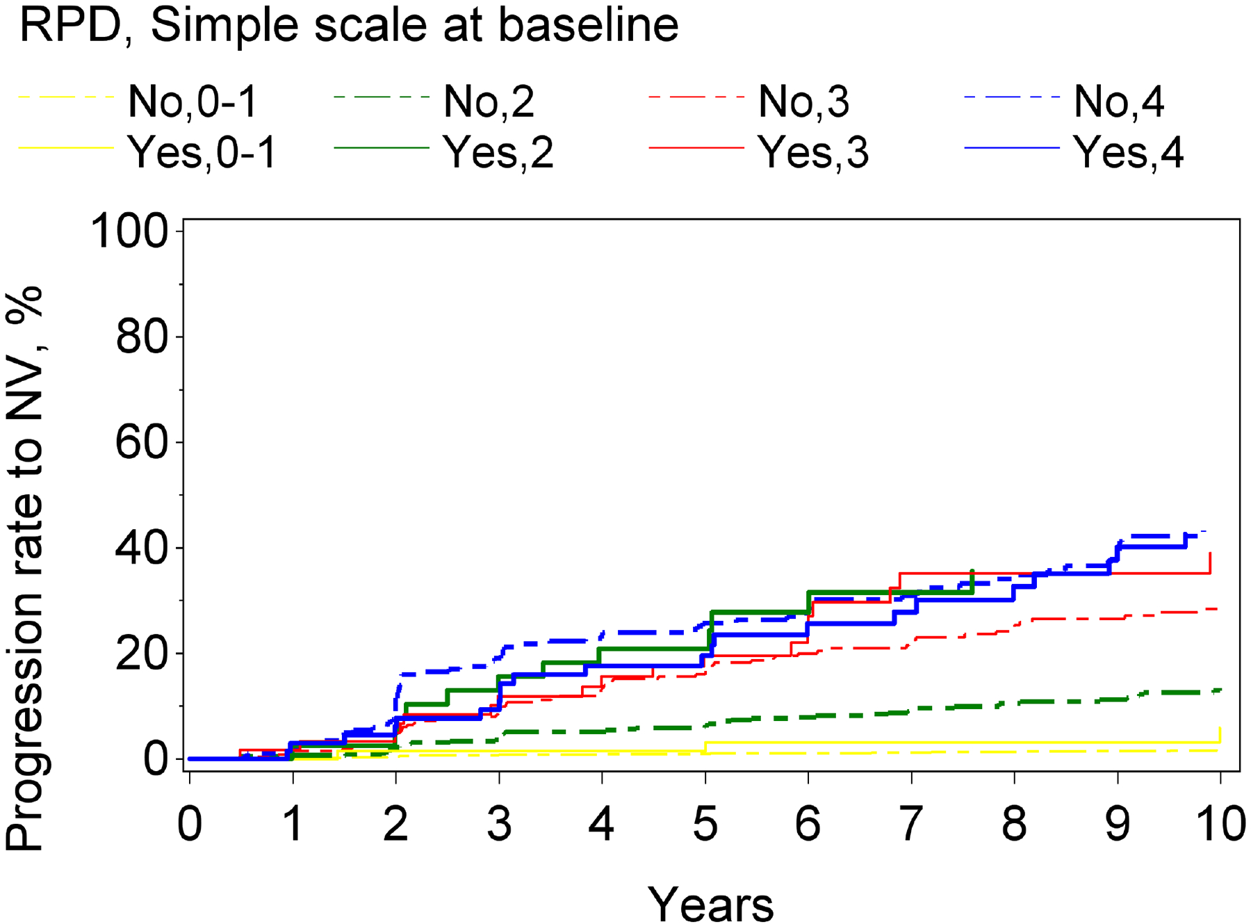
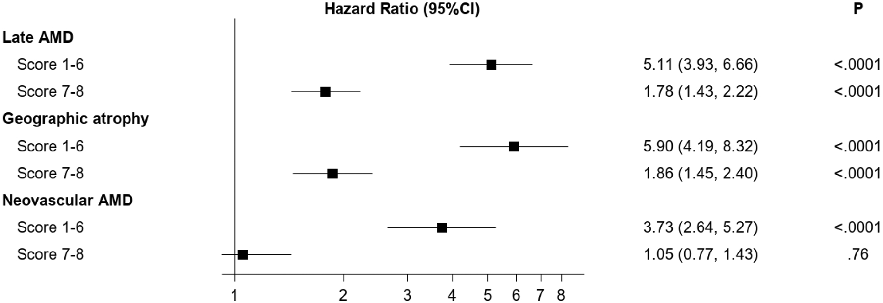
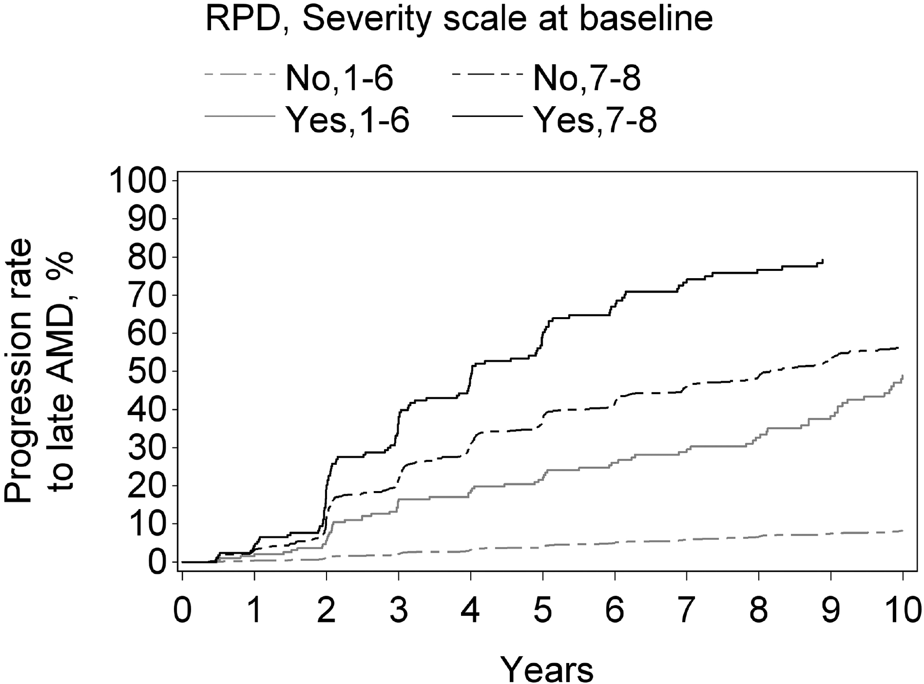
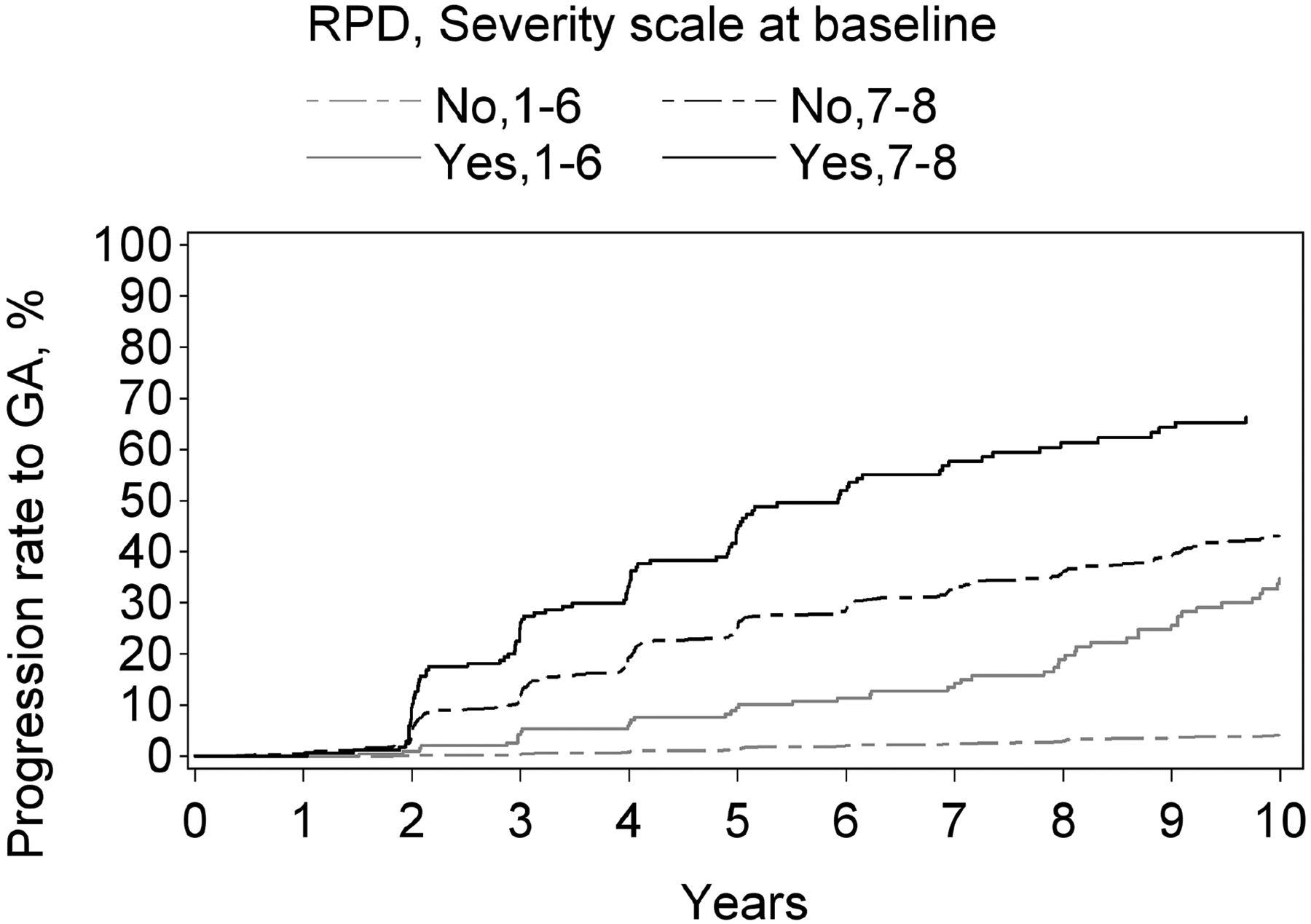
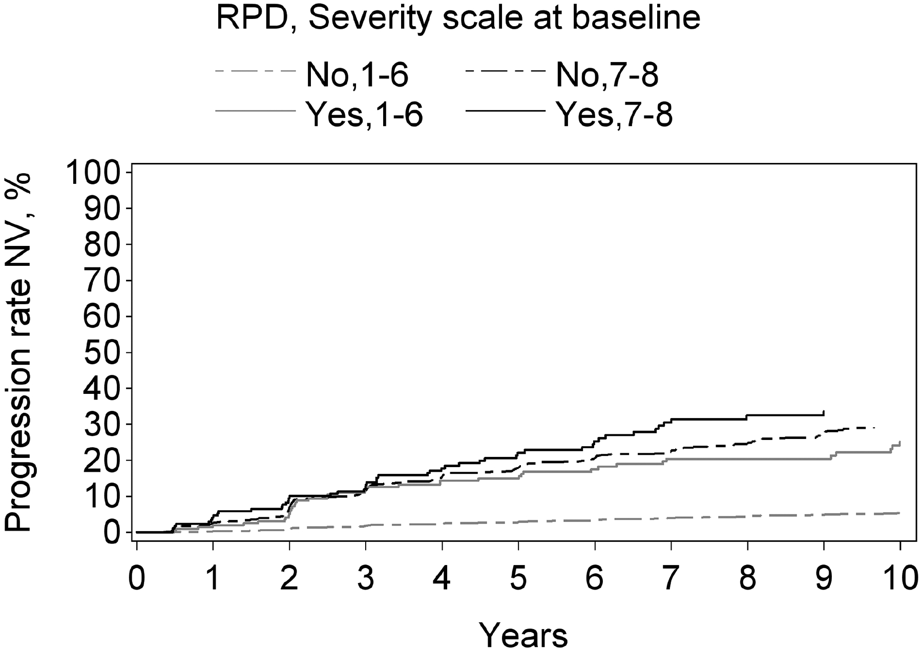
Results: In AREDS, for late AMD analyses by person, in a model considering the simplified severity scale simultaneously, RPD presence was associated with a higher risk of progression: hazard ratio (HR), 2.15 (95% confidence interval [CI], 1.75-2.64). However, the risk associated with RPD presence differed at different severity scale levels: HR, 3.23 (95% CI, 1.60-6.51), HR, 3.81 (95% CI, 2.38-6.10), HR, 2.28 (95% CI, 1.59-3.27), and HR, 1.64 (95% CI, 1.20-2.24), at levels 0-1, 2, 3, and 4, respectively. Considering the 9-step scale (by eye), RPD presence was associated with higher risk: HR, 2.54 (95% CI, 2.07-3.13). The HRs were 5.11 (95% CI, 3.93-6.66) at levels 1-6 and 1.78 (95% CI, 1.43-2.22) at levels 7 and 8. In AREDS2, by person, RPD presence was not associated with higher risk: HR, 1.18 (95% CI, 0.90-1.56); by eye, it was HR, 1.57 (95% CI, 1.31-1.89). In both cohorts, RPD presence carried a higher risk for GA than neovascular AMD.
Conclusions: Reticular pseudodrusen represent an important risk factor for progression to late AMD, particularly GA. However, the added risk varies markedly by severity level, with highly increased risk at lower/moderate levels and less increased risk at higher levels. Reticular pseudodrusen status should be included in updated AMD classification systems, risk calculators, and clinical trials.
Keywords: Age-Related Eye Disease Study; Age-Related Eye Disease Study 2; Age-related macular degeneration; Choroidal neovascularization; Disease progression; Geographic atrophy; Reticular pseudodrusen; Risk calculator; Risk factor; Severity scale; Subretinal drusenoid deposits.
Published by Elsevier Inc.
Conflict of interest statement
Conflicts of interest
No conflicting relationship exists for any author.
Figures
References
-
- Age-Related Eye Disease Study Research Group. A randomized, placebo-controlled, clinical trial of high-dose supplementation with vitamins C and E, beta carotene, and zinc for age-related macular degeneration and vision loss: AREDS report no. 8. Arch Ophthalmol. 2001;119(10):1417–1436. - PMC - PubMed
-
- Age-Related Eye Disease Study 2 Research Group. Lutein + zeaxanthin and omega-3 fatty acids for age-related macular degeneration: the Age-Related Eye Disease Study 2 (AREDS2) randomized clinical trial. JAMA. 2013;309(19):2005–2015. - PubMed
-
- Hogg RE, Woodside JV. Mediterranean Diet and Age-Related Macular Degeneration: Is It Time to Attempt Dietary Modification? Ophthalmology. 2019;126(3):391–392. - PubMed
Publication types
MeSH terms
Substances
Grants and funding
LinkOut - more resources
Full Text Sources
