. 2022 Jun 4;13(6):526.
doi: 10.1038/s41419-022-04929-z.

Nuclear p120 catenin is a component of the perichromosomal layer and coordinates sister chromatid segregation during mitosis in lung cancer cells

Affiliations

Nuclear p120 catenin is a component of the perichromosomal layer and coordinates sister chromatid segregation during mitosis in lung cancer cells

Shu-Er Chow et al. Cell Death Dis. .

Abstract

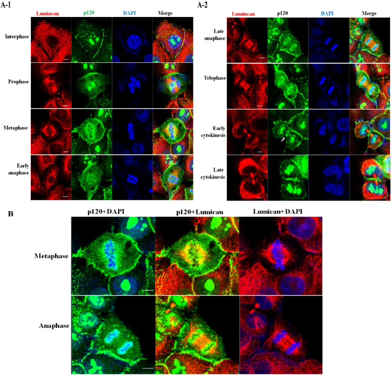
Abnormal expression of p120 catenin is associated with the malignant phenotype in human lung cancer. Numerous studies have focused on the function of p120 catenin in the juxta-membrane compartment. However, the role of nuclear p120 catenin remains unclear. In this study, the dynamic changes in nuclear p120 catenin localization during cell cycle progression were investigated. Immunofluorescent staining, FACS analysis, and western blotting revealed that nuclear p120 catenin is a major architectural constituent of the chromosome periphery during mitosis. During mitosis, granule-like p120 catenin dispersed into a cloudy-like structure and formed cordon-like structures surrounding the condensed chromosomes to create the peri-chromosomal layer. Interestingly, lumican and p120 catenin colocalized at the spindle fiber where the perichromosomal layer connects to the condensed chromosomes during mitosis. Furthermore, downregulation of p120 catenin using a specific siRNA induced cell cycle stalling in the G2/M phase and promoted aneuploidy. This study validates the role of nuclear p120 catenin in the formation of the chromosome periphery and reveals the p120 catenin-lumican interaction may couple orientation of cell division with the segregation of sister chromatids during mitosis. Our data suggest the protective role of p120 catenin in maintaining the integrity of chromosomes, and also warrants further studies to evaluate the contribution of the loss of p120 catenin to the creation of gene rearrangement in cancer evolution and tumor progression.


Conflict of interest statement

The authors declare no competing interests.

Figures

Fig. 1
Fig. 1. Downregulation of p120 catenin leads to G2/M arrest in H460/A549 cells.
Silencing of p120 catenin by transfection of a specific siRNA decreased the expression of p120 catenin. Lung cancer cells H460/A549 were transfected with NCi (negative control siRNA) or p120si (p120 catenin siRNA) and harvested at 24 h. A Western blotting of cell lysates with the indicated antibodies confirmed silencing of p120 catenin using p120si effectively decreased the expression of p120 catenin. The expression levels of p120 catenin and GAPDH were determined by densitometry and are expressed in arbitrary units (RLU, relative luminescent units). B FACS analysis of the cell cycle profile of transfected cells arrested at the beginning of the S phase using a double thymidine block. Cell synchronization was confirmed by flow cytometry of propidium iodide-stained cells. Representative flow cytometry analysis of the DNA content of NCi/p120si-transfected H460/A549 cells (lower panel). The number of cells (arbitrary units) is plotted against DNA content after release from thymidine block and was quantified using FloJo (right panel); N = 3. *p < 0.05 compared with the cells transfected with the control siRNA.
Fig. 2
Fig. 2. Dynamic morphology of p120 catenin during the processes of cell division.
A Subcellular distribution of p120 catenin in lung cancer cells. H460 cells were fixed and immunostained using anti-p120 catenin (green) and anti-lumican (red) antibodies. Cell nuclei were visualized by DAPI staining (blue). The arrows indicate the interactions of p120 catenin and lumican at the juxta-membrane region. The relative fluorescence intensity of p120 catenin (green) was quantified using Image J. Scale bar, 20 µm. B Distribution and morphology of p120 catenin during cell division. Cells were immunostained using anti-p120 catenin (green) and anti-α-tubulin antibodies (red). DNA was stained with DAPI (blue). Confocal images of cells in interphase, prophase, metaphase, and telophase (cytokinesis) are shown. C Dynamic changes in the distribution of p120 catenin were observed during the mitotic stage.
Fig. 3
Fig. 3. P120 catenin colocalizes with lumican at the boundary of the chromosome periphery.
H460 cells were immunostained using anti-lumican (red) and anti-p120 catenin (green) antibodies for 24 h. A Confocal images of cells in interphase, prophase, metaphase, and telophase (cytokinesis). B High-power merged images of p120 catenin + lumican and p120 catenin + DAPI in the cells in metaphase/anaphase are shown in (A).
Fig. 4
Fig. 4. Depletion of p120 catenin decreases formation of the perichromosomal layer.
A Depletion of p120si effectively decreased the expression of lumican. H460 cells were transfected with p120si and harvested at 24 h. The cell lysates were subjected to western blotting using anti-lumican and anti-p120 catenin antibodies. B Depletion of p120 catenin decreased the formation of the perichromosomal layer. The transfected cells were fixed and co-incubated with anti-lumican and anti-p120 catenin antibodies. Lumican (red) and p120 catenin (green) were detected by immunofluorescent staining in H460 cells. Cell nuclei were visualized by DAPI staining (blue). In addition, cells in the metaphase–telophase stage were observed.
Fig. 5
Fig. 5. Downregulation of p120 catenin induces aberrant mitosis.
P120 siRNA-transfected cells were stained using anti-p120 catenin and anti-lumican antibodies and DAPI to visualize the cell boundary and nuclei, respectively. A Multi-nucleated cells immunostained using the anti-p120 catenin antibody (green) and DAPI. Representative images of micro-nuclei/aneuploid cells (marked by the white arrows) in NCi- and p120si-transfected H460 cells. The letters and numbers in the panel represent the images captured in the multi-nucleated cells. Multi-nucleated cells are quantified in the lower panel. B Micronuclei were detected in p120si-transfected cells. C Immunostaining with anti-p120 (green) and anti-α tubulin (red) antibodies revealed a p120si-transfected cell with a chromatin bridge (indicated by an arrow) during anaphase.

References

    1. Varella-Garcia M. Chromosomal and genomic changes in lung cancer. Cell Adh Migr. 2010;4:100–6. doi: 10.4161/cam.4.1.10884. - DOI - PMC - PubMed
    1. Ben-David U, Amon A. Context is everything: aneuploidy in cancer. Nat Rev Genet. 2020;21:44–62. doi: 10.1038/s41576-019-0171-x. - DOI - PubMed
    1. Petersen I. Chromosomes, ploidy and differentiation of lung cancer. Cancer Res. 2004;64(7 Suppl):238.
    1. Ohta S, Wood L, Bukowski-Wills J-C, Rappsilber J, Earnshaw WC. Building mitotic chromosomes. Curr Opin Cell Biol. 2011;23:114–21. doi: 10.1016/j.ceb.2010.09.009. - DOI - PMC - PubMed
    1. Van Hooser AA, Yuh P, Heald R. The perichromosomal layer. Chromosoma. 2005;114:377–88. doi: 10.1007/s00412-005-0021-9. - DOI - PubMed

Publication types