. 2022 Jul 1;149(13):dev200426.
doi: 10.1242/dev.200426. Epub 2022 Jun 30.

Orb-dependent polyadenylation contributes to PLP expression and centrosome scaffold assembly

Affiliations

Orb-dependent polyadenylation contributes to PLP expression and centrosome scaffold assembly

Junnan Fang et al. Development. .

Abstract

As the microtubule-organizing centers of most cells, centrosomes engineer the bipolar mitotic spindle required for error-free mitosis. Drosophila Pericentrin-like protein (PLP) directs formation of a pericentriolar material (PCM) scaffold required for PCM organization and microtubule-organizing center function. Here, we investigate the post-transcriptional regulation of Plp mRNA. We identify conserved binding sites for cytoplasmic polyadenylation element binding (CPEB) proteins within the Plp 3'-untranslated region and examine the role of the CPEB ortholog Oo18 RNA-binding protein (Orb) in Plp mRNA regulation. Our data show that Orb interacts biochemically with Plp mRNA to promote polyadenylation and PLP protein expression. Loss of orb, but not orb2, diminishes PLP levels in embryonic extracts. Consequently, PLP localization to centrosomes and its function in PCM scaffolding are compromised in orb mutant embryos, resulting in genomic instability and embryonic lethality. Moreover, we find that PLP overexpression restores centrosome scaffolding and rescues the cell division defects caused by orb depletion. Our data suggest that Orb modulates PLP expression at the level of Plp mRNA polyadenylation and demonstrates that the post-transcriptional regulation of core, conserved centrosomal mRNAs is crucial for centrosome function.

Keywords: CPEB; Centrosome; PCM; Polyadenylation; Post-transcriptional regulation; RNA localization.


Conflict of interest statement

Competing interests The authors declare no competing or financial interests.

Figures

Fig. 1.
Fig. 1.
Plp mRNA localizes to centrosomes. (A-D) Maximum intensity projections of NC 12 control embryos expressing GFP-γTub (green) labeled with Plp smFISH probes (red) in interphase (A) and metaphase (B) or Gapdh1 smFISH probes (red) in interphase (C) and metaphase (D). Boxed regions are enlarged below. Arrowheads mark mRNA overlapping with centrosomes. (E) Quantification of Plp versus Gapdh1 mRNA localization to centrosomes at different NC stages. Table S1 lists the number of embryos, centrosomes and RNA objects quantified per condition. (F) Quantification of RNA localization to centrosomes in NC 13 interphase embryos reproduced from (E) then re-measured following 90° rotation of the RNA channel. (G) NC 13 Drosophila embryo expressing GFP-PLP (green) and labeled with Plp smFISH probes (red). Dashed ovals mark the pseudo-cells, and insets show Plp mRNA overlapping PLP protein (arrowheads). Mean±s.d. are displayed. n.s., not significant; ***P<0.001; ****P<0.0001 by one-way ANOVA followed by Dunnett's T3 multiple comparisons test. Scale bars: 5 μm (main panels); 1 μm (insets).
Fig. 2.
Fig. 2.
Plp mRNA associates with Orb. (A) Venn diagram showing the common mRNA targets of CPEB1 and Orb (Stepien et al., 2016; Pascual et al., 2021). Table S2 lists overlapping mRNA targets. (B) Alignment of the Plp 3′UTRs from Drosophila melanogaster (Dmel), D. simulans (Dsim), D. sechellia (Dsec), D. yakuba (Dyak), D. erecta (Dere), D. biarmipes (Dbii) and human (Hsap) PCNT 3′UTRs (Goldman-Huertas et al., 2015; Larkin et al., 2021). Consensus (red solid boxes) and non-consensus (red dashed boxes) CPE motifs, and canonical CPSF A(A/U)UAAA hexamers (green boxes) are indicated. The nucleotide number of D. mel Plp and human PCNT 3′UTRs are listed above. (C) RNA immunoprecipitation from GFP-Trap beads using WT and GFP-Orb ovarian extracts. Input shows 10% of the total RNA. cDNAs were amplified with the indicated primers: orb is a positive control (Tan et al., 2001), Gapdh1 is a negative control and Plp mRNA is 7.5-fold enriched in GFP-Orb relative to WT immunoprecipitated samples. Uncropped gels are available to view on Figshare: https://doi.org/10.6084/m9.figshare.16900417.v1.
Fig. 3.
Fig. 3.
Orb is dispensable for Plp mRNA localization. (A,B,D,E,G,H) Maximum intensity projections of NC 10 (A,B) or NC 12 (D,E,G,H) embryos of the indicated genotypes expressing GFP-γTub (green) and labeled with Plp or Gapdh1 smFISH probes (red). (C,F,I) Quantification of Plp or Gapdh1 mRNA localization to the centrosome surface in NC 10 or NC 12 embryos of the indicated genotypes. Table S1 lists the number of embryos, centrosomes and RNA objects quantified per condition. orb mutant: orbF343/orbmel mutant. Mean±s.d. are displayed. n.s., not significant by unpaired t-test. Scale bars: 5 μm.
Fig. 4.
Fig. 4.
Orb specifically promotes PLP protein expression. (A-D) Immunoblots showing PLP levels relative to the Khc (anti-SUK4) load control in 0- to 2-h-old Drosophila embryo extracts from the indicated genotypes. orb mutant: orbF343/orbmel; Plp/+, orb mutant: Plp2172/+, orbF343/orbmel. (E,F) Quantification of the relative levels of the upper MW PLP bands (carets) (E) and middle (mid-) MW PLP bands (arrowheads) (F) normalized to WT. Mean±s.d. are displayed. Significance was determined by one-way ANOVA with Dunnett's T3 multiple comparisons test relative to WT unless otherwise noted. n.s., not significant; *P<0.05; **P<0.01; ***P<0.001; ****P<0.0001. Uncropped blots are available to view on Figshare: https://doi.org/10.6084/m9.figshare.16900423.v1.
Fig. 5.
Fig. 5.
Orb promotes Plp polyadenylation. (A) Diagram showing the PCR primers and EcoRI and BmrI restriction sites on the short and long Plp 3′UTRs. The predicted sizes of internal PCR products are listed below. (B) cDNA products amplified from G/I-tailed RNA extracted from 0- to 2-h-old control (iso-1) and orbF343/orbmel embryos. PCR products were digested with EcoRI or BmrI, as indicated. The approximate lengths of poly(A)-tails are noted (arrowheads). (C) Relative levels of total Plp mRNA and PlpRM mRNA normalized to RP49 mRNA were examined by qRT-PCR in 0- to 2-h-old WT embryos; ****P<0.0001 by unpaired t-test. a.u., arbitrary units. (D) Line profiles from undigested (solid lines; lanes 9, 10) and EcoR1-digested (dashed lines; lanes 13, 14) PAT products. Poly(A)-tail length was calculated by subtracting the internal PCR product plus G/I tail length from the total PAT product length. Uncropped gel is available to view on Figshare: https://doi.org/10.6084/m9.figshare.16900471.v4.
Fig. 6.
Fig. 6.
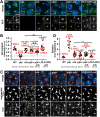
Formation of the PLP-Cnn PCM scaffold requires Orb. (A) Maximum intensity projections of NC 13 embryos of the indicated genotypes stained with anti-PLP antibodies. The same LUT was used to display PLP signals in all genotypes to highlight PLP within the flare-zone. Boxed regions are enlarged in the insets, and less-saturated images are shown in the yellow boxes. (B) Quantification of PLP intensity within 2 μm of the centrosome. Values were normalized to the mean intensity from WT embryos. Each circle represents the PLP intensity of one embryo averaged from all centrosomes in a 65 μm2 region. (C) Top and bottom: Maximum intensity projections of NC 13 embryos stained with anti-Cnn antibodies (red). Yellow arrowheads highlight Cnn fragments. Boxed regions are enlarged in the insets. Middle: Segmented Cnn images. (D) Quantification of the number of Cnn fragments normalized to the WT mean. Each circle represents the relative number of Cnn fragments of one embryo averaged from all centrosomes in a 65 μm2 region. Significance was determined by one-way ANOVA followed by Dunnett's T3 multiple comparisons test relative to WT. n.s., not significant; *P<0.05; **P<0.01; ***P<0.001; ****P<0.0001. Genotypes used were: orb mutant: orbF343/orbmel; Plp/+ hemizygote: Plp2172/+; Plp/+, orb mutant: Plp2172/+, orbF343/orbmel; PLP-GFP; orb mutant: PLP-GFP; orbF343/orbmel. Mean±s.d. are displayed. Scale bars: 5 μm (main panels); 2 μm (insets).
Fig. 7.
Fig. 7.
Orb functions upstream of PLP to support mitotic fidelity. (A) Maximum intensity projections of NC 12 telophase embryos labeled with anti-pH3 (red) and Asl (magenta) antibodies to visualize chromosome separation. Boxed regions are enlarged in the insets. Yellow arrowhead indicates an anaphase bridge. (B) Percentage of embryos with CIN. (C) Quantification of the proportion of CIN per embryo. Error bars show mean±s.d. (D,E) Maximum intensity projections of NC 13 interphase embryos labeled with DAPI to reveal NUF (D), with quantification in E. Significance was determined by χ2 analysis (B,E) or one-way ANOVA followed by Kruskal–Wallis multiple comparisons test (C). n.s., not significant; *P<0.05; **P<0.01; ****P<0.0001. orb mutant: orbF343/orbmel; PLP-GFP; orb mutant: PLP-GFP; orbF343/orbmel. Scale bars: 20 μm.
Fig. 8.
Fig. 8.
Orb promotes genome stability via PLP. Maximum intensity projected stills from time-lapse imaging of GFP-γTub or PLP-GFP with H2A-RFP in control (A), orbF303/orbmel mutant (B) and PLP-GFP; orbF303/orbmel (C) embryos. Time is in min:s. Dashed lines mark NUF. Stills correspond to Movies 1-3. (D) Proposed model for Orb-mediated regulation of Plp mRNA by promotion of Plp polyadenylation. Orb associates with Plp mRNA; this interaction may be direct through CPE motifs in the 3′UTR or indirect. Orb promotes the polyadenylation of Plp mRNA, which contributes to PLP protein synthesis. PLP expression supports the PLP-Cnn scaffold assembly necessary for centrosome function and error-free mitosis. Scale bar: 10 μm.

References

    1. Barkoff, A. F., Dickson, K. S., Gray, N. K. and Wickens, M. (2000). Translational control of cyclin B1 mRNA during meiotic maturation: coordinated repression and cytoplasmic polyadenylation. Dev. Biol. 220, 97-109. 10.1006/dbio.2000.9613 - DOI - PubMed
    1. Benoit, P., Papin, C., Kwak, J. E., Wickens, M. and Simonelig, M. (2008). PAP- and GLD-2-type poly(A) polymerases are required sequentially in cytoplasmic polyadenylation and oogenesis in Drosophila. Development 135, 1969-1979. 10.1242/dev.021444 - DOI - PMC - PubMed
    1. Blower, M. D., Feric, E., Weis, K. and Heald, R. (2007). Genome-wide analysis demonstrates conserved localization of messenger RNAs to mitotic microtubules. J. Cell Biol. 179, 1365-1373. 10.1083/jcb.200705163 - DOI - PMC - PubMed
    1. Brizuela, B. J., Elfring, L., Ballard, J., Tamkun, J. W. and Kennison, J. A. (1994). Genetic analysis of the brahma gene of Drosophila melanogaster and polytene chromosome subdivisions 72AB. Genetics 137, 803-813. 10.1093/genetics/137.3.803 - DOI - PMC - PubMed
    1. Burns, D. M. and Richter, J. D. (2008). CPEB regulation of human cellular senescence, energy metabolism, and p53 mRNA translation. Genes Dev. 22, 3449-3460. 10.1101/gad.1697808 - DOI - PMC - PubMed

Publication types

LinkOut - more resources