. 2022 Jun 4;13(1):99.
doi: 10.1186/s13244-022-01236-1.

Imaging in medically refractory epilepsy at 3 Tesla: a 13-year tertiary adult epilepsy center experience

Affiliations

Imaging in medically refractory epilepsy at 3 Tesla: a 13-year tertiary adult epilepsy center experience

Nicolin Hainc et al. Insights Imaging. .

Abstract

Objectives: MRI negative epilepsy has evolved through increased usage of 3 T and insights from surgically correlated studies. The goal of this study is to describe dedicated 3 T epilepsy MRI findings in medically refractory epilepsy (MRE) patients at a tertiary epilepsy center to familiarize radiologists with an updated spectrum and frequency of potential imaging findings in the adult MRE population.

Methods: Included were all patients with MRE admitted to the epilepsy monitoring unit who were discussed at weekly interdisciplinary imaging conferences at Toronto Western Hospital with MRI studies (3 T with dedicated epilepsy protocol) performed between January 2008 and January 2021. Lesion characterization was performed by two readers based on most likely imaging diagnosis in consensus. Lobes involved per case were recorded.

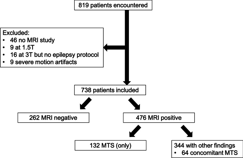
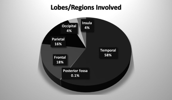
Results: A total of 738 patients (386 female; mean age 35 years, range 15-77) were included. A total of 262 patients (35.5%) were MRI negative. The most common imaging finding was mesial temporal sclerosis, seen in 132 patients (17.9%), followed by encephalomalacia and gliosis, either posttraumatic, postoperative, postischemic, or postinfectious in nature, in 79 patients (10.7%). The most common lobar involvement (either partially or uniquely) was temporal (341 cases, 58.6%). MRE patients not candidates for surgical resection were included in the study, as were newly described pathologies from surgically correlated studies revealing findings seen retrospectively on reported MRI negative exams (isolated enlargement of the amygdala, temporal pole white matter abnormality, temporal encephalocele).

Conclusion: This study provides an updated description of the spectrum of 3 T MRI findings in adult MRE patients from a tertiary epilepsy center.

Keywords: Drug resistant epilepsy; Epilepsy; Magnetic resonance imaging.


Conflict of interest statement

The authors of this manuscript declare no relationships with any companies, whose products or services may be related to the subject matter of the article.

Figures

Fig. 1
Fig. 1
Patients included in study including number of mesial temporal sclerosis (MTS) diagnoses
Fig. 2
Fig. 2
Pie chart of brain lobes/regions involved by the imaging diagnosis
Fig. 3
Fig. 3
Temporal lobe findings. a Subtle sign of left sided MTS with loss of the interdigitations of the pes hippocampi (arrow). b Right sided isolated enlargement of the amygdala (arrow). c White matter abnormality in the anterior left temporal lobe (arrow). d Suprasellar arachnoid cyst with compression of the left hippocampus (arrow). e Right temporopolar encephalocele with herniation of brain parenchyma (arrow) through a bony defect in the sphenoid (f, arrow, same patient as e). g Left sided MCA aneurysm with lamellated onion skin appearance (arrow) resulting in inferior displacement and compression of the left hippocampus (h, arrow, same patient as g)
Fig. 4
Fig. 4
Extratemporal findings: a Chronic left MCA infarct with concomitant left MTS (arrow, dual pathology). b Adult patient with birth-related findings of biparietal thinning of the white and gray matter with associated gliosis and ulegyria (arrow). c Cortically based, right parietal cavernoma with associated hemosiderin staining of the white matter. d and e (same patient) Focal cortical dysplasia with cortical thickening, blurring and FLAIR Hyperintensity of the gray-white matter junction (arrows)

References

    1. Ngugi AK, Kariuki SM, Bottomley C, et al. Incidence of epilepsy: a systematic review and meta-analysis. Neurology. 2011;77:1005–1012. doi: 10.1212/WNL.0b013e31822cfc90. - DOI - PMC - PubMed
    1. Picot M-C, Baldy-Moulinier M, Daurès J-P, et al. The prevalence of epilepsy and pharmacoresistant epilepsy in adults: a population-based study in a Western European country. Epilepsia. 2008;49:1230–1238. doi: 10.1111/j.1528-1167.2008.01579.x. - DOI - PubMed
    1. Kwan P, Brodie MJ. Early identification of refractory epilepsy. N Engl J Med. 2000;342:314–319. doi: 10.1056/NEJM200002033420503. - DOI - PubMed
    1. Téllez-Zenteno JF, Hernández Ronquillo L, Moien-Afshari F, Wiebe S. Surgical outcomes in lesional and non-lesional epilepsy: a systematic review and meta-analysis. Epilepsy Res. 2010;89:310–318. doi: 10.1016/j.eplepsyres.2010.02.007. - DOI - PubMed
    1. Phal PM, Usmanov A, Nesbit GM, et al. Qualitative comparison of 3-T and 1.5-T MRI in the evaluation of epilepsy. AJR Am J Roentgenol. 2008;191:890–895. doi: 10.2214/AJR.07.3933. - DOI - PubMed

LinkOut - more resources