Review
. 2022 Jun 5;79(6):345.
doi: 10.1007/s00018-022-04377-0.

Early human trophoblast development: from morphology to function

Affiliations
Review

Early human trophoblast development: from morphology to function

Martin Gauster et al. Cell Mol Life Sci. .

Abstract

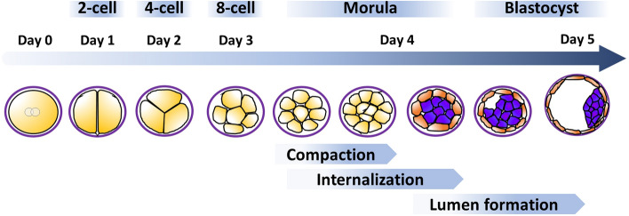
Human pregnancy depends on the proper development of the embryo prior to implantation and the implantation of the embryo into the uterine wall. During the pre-implantation phase, formation of the morula is followed by internalization of blastomeres that differentiate into the pluripotent inner cell mass lineage, while the cells on the surface undergo polarization and differentiate into the trophectoderm of the blastocyst. The trophectoderm mediates apposition and adhesion of the blastocyst to the uterine epithelium. These processes lead to a stable contact between embryonic and maternal tissues, resulting in the formation of a new organ, the placenta. During implantation, the trophectoderm cells start to differentiate and form the basis for multiple specialized trophoblast subpopulations, all of which fulfilling specific key functions in placentation. They either differentiate into polar cells serving typical epithelial functions, or into apolar invasive cells that adapt the uterine wall to progressing pregnancy. The composition of these trophoblast subpopulations is crucial for human placenta development and alterations are suggested to result in placenta-associated pregnancy pathologies. This review article focuses on what is known about very early processes in human reproduction and emphasizes on morphological and functional aspects of early trophoblast differentiation and subpopulations.

Keywords: Blastocyst; Extravillous trophoblast; Syncytialization; Trophectoderm; Villous trophoblast.


Conflict of interest statement

The authors declare that there are no competing interests.

Figures

Fig. 1
Fig. 1
Blastocyst formation. Fertilization of the oocyte gives rise to a diploid zygote (day 0) that undergoes three rounds of cleavage divisions until day 3 post fertilization. Thereafter, compaction of the arising blastomeres starts to arrange cells in a compact cell cluster. By day 4 post fertilization, the morula stage becomes finalized when the embryo is compacted and contains internalized cells (purple), giving rise to the inner cell mass. The outer cells (orange) become the trophectoderm and fluidic uptake leads to a growing lumen (blastocoel), flattening of the outer cell layer and arrangement of the inner cell mass on one pole (embryonic pole). Note that the blastocyst is still surrounded by the zona pellucida, a glycoprotein-rich membrane formed already during oogenesis. Adapted from [60]
Fig. 2
Fig. 2
Implantation and initial syncytium formation. A The blastocyst is attached to the uterine epithelium. This epithelium is covered by a massive glycocalyx. At the site of blastocyst attachment, the pinopodes of the uterine epithelial cells allow direct interaction between these cells and the trophectoderm cells of the embryonic pole of the blastocyst, bypassing the glycocalyx. B At the embryonic pole of the blastocyst, the trophectoderm cells start to proliferate and form multiple cell layers of daughter cells, the primitive cytotrophoblasts. Some of the primitive cytotrophoblasts further differentiate and fuse with each other to generate the primitive syncytiotrophoblast, a multinucleated trophoblast structure. Only this primitive syncytiotrophoblast seems to be able to penetrate the uterine epithelium, allowing invasion into the uterine stroma. C By means of the invading primitive syncytiotrophoblast, the embryo has passed the uterine epithelium and is now fully embedded in the uterine stroma. The primitive syncytiotrophoblast fully surrounds the underlying mono-nucleated cytotrophoblasts as well as the embryo and thus is the only embryonic layer in contact with maternal tissues. Already at this stage of placental development, the primitive syncytiotrophoblast invades into a uterine gland to allow nutritive support of the embryo using glandular secretion products, called uterine milk
Fig. 3
Fig. 3
Extravillous trophoblast invasion: The interstitial, endoglandular and endovascular trophoblast subtypes. Rows (a, b and c, d): Serial sections from first trimester decidua basalis (gestational age 7 weeks). Columns (a, c, b, d): Immuno double staining for HLA-G (dark brown) and keratin 7 (blue) (a, c) or HLA-G (brown) and von Willebrand factor (blue) (b, d). EVTs (brown) appear spindle-shaped or polygonal within the decidual stroma (ad). Glands (red asterisks) are invaded by endoglandular EVTs (arrows) from the interstitial side, glandular epithelium (blue in a, c) looks disintegrated in the invaded regions (a, c). EVTs (arrows) are also present within all types of uterine vessels (circles, endothelium stained in blue) (b, d). A clear differentiation of endovascular subtypes is not possible in such small vessels. Scale bar represents 50 µm, no nuclear counterstain

References

    1. Aberkane A, Essahib W, Spits C, De Paepe C, Sermon K, Adriaenssens T, Mackens S, Tournaye H, Brosens JJ, Van de Velde H. Expression of adhesion and extracellular matrix genes in human blastocysts upon attachment in a 2D co-culture system. Mol Hum Reprod. 2018;24:375–387. - PubMed
    1. Adam S, Elfeky O, Kinhal V, Dutta S, Lai A, Jayabalan N, Nuzhat Z, Palma C, Rice GE, Salomon C. Fetal-maternal communication via extracellular vesicles: implications for complications of pregnancies. Placenta. 2017;54:83–88. doi: 10.1016/j.placenta.2016.12.001. - DOI - PubMed
    1. Alikani M. Epithelial cadherin distribution in abnormal human pre-implantation embryos. Hum Reprod. 2005;20:3369–3375. doi: 10.1093/humrep/dei242. - DOI - PubMed
    1. Allerkamp HH, Clark AR, Lee TC, Morgan TK, Burton GJ, James JL. Something old, something new: digital quantification of uterine vascular remodelling and trophoblast plugging in historical collections provides new insight into adaptation of the utero-placental circulation. Hum Reprod. 2021;36:571–586. doi: 10.1093/humrep/deaa303. - DOI - PubMed
    1. Aoki R, Fukuda MN. Recent molecular approaches to elucidate the mechanism of embryo implantation: trophinin, bystin, and tastin as molecules involved in the initial attachment of blastocysts to the uterus in humans. Semin Reprod Med. 2000;18:265–271. doi: 10.1055/s-2000-12564. - DOI - PubMed

LinkOut - more resources