. 2022 May 19:10:899293.
doi: 10.3389/fbioe.2022.899293. eCollection 2022.

Biomimetic Design and Fabrication of Sericin-Hydroxyapatite Based Membranes With Osteogenic Activity for Periodontal Tissue Regeneration

Affiliations

Biomimetic Design and Fabrication of Sericin-Hydroxyapatite Based Membranes With Osteogenic Activity for Periodontal Tissue Regeneration

Piaoye Ming et al. Front Bioeng Biotechnol. .

Abstract

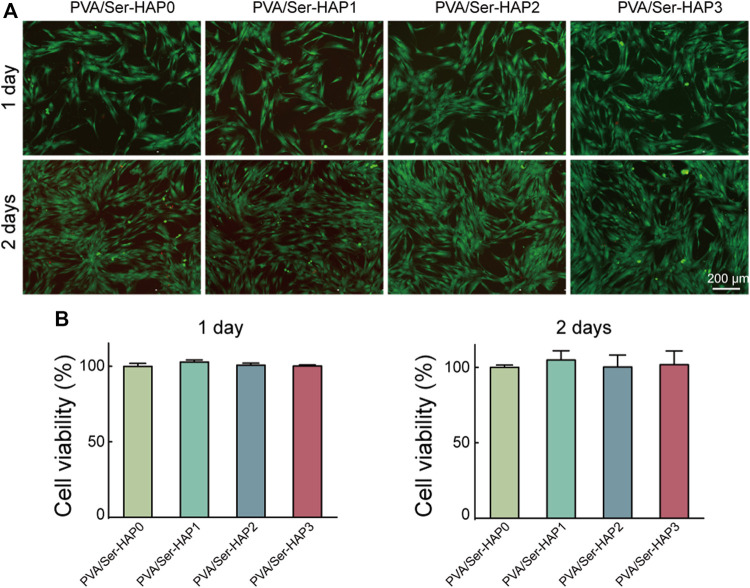
The guided tissue regeneration (GTR) technique is a promising treatment for periodontal tissue defects. GTR membranes build a mechanical barrier to control the ingrowth of the gingival epithelium and provide appropriate space for the regeneration of periodontal tissues, particularly alveolar bone. However, the existing GTR membranes only serve as barriers and lack the biological activity to induce alveolar bone regeneration. In this study, sericin-hydroxyapatite (Ser-HAP) composite nanomaterials were fabricated using a biomimetic mineralization method with sericin as an organic template. The mineralized Ser-HAP showed excellent biocompatibility and promoted the osteogenic differentiation of human periodontal membrane stem cells (hPDLSCs). Ser-HAP was combined with PVA using the freeze/thaw method to form PVA/Ser-HAP membranes. Further studies confirmed that PVA/Ser-HAP membranes do not affect the viability of hPDLSCs. Moreover, alkaline phosphatase (ALP) staining, alizarin red staining (ARS), and RT-qPCR detection revealed that PVA/Ser-HAP membranes induce the osteogenic differentiation of hPDLSCs by activating the expression of osteoblast-related genes, including ALP, Runx2, OCN, and OPN. The unique GTR membrane based on Ser-HAP induces the differentiation of hPDLSCs into osteoblasts without additional inducers, demonstrating the excellent potential for periodontal regeneration therapy.

Keywords: biomimetic membranes; human periodontal membrane stem cells; nano-hydroxyapatite; osteogenic differentiation; sericin.


Conflict of interest statement

The authors declare that the research was conducted in the absence of any commercial or financial relationships that could be construed as a potential conflict of interest.

Figures

SCHEME 1
SCHEME 1
A schematic illustration of the fabrication and application of PVA/Ser-HAP membrane. (A) The preparation process of PVA/Ser-HAP membrane; (B) Proposed schematic diagram depicting sericin-mediated nucleation of HAPs and preparation of PVA/Ser-HAP membrane; (C) The osteogenic differentiation of hPDLSCs on mineralized PVA/Ser-HAP membrane and potential periodontal tissue regeneration application for PVA/Ser-HAP membrane.
FIGURE 1
FIGURE 1
Surface morphology and characterization of Ser-HAP. (A) TEM micrographs of Ser-HAP particles; (B) TEM and HRTEM images of Ser100-HAP particles; (C) EDS analysis of Ser100-HAP particles; (D) XPS spectra; (E) FTIR spectra; and (F) XRD patterns of Ser-HAP particles.
FIGURE 2
FIGURE 2
Culture and identification of hPDLSCs. (A) Morphology of hPDLSCs; (B) Flow cytometry analysis of the indicated cell surface marker expression in hPDLSCs.
FIGURE 3
FIGURE 3
Cytocompatibility of sericin solution and the Ser-HAP particles. (A) Live/Dead staining and (B) CCK-8 assay of sericin at 1 and 2 days; (C) Live/Dead staining and (D) CCK-8 assay of Ser-HAP particles at 1 and 2 days.
FIGURE 4
FIGURE 4
ALP staining of hPDLSCs after culture with Ser-HAP for 3 days (A) and 5 days (B); Quantitative analysis of ALP staining at 3 (C) and 5 days (D).
FIGURE 5
FIGURE 5
Surface morphology and characterization of PVA/Ser-HAP membranes. (A) SEM micrographs of PVA/Ser-HAP membranes; (B) Elemental mapping of P and Ca in the PVA/Ser-HAP3 membrane; (C) Swelling and (D) degradation of the PVA/Ser-HAP membranes.
FIGURE 6
FIGURE 6
Biological compatibility of PVA/Ser-HAP membranes. Live/Dead staining (A) and CCK-8 assay (B) of hPDLSCs on the PVA/Ser-HAP membranes.
FIGURE 7
FIGURE 7
Morphology of hPDLSCs after seeding on PVA/Ser-HAP membranes for 3 days (A) and 5 days (B). Green, cytoskeleton stained with phalloidin; Blue, nuclei stained with DAPI.
FIGURE 8
FIGURE 8
(A) ALP staining of the hPDLSCs following cultured by extracts of PVA/Ser-HAP membranes for 3 days; (B) Alizarin red staining of the hPDLSCs after cultured by extracts of the membranes for 21 days; (C) Quantitative analysis of ALP staining at 3 days; (D) Quantitative analysis of Alizarin red staining at 21 days.
FIGURE 9
FIGURE 9
RT-qPCR analysis of the ALP, OCN, OPN, and Runx2 expression of hPDLSCs following cultured by extracts of the PVA/Ser-HAP membranes for 3 days (A) and 5 days (B).

References

    1. Aral K., Milward M. R., Kapila Y., Berdeli A., Cooper P. R. (2020). Inflammasomes and Their Regulation in Periodontal Disease: A Review. J. Periodont Res. 55 (4), 473–487. 10.1111/jre.12733 - DOI - PubMed
    1. Bal Z., Korkusuz F., Ishiguro H., Okada R., Kushioka J., Chijimatsu R., et al. (2021). A Novel Nano-Hydroxyapatite/synthetic Polymer/bone Morphogenetic Protein-2 Composite for Efficient Bone Regeneration. Spine J. 21 (5), 865–873. 10.1016/j.spinee.2021.01.019 - DOI - PubMed
    1. Cai Y., Mei D., Jiang T., Yao J. (2010). Synthesis of Oriented Hydroxyapatite Crystals: Effect of Reaction Conditions in the Presence or Absence of Silk Sericin. Mater. Lett. 64 (24), 2676–2678. 10.1016/j.matlet.2010.08.071 - DOI
    1. Catori D. M., Fragal E. H., Messias I., Garcia F. P., Nakamura C. V., Rubira A. F. (2021). Development of Composite Hydrogel Based on Hydroxyapatite Mineralization over Pectin Reinforced with Cellulose Nanocrystal. Int. J. Biol. Macromol. 167, 726–735. 10.1016/j.ijbiomac.2020.12.012 - DOI - PubMed
    1. Chen X., Liu Y., Yang J., Wu W., Miao L., Yu Y., et al. (2016). The Synthesis of Hydroxyapatite with Different Crystallinities by Controlling the Concentration of Recombinant CEMP1 for Biological Application. Mater. Sci. Eng. C 59, 384–389. 10.1016/j.msec.2015.10.029 - DOI - PubMed