Altered thalamic subregion functional networks in patients with treatment-resistant schizophrenia
- PMID: 35663295
- PMCID: PMC9150031
- DOI: 10.5498/wjp.v12.i5.693
Altered thalamic subregion functional networks in patients with treatment-resistant schizophrenia
Abstract
Background: The thalamus plays a key role in filtering information and has extensive interconnectivity with other brain regions. A large body of evidence points to impaired functional connectivity (FC) of the thalamocortical pathway in schizophrenia. However, the functional network of the thalamic subregions has not been investigated in patients with treatment-resistant schizophrenia (TRS).
Aim: To identify the neural mechanisms underlying TRS, we investigated FC of thalamic sub-regions with cortical networks and voxels, and the associations of this FC with clinical symptoms. We hypothesized that the FC of thalamic sub-regions with cortical networks and voxels would differ between TRS patients and HCs.
Methods: In total, 50 patients with TRS and 61 healthy controls (HCs) matched for age, sex, and education underwent resting-state functional magnetic resonance imaging (rs-fMRI) and clinical evaluation. Based on the rs-fMRI data, we conducted a FC analysis between thalamic subregions and cortical functional networks and voxels, and within thalamic subregions and cortical functional networks, in the patients with TRS. A functional parcellation atlas was used to segment the thalamus into nine subregions. Correlations between altered FC and TRS symptoms were explored.
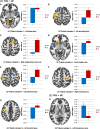
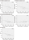
Results: We found differences in FC within thalamic subregions and cortical functional networks between patients with TRS and HCs. In addition, increased FC was observed between thalamic subregions and the sensorimotor cortex, frontal medial cortex, and lingual gyrus. These abnormalities were associated with the pathophysiology of TRS.
Conclusion: Our findings suggest that disrupted FC within thalamic subregions and cortical functional networks, and within the thalamocortical pathway, has potential as a marker for TRS. Our findings also improve our understanding of the relationship between the thalamocortical pathway and TRS symptoms.
Keywords: Functional connectivity; Rs-fMRI; Thalamocortical pathway; Thalamus; Treatment-resistant schizophrenia.
©The Author(s) 2022. Published by Baishideng Publishing Group Inc. All rights reserved.
Conflict of interest statement
Conflict-of-interest statement: No benefits in any form have been received or will be received from a commercial party related directly or indirectly to the subject of this article.
Figures


Similar articles
-
Altered functional connectivity in anterior cingulate cortex subregions in treatment-resistant schizophrenia patients.Neurosci Lett. 2023 Sep 25;814:137445. doi: 10.1016/j.neulet.2023.137445. Epub 2023 Aug 18. Neurosci Lett. 2023. PMID: 37597741
-
Subregional thalamic functional connectivity abnormalities and cognitive impairments in first-episode schizophrenia.Asian J Psychiatr. 2024 Jun;96:104042. doi: 10.1016/j.ajp.2024.104042. Epub 2024 Apr 2. Asian J Psychiatr. 2024. PMID: 38615577
-
Exploring the thalamus: a crucial hub for brain function and communication in patients with bulimia nervosa.J Eat Disord. 2023 Nov 20;11(1):207. doi: 10.1186/s40337-023-00933-6. J Eat Disord. 2023. PMID: 37986127 Free PMC article.
-
Resting-state functional connectivity in treatment response and resistance in schizophrenia: A systematic review.Schizophr Res. 2019 Sep;211:10-20. doi: 10.1016/j.schres.2019.07.020. Epub 2019 Jul 19. Schizophr Res. 2019. PMID: 31331784
-
Thalamic pathology in schizophrenia.Curr Top Behav Neurosci. 2010;4:509-28. doi: 10.1007/7854_2010_55. Curr Top Behav Neurosci. 2010. PMID: 21312411 Review.
Cited by
-
Consistent frontal-limbic-occipital connections in distinguishing treatment-resistant and non-treatment-resistant schizophrenia.Neuroimage Clin. 2025;45:103726. doi: 10.1016/j.nicl.2024.103726. Epub 2024 Dec 12. Neuroimage Clin. 2025. PMID: 39700898 Free PMC article.
-
Treatment-resistant schizophrenia: How far have we traveled?Front Psychiatry. 2022 Aug 30;13:994425. doi: 10.3389/fpsyt.2022.994425. eCollection 2022. Front Psychiatry. 2022. PMID: 36111312 Free PMC article.
-
Resting-state functional MRI in treatment-resistant schizophrenia.Front Neuroimaging. 2023 Apr 6;2:1127508. doi: 10.3389/fnimg.2023.1127508. eCollection 2023. Front Neuroimaging. 2023. PMID: 37554635 Free PMC article. Review.
References
-
- Janssen J, Alemán-Gómez Y, Reig S, Schnack HG, Parellada M, Graell M, Moreno C, Moreno D, Mateos-Pérez JM, Udias JM, Arango C, Desco M. Regional specificity of thalamic volume deficits in male adolescents with early-onset psychosis. Br J Psychiatry . 2012;200:30–36. - PubMed
LinkOut - more resources
Full Text Sources

