. 2022 May 27;11(6):e1396.
doi: 10.1002/cti2.1396. eCollection 2022.

Increased amphiregulin expression by CD4+ T cells from individuals with asymptomatic Leishmania donovani infection

Affiliations

Increased amphiregulin expression by CD4+ T cells from individuals with asymptomatic Leishmania donovani infection

Siddharth Sankar Singh et al. Clin Transl Immunology. .

Abstract

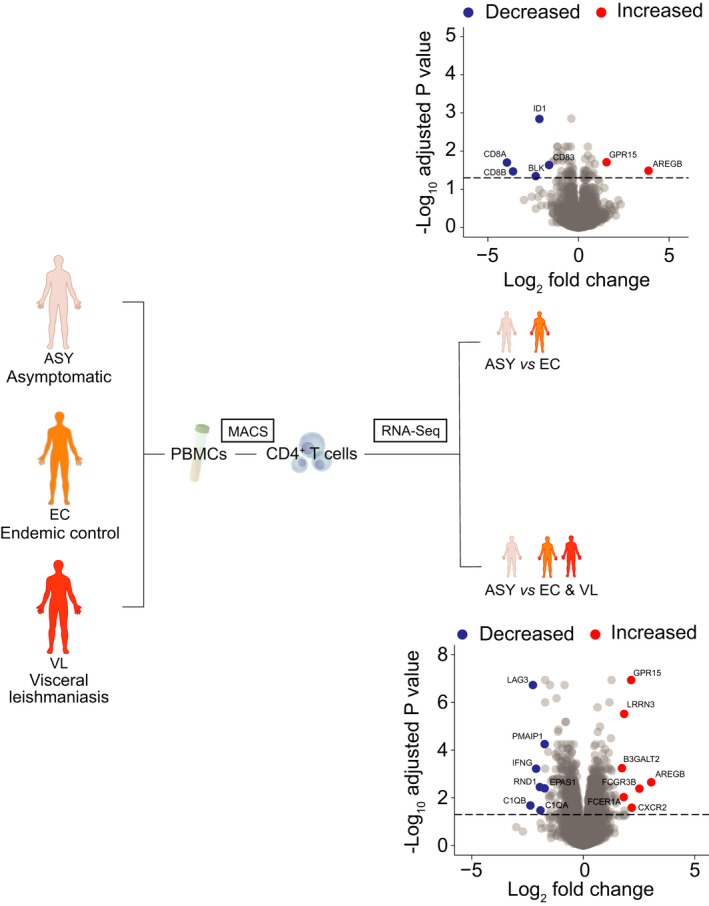
Objectives: There is an urgent need to be able to identify individuals with asymptomatic Leishmania donovani infection, so their risk of progressing to VL and transmitting parasites can be managed. This study examined transcriptional markers expressed by CD4+ T cells that could distinguish asymptomatic individuals from endemic controls and visceral leishmaniasis (VL) patients.

Methods: CD4+ T cells were isolated from individuals with asymptomatic L. donovani infection, endemic controls and VL patients. RNA was extracted and RNAseq employed to identify differentially expressed genes. The expression of one gene and its protein product during asymptomatic infection were evaluated.

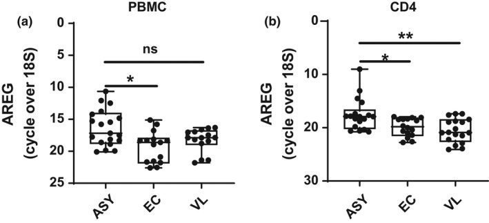
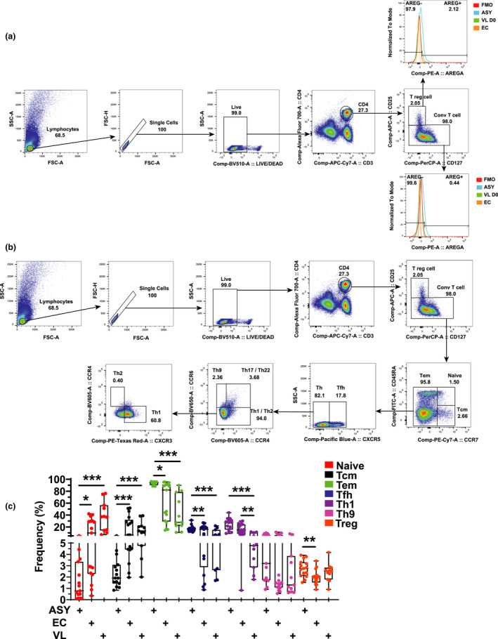
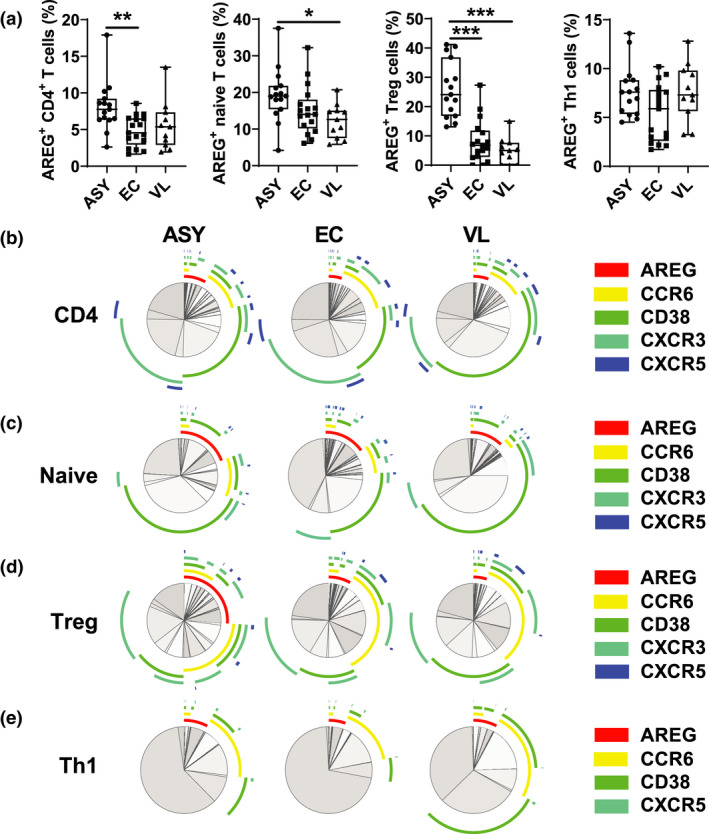
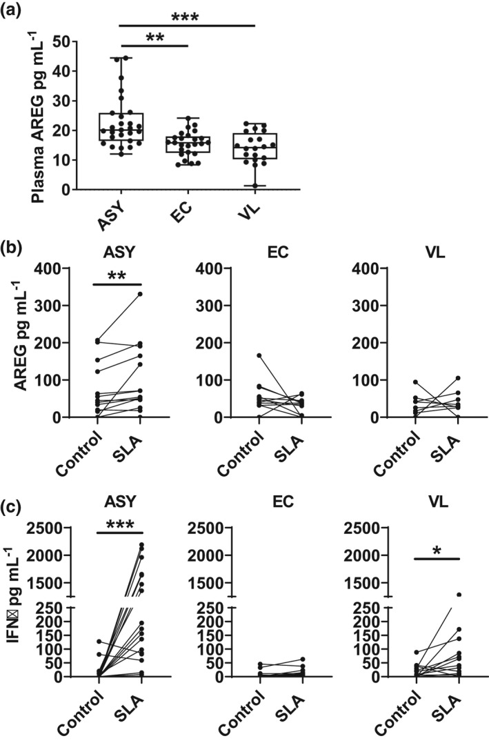
Results: Amphiregulin (AREG) was identified as a distinguishing gene product in CD4+ T cells from individuals with asymptomatic L. donovani infection, compared to VL patients and healthy endemic control individuals. AREG levels in plasma and antigen-stimulated whole-blood assay cell culture supernatants were significantly elevated in asymptomatic individuals, compared to endemic controls and VL patients. Regulatory T (Treg) cells were identified as an important source of AREG amongst CD4+ T-cell subsets in asymptomatic individuals.

Conclusion: Increased Treg cell AREG expression was identified in individuals with asymptomatic L. donovani infection, suggesting the presence of an ongoing inflammatory response in these individuals required for controlling infection and that AREG may play an important role in preventing inflammation-induced tissue damage and subsequent disease in asymptomatic individuals.

Keywords: CD4+ T cell; Leishmania donovani; amphiregulin; regulatory T cell; visceral leishmaniasis.


Conflict of interest statement

The authors declare no conflict of interest.

Figures

Figure 1
Figure 1
Defining a transcriptomic signature of peripheral blood CD4+ T cells from asymptomatic (ASY) individuals infected with Leishmania donovani. A schematic showing the approach taken to identify transcriptomic signatures of peripheral blood CD4+ T cells from ASY individuals, endemic controls (EC) and visceral leishmaniasis (VL) patients (n = 12 biological replicates in each group). A volcano plot of the differentially expressed genes identified in the comparison of asymptomatic versus endemic controls (upper plot) and ASY versus the average of VL patients and EC. Genes are coloured red (upregulated) or blue (downregulated) according to a false discovery rate < 0.05. Gene labels are shown for selected genes.
Figure 2
Figure 2
Amphiregulin (AREG) is elevated in CD4+ T cells from asymptomatic individuals infected with Leishmania donovani. RNA was isolated from peripheral blood mononuclear cells (PBMCs) (a) and CD4+ T cells (b) isolated from asymptomatic (ASY; n = 19) individuals, endemic controls (EC; n = 15) and visceral leishmaniasis (VL; n = 15) patients and subjected to qPCR to measure AREG mRNA levels. Median + minimum and maximum are shown; ns = not significant, *P < 0.05, **P < 0.01. Significance was assessed by the Kruskal–Wallis test.
Figure 3
Figure 3
Changes in CD4+ T‐cell subset frequencies and expression of AREG. CD4+ T cells were identified by CD3ε and CD4 expression prior to being divided into regulatory T (Treg) cells and conventional T cells, based on CD25 and CD127 expression, before assessing AREG expression (a). CD4+ T cells were divided into T helper cell subsets based on chemokine receptor expression (b) and frequencies measured in peripheral blood from asymptomatic (ASY; n = 15) individuals, endemic controls (EC; n = 17) visceral leishmaniasis patients (VL; n = 11). The box shows the extent of the lower and upper quartiles plus the median, while the whiskers indicate the minimum and maximum data points (c). *P < 0.05, **P < 0.01 and ***P < 0.001. Significance was assessed by the Kruskal–Wallis test.
Figure 4
Figure 4
Amphiregulin (AREG) expression by CD4+ T‐cell subsets. The frequency of AREG+ CD4+ T‐cell subsets was measured by FACS. The box shows the extent of the lower and upper quartiles plus the median, while the whiskers indicate the minimum and maximum data points (a). A simplified presentation of incredibly complex evaluations (SPICE) was used to establish overlap in expression of AREG, CD38 and indicated chemokine receptors by all CD4+ T cells (b), naive CD4+ T cells (c), regulatory T (Treg) cells (d) and Th1 cells (e) from asymptomatic (ASY; n = 15) individuals, endemic controls (EC; n = 17) and visceral leishmaniasis (V; n = 11) patients, as indicated. *P < 0.05, **P < 0.01 and ***P < 0.001. Significance was assessed by a one‐way ANOVA with a Tukey’s multiple comparisons test (a).
Figure 5
Figure 5
Amphiregulin (AREG) is a marker of asymptomatic L. donovani infection. AREG was measured in plasma from (ASY; n = 27) individuals, endemic controls (EC; n = 25) and visceral leishmaniasis (VL; n = 20) patients (a), as well as from supernatants from whole blood assays stimulated with soluble Leishmania antigen (SLA) or media alone (control), as indicated, from ASY (n = 15), EC (n = 12) and VL (n = 12) (b). IFNγ was measured from the same WBA cell culture supernatants (c). The box shows the extent of the lower and upper quartiles plus the median, while the whiskers indicate the minimum and maximum data points (a). Paired samples are shown in b and c; *P < 0.05, **P < 0.01 and ***P < 0.001. Significance was assessed by the Kruskal–Wallis test (a) or the Wilcoxon matched‐pairs signed rank test (b, c).

References

    1. Stauch A, Sarkar RR, Picado A et al. Visceral leishmaniasis in the Indian subcontinent: modelling epidemiology and control. PLoS Negl Trop Dis 2011; 5: e1405. - PMC - PubMed
    1. Ismael A, Zijlstra EE, Ghalib HW, El‐Hassan AM. Endemic kala‐azar in eastern Sudan: a longitudinal study on the incidence of clinical and subclinical infection and post‐kala‐azar dermal leishmaniasis. Am J Trop Med Hyg 1994; 51: 826–836. - PubMed
    1. Schaefer KU, Kurtzhals JA, Gachihi GS, Muller AS, Kager PA. A prospective sero‐epidemiological study of visceral leishmaniasis in Baringo District, Rift Valley Province, Kenya. Trans R Soc Trop Med Hyg 1995; 89: 471–475. - PubMed
    1. Ali A, Ashford RW. Visceral leishmaniasis in Ethiopia. IV. Prevalence, incidence and relation of infection to disease in an endemic area. Ann Trop Med Parasitol 1994; 88: 289–293. - PubMed
    1. Evans TG, Teixeira MJ, McAuliffe IT et al. Epidemiology of visceral leishmaniasis in northeast Brazil. J Infect Dis 1992; 166: 1124–1132. - PubMed