. 2022 May 18:13:861486.
doi: 10.3389/fimmu.2022.861486. eCollection 2022.

RGMa Participates in the Blood-Brain Barrier Dysfunction Through BMP/BMPR/YAP Signaling in Multiple Sclerosis

Affiliations

RGMa Participates in the Blood-Brain Barrier Dysfunction Through BMP/BMPR/YAP Signaling in Multiple Sclerosis

Lei Zhang et al. Front Immunol. .

Abstract

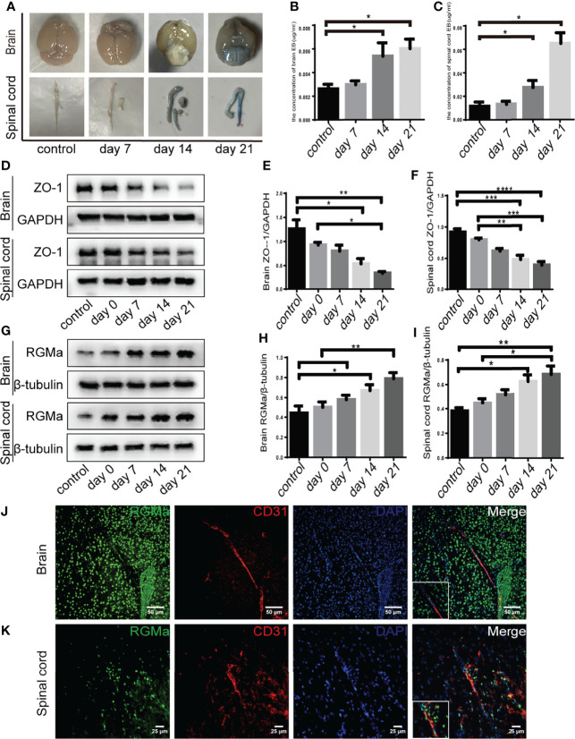
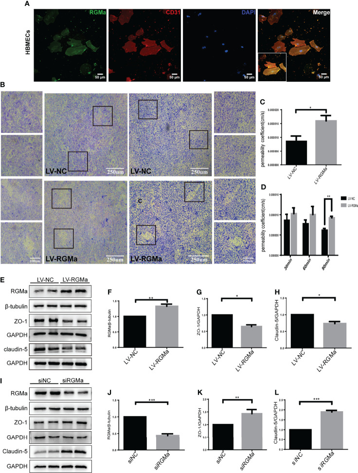
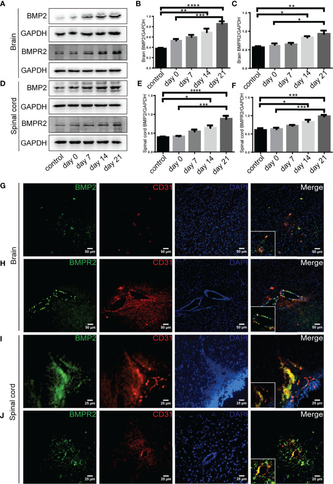
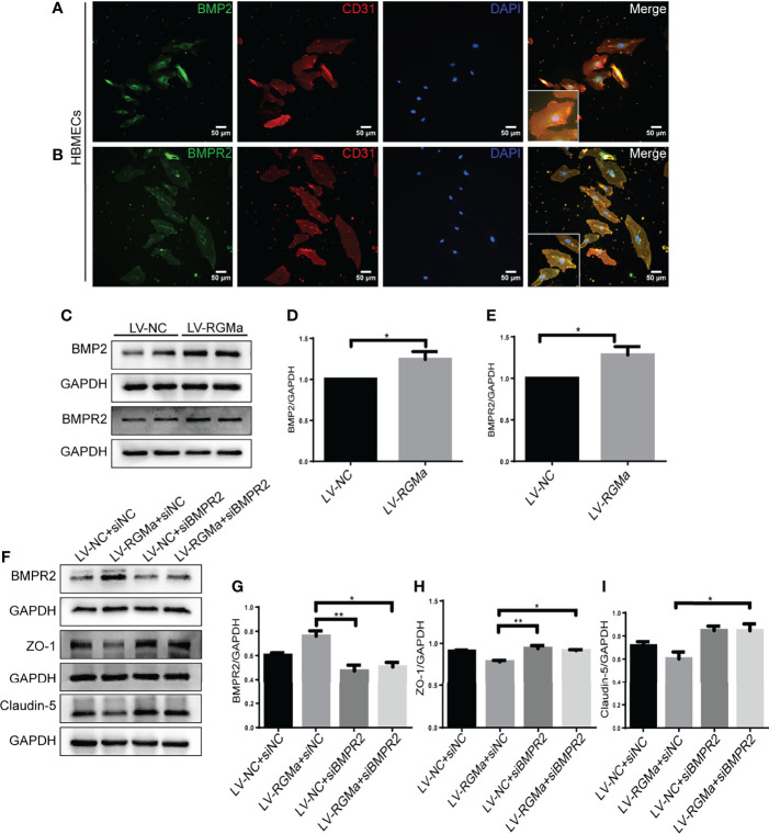
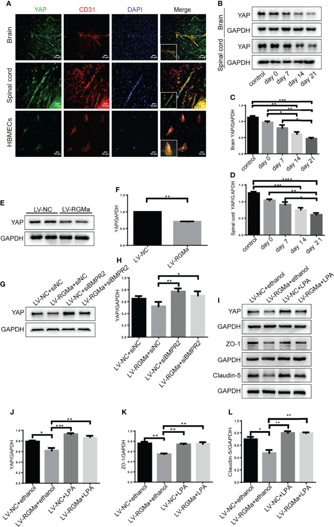
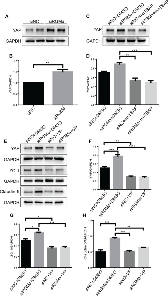
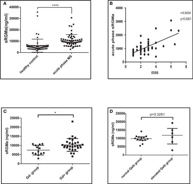
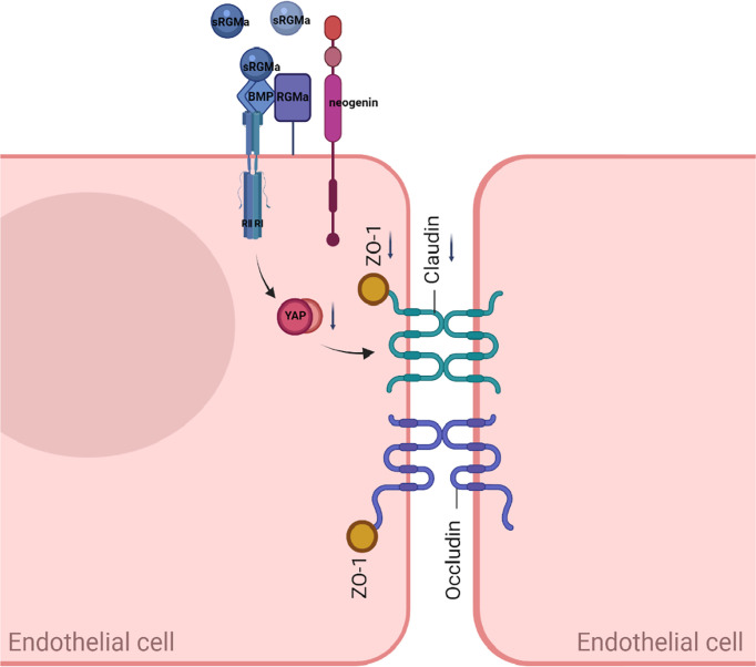
The infiltration of inflammatory cells into the central nervous system (CNS) through the dysfunctional blood-brain barrier (BBB) was critical in the early stages of MS. However, the mechanisms underlying BBB dysfunction remain unknown. Repulsive guidance molecule-a (RGMa) is involved in the pathogenesis of multiple sclerosis (MS), but its role needs to be further explored. This study aimed to evaluate whether RMGa regulates BBB permeability in endothelial cells and MS, and if so, what mechanism may be involved. We created an experimental autoimmune encephalomyelitis (EAE) model in C57BL/6 mice and a human brain microvascular endothelial cell (HBMEC) culture. The permeability of the BBB is measured in response to various interventions. Our results showed that RGMa is expressed in the endothelial cells in HBMECs and EAE mice. RGMa and its signaling counterpart, bone morphogenetic protein 2 (BMP2)/bone morphogenetic protein receptor type II (BMPRII), were gradually increased as the disease progressed. Moreover, as EAE progressed and the BBB was disrupted, the downstream effector, yes-associated protein (YAP), as well as the tight junctional proteins zonula occludens 1 (ZO-1) and claudin-5, decreased significantly. The permeability assay revealed that lentivirus-induced RGMa overexpression in HBMECs caused a significant breakdown of the BBB, whereas RGMa knockdown significantly strengthens the integrity of the BBB. Furthermore, specifically activating BMPR II or inhibiting YAP based on RGMa knockdown results in a significant decrease of ZO-1 and claudin-5 in vitro. On the contrary, inhibition of BMPR II or activation of YAP after upregulating RGMa prevents the downregulation of ZO-1 and claudin-5 in HBMECs. In addition, serum-soluble RGMa (sRGMa) levels were significantly higher in MS patients, particularly in MS patients with Gd+ lesions, indicating that the BBB has been disrupted. In conclusion, this study shows that RGMa causes BBB dysfunction in endothelial cells via BMP2/BMPR II/YAP, resulting in BBB integrity disruption in MS and that it could be a novel therapeutic target for BBB permeability in MS.

Keywords: blood–brain barrier; bone morphogenetic protein; multiple sclerosis; repulsive guidance molecule-a; yes-associated protein.


Conflict of interest statement

The authors declare that the research was conducted in the absence of any commercial or financial relationships that could be construed as a potential conflict of interest.

Figures

Figure 1
Figure 1
RGMa is associated with blood–brain barrier disruption in EAE mice. (A–C) Quantitative analysis of Evans Blue Dye staining showed BBB disruption was significantly higher in both the 14-day group and the 21-day group compared with the control group (control vs. 14 days: brain, * p < 0.05; spinal cord, **** p < 0.0005; control vs. day 21: brain, * p < 0.05; spinal cord, * p < 0.05). (D–F) In both the brain and spinal cord, ZO-1 expression levels were decreased at both 14 and 21 days compared with the control group or 0-day group. (G–I) RGMa expression was significantly increased in the brain and spinal cord at 14 and 21 days compared with the control or 0-day groups, and significantly increased at the 21-day group compared with 7-day group (n = 4, error bar: SEM; * p < 0.05, ** p < 0.01, *** p < 0.001, **** p < 0.0001, one-way ANOVA with Bonferroni). (J–K) RGMa and CD31+ endothelial cells were found colocalized in the brain cortex and spinal cord white matter. Scale bar: 50 µm (brain), 25 µm (spinal cord).
Figure 2
Figure 2
RGMa deteriorates the integrity of the in vitro BBB by HBMECs. (A) RGMa was expressed in CD31+ HBMECs. Scale bar: 50 µm. (B) The permeability of in vitro BBB was detected by crystal violet staining and FITC-dextran-4 kDa (right) or 70 kDa (left) assay. Overexpression of RGMa significantly exacerbates BBB permeability (error bar: SEM; ** p < 0.01, scale bar: 250 µm). (C) After 90 min, the permeability coefficient of FITC-dextran-70 kDa in the LV-NC and LV-RGMa groups. (D) After 90 min, the permeability coefficient of FITC-dextran-4 kDa in the LV-NC and LV-RGMa groups. (E–L) By specifically upregulating or downregulating RGMa, the expression of ZO-1 and claudin-5 were accordingly decreased or increased (n = 3, error bar: SEM; * p < 0.05, ** p < 0.01, *** p < 0.001, t-test).
Figure 3
Figure 3
BMP2 and BMPR II expression in EAE mice. (A–F) BMP2 and BMPR II in the brain and spinal cord were both upregulated in the 14- and 21-day group compared with the control group. BMP2 and BMPR II in both the brain and spinal cord were significantly higher in the 21-day than the 0-day groups (n = 4, error bar: SEM; * p < 0.05, ** p < 0.01, *** p < 0.001, **** p < 0.0001, one-way ANOVA with Bonferroni). (G–J) Immunofluorescence showed BMP2 and BMPR II were located in CD31+ endothelial cells in both the brain cortex and the white matter of the spinal cord. Scale bar: 50 µm (brain), 25 µm (spinal cord).
Figure 4
Figure 4
BMPR II mediates the integrity of the BBB induced by RGMa. (A, B) Immunofluorescence showed BMP2 and BMPR II were expressed in HBMECs. Scale bar: 50 µm. (C–E) Both BMP2 and BMPR II expression levels were significantly increased after overexpressing RGMa (n = 3, * p < 0.05, t-test). (F–I) On the basis of overexpressing RGMa, specifically silencing BMPR II significantly augments ZO-1 and claudin-5 expression (n = 3, * p < 0.05, t-test). **p < 0.01.
Figure 5
Figure 5
RGMa regulates BMP2 and thereby tight junctional protein expression. (A–C) BMP2 and BMPR II expression were significantly decreased after RGMa knockdown in vitro BBB (n = 3, * p < 0.05, t-test). (D–G) After activating BMPR II by mnTBAP 100 µM for 1 h based on inhibiting RGMa, ZO-1 and claudin-5 expression levels were significantly reduced (n = 3, * p < 0.05, t-test). ****p < 0.0001.
Figure 6
Figure 6
YAP regulates RGMa-induced integrity of the BBB. (A) Immunofluorescence showed that YAP was expressed in CD31+ endothelial cell both in EAE mice and HBMECs. Scale bar: 50 µm (brain cortex and HBMECs) and 25 µm (spinal cord). (B–D) YAP expression gradually decreased with the progression of EAE. (n = 4, error bar: SEM; * p < 0.05, ** p < 0.01, *** p < 0.001, **** p < 0.0001, one-way ANOVA with Bonferroni). (E, F) YAP expression was significantly diminished when overexpressing RGMa in vitro (n = 3, ** p < 0.01, t-test). (G, H) The YAP expression level was ameliorated when blocking BMPR II (n = 3, * p < 0.05, t-test). (I–K) By overexpressing YAP by LPA 20 µM for 2 h after overexpressing RGMa, both ZO-1 and claudin-5 were significantly increased (n = 3, ** p < 0.01, t-test).
Figure 7
Figure 7
Inhibiting YAP augments the permeability of the BBB and mediates via RGMa/BMP pathway. (A–D) Western blot analysis showed that YAP was significantly upregulated in the siRGMa group (n = 3, **p < 0.01, t-test) but significantly decreased when BMPR II was activated for 1 h with mnTBAP 100 µM (n = 3, ***p < 0.001). (E–H) YAP, ZO-1, and claudin-5 levels were significantly reduced when YAP expression was specifically inhibited by VP 2 µM for 24 h on the basis of RGMa knockdown (n = 3, *p < 0.05, ** p < 0.01, *** p < 0.001, **** p < 0.0001, t-test).
Figure 8
Figure 8
Serum RGMa expression and correlation with BBB permeability in MS patients. (A) ELISA detected a significantly higher level of sRGMa expression in acute phase MS patients than in the HC group (10,706 ± 689.3 ng/ml vs. 6,081 ± 723.0 ng/ml, error bar: SD; **** p < 0.0001). (B) Pearson correlation coefficient showed a significant correlation between EDSS at admission and sRGMa in acute phase MS (p < 0.0001, r 2 = 0.6434). (C) ELISA detected significantly higher levels of sRGMa expression in the Gd+ group than in the Gd group by (7,448 ± 734.5 ng/ml vs. 10,283 ± 650.6 ng/ml, error bar: SD; * p < 0.05). (D) ELISA detected a slightly increased level of sRGMa expression in the elevated Qalb group than in the normal Qalb group (9,883 [8,592, 10,993] vs. 11,956 [5,499, 17,517] ng/ml, median, 95% CI, p = 0.3281, Kolmogorov–Smirnov test).
Figure 9
Figure 9
A proposed model of RGMa regulation of BMP2/BMPR II/YAP signaling in endothelial cells. The overexpression of RGMa forms a complex by binding BMP2 and BMPR II, then attenuates YAP expression, which promotes the disruption of the BBB. By regulating the BMP2/BMPR II/YAP pathway, RGMa mediates dysfunction of the blood–brain barrier.

References

    1. Dobson R, Giovannoni G. Multiple Sclerosis - A Review. Eur J Neurol (2019) 26:27–40. doi: 10.1111/ene.13819 - DOI - PubMed
    1. Chan VS-F. Epigenetics in Multiple Sclerosis. In Epigenetics in Allergy and Autoimmunity. Lu CCQ, editor. Singapore: Springer Nature Singapore Pte Ltd; (2020) p. 309–74.
    1. Lassmann H, van Horssen J, Mahad D. Progressive Multiple Sclerosis: Pathology and Pathogenesis. Nat Rev Neurol (2012) 8:647–56. doi: 10.1038/nrneurol.2012.168 - DOI - PubMed
    1. Gaitan MI, Shea CD, Evangelou IE, Stone RD, Fenton KM, Bielekova B, et al. Evolution of the Blood-Brain Barrier in Newly Forming Multiple Sclerosis Lesions. Ann Neurol (2011) 70:22–9. doi: 10.1002/ana.22472 - DOI - PMC - PubMed
    1. Tassew NG, Mothe AJ, Shabanzadeh AP, Banerjee P, Koeberle PD, Bremner R, et al. Modifying Lipid Rafts Promotes Regeneration and Functional Recovery. Cell Rep (2014) 8:1146–59. doi: 10.1016/j.celrep.2014.06.014 - DOI - PubMed

Publication types