Review
. 2022 May 19:13:820442.
doi: 10.3389/fgene.2022.820442. eCollection 2022.

Evolutionary Genomics Reveals Multiple Functions of Arylalkylamine N-Acetyltransferase in Fish

Affiliations
Review

Evolutionary Genomics Reveals Multiple Functions of Arylalkylamine N-Acetyltransferase in Fish

Yu Huang et al. Front Genet. .

Abstract

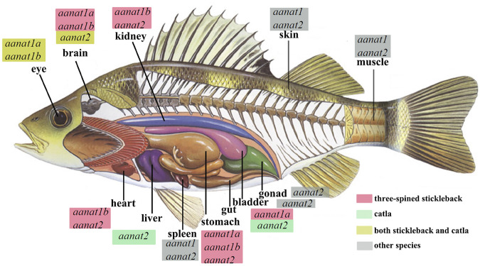
As an important hormone, melatonin participates in endocrine regulation of diverse functions in vertebrates. Its biosynthesis is catalyzed by four cascaded enzymes, among them, arylalkylamine N-acetyltransferase (AANAT) is the most critical one. Although only single aanat gene has been identified in most groups of vertebrates, researchers including us have determined that fish have the most diverse of aanat genes (aanat1a, aanat1b, and aanat2), playing various potential roles such as seasonal migration, amphibious aerial vision, and cave or deep-sea adaptation. With the rapid development of genome and transcriptome sequencing, more and more putative sequences of fish aanat genes are going to be available. Related phylogeny and functional investigations will enrich our understanding of AANAT functions in various fish species.

Keywords: arylalkylamine N-acetyltransferase (AANAT); fish; melatonin biosynthesis; phylogeny; physiological function.


Conflict of interest statement

The authors YH, CB, RL, XY, and QS were employed by BGI Academy of Marine Sciences. The remaining author declares that the research was conducted in the absence of any commercial or financial relationships that could be construed as a potential conflict of interest.

Figures

FIGURE 1
FIGURE 1
The substrates of AANAT and one of its products (melatonin; Mel) synthesized in fish. (A) AANAT can catalyze arylalkylamines, including phenylethylamines (dopamine, octopamine, tyramine, and phenylethylamine) and indolethylamines (serotonin and tryptamine). (B) The fish pineal organ. Upper left: the depigmented area around the pineal window (circle) of a polar cod (Boreogadus saida) head (adopted from Falcón et al., 2011). Upper right: zebrafish pineal gland with enhanced green fluorescent protein (EGFP) expressed under the control of aanat2 promoter (Ben-Moshe et al., 2014). Down: melatonin synthesis in the fish pineal gland through regulation of light. (C) Melatonin biosynthesis pathway and related enzymes including AANAT. (D) Multiple important functions of melatonin in various fishes.
FIGURE 2
FIGURE 2
Phylogenetics of fish aanat genes. (A) The phylogenetic model of aanats. aanat genes from the same species are marked as the same color and shape. Red, purple, and pink dots represent vertebrate, teleost-specific, and lineage-specific genome duplication events, respectively. Red crosses mark the loss of aanat in corresponding clades. (B) Syntenic relationship among aanats in representative species. Twenty genes at the up-stream and down-stream around aanats were chosen to establish the syntenic relationship. Blue (+) and green (-) boxes represent genes with different coding directions, and the brown synteny blocks along various species indicate the aanat genes.
FIGURE 3
FIGURE 3
Sequence alignment of AANAT proteins. Conserved motifs and secondary structures of alpha helix (α) and beta strand (β) are shown. Catalytic residues (red) and substrate binding sites (green) are marked with asterisks. BP (Boleophthalmus pectinirostris) and PM (Periophthalmus magnuspinnatus) are two representative amphibious mudskippers (You et al., 2014).
FIGURE 4
FIGURE 4
Structures of human (validated) and zebrafish (predicted by AlphaFold) AANATs. (A) X-ray structure of human AANAT bound to bisubstrate analog, highlighting the four GNAT family motifs (marked as motifs A–D), and catalytic histidine (H120 and H122) residues. It was adopted from Scheibner et al. (2002). (B) Zebrafish AANAT1 (UniProt accession Q6V7J8). (C) Zebrafish AANAT2 (UniProt accession Q9PVD7).
FIGURE 5
FIGURE 5
Endogenous sources of melatonin and aanat expression in various fishes, including carp (Catla; Maitra and Pal, 2017) and three-spined stickleback (Gasterosteus aculeatus; Kulczykowska et al., 2017). Detection of aanat mRNA in different tissues is according to the references. aanat with a red box marks its existence in the corresponding tissue of the stickleback, aanat with a green box refers to its existence in the catla carp, while those with a yellow box means aanat(s) expressed both in stickleback and catla. “Other species” are the goldlined spinefoot (Siganus guttatus), rainbow trout (Oncorhynchus mykiss), goldfish (Carassius auratus), and sea bass (Dicentrarchus labrax) (Kulczykowska et al., 2017).
FIGURE 6
FIGURE 6
Detection of aanat (aanat1a, aanat1b, and aanat2) transcriptions in fish pineal gland (A) and retina (B) (adopted from Paulin et al., 2015). (A) Pinealocytes (arrows) are labeled with the antisense aanat2 probe, but not with either aanat1a or aanat1b probes. Cells of dorsal sac (arrow heads) are stained with aanat1b antisense probes but not with aanat2 probe. ds: dorsal sac; P: pineal gland. (B) Photoreceptor cells of the ONL are stained (double white arrow). Bipolar (arrow heads) and amacrine (upward red arrow) cells in the INL are also labeled. A few cells are labeled in the GCL (downward red arrow) and RPE (downward yellow arrows). GCL: ganglion cell layer; INL: inner nuclear layer; IPL: inner plexiform layer; ONL: outer nuclear layer; OPL: outer plexiform layer; RPE: retinal pigment epithelium.
FIGURE 7
FIGURE 7
Transcription of aanat1 and aanat2 mRNAs in the eye (A–C) and brain (B–D) of chum salmon (adopted from Shi et al., 2004). Juveniles begin to migrate downstream from day +100 (boxed). Duncan’s multiple range test (different letters show p < 0.05) and Student’s t test (*, p < 0.05; **, p < 0.01; ***, and p < 0.001) were used to compare the significance between mid-light or mid-dark values at different developmental stages and within the same date, respectively.

References

    1. Albrecht M. (2009). Insights into the Nature of Human Testicular Peritubular Cells. Ann. Anat. - Anatomischer Anzeiger 191, 532–540. 10.1016/j.aanat.2009.08.002 - DOI - PubMed
    1. Aripin S. A., Jintasataporn O., Yoonpundh R. (2015). Effects of Exogenous Melatonin in Clarias Macrocephalus Male Broodstock First Puberty Stage. J. Aquac. Res. Develop. 6, 307. 10.4172/2155-9546.1000307 - DOI
    1. Badruzzaman M., Ikegami T., Amin A. K. M. R., Shahjahan M. (2020). Melatonin Inhibits Reproductive Activity through Changes of Serotonergic Activity in the Brain of Freshwater Catfish (Mystus Cavasius). Aquaculture 526, 735378. 10.1016/j.aquaculture.2020.735378 - DOI
    1. Ben-Moshe Z., Foulkes N. S., Gothilf Y. (2014). Functional Development of the Circadian Clock in the Zebrafish Pineal Gland. Biomed. Res. Int. 2014, 1–8. 10.1155/2014/235781 - DOI - PMC - PubMed
    1. Berthelot C., Brunet F., Chalopin D., Juanchich A., Bernard M., Noël B., Bento P., Da Silva C., Labadie K., Alberti A., Aury J.-M., Louis (2014). The Rainbow trout Genome Provides Novel Insights into Evolution after Whole-Genome Duplication in Vertebrates. Nat. Commun. 5, 3657. 10.1038/ncomms4657 - DOI - PMC - PubMed