. 2022 May 30:31:101284.
doi: 10.1016/j.bbrep.2022.101284. eCollection 2022 Sep.

Revisiting misfolding propensity of serum amyloid A1: Special focus on the signal peptide region

Affiliations

Revisiting misfolding propensity of serum amyloid A1: Special focus on the signal peptide region

Morgan S Haines et al. Biochem Biophys Rep. .

Erratum in

Abstract

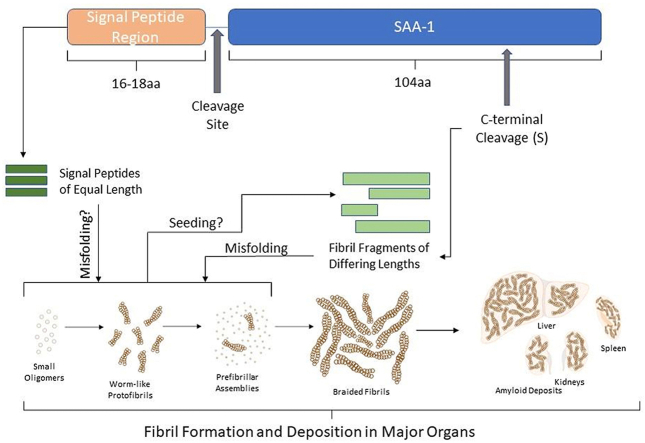
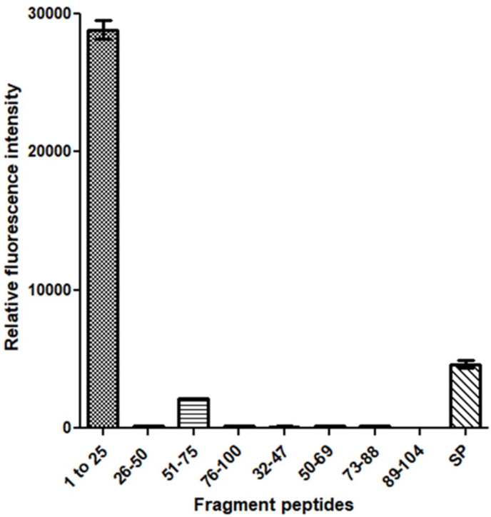
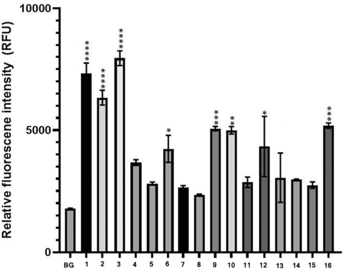
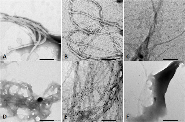
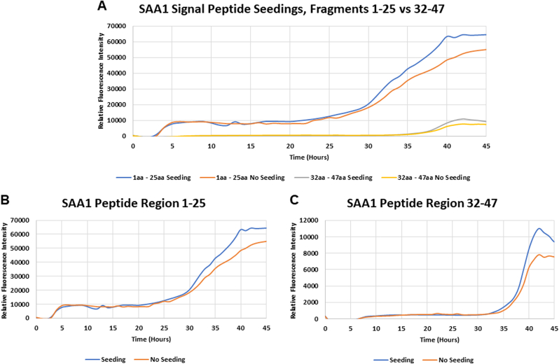
AA amyloidosis is the result of overproduction and aberrant processing of acute-phase serum amyloid A1 (SAA1) by hepatocytes. Proteolytic cleavage of SAA1 is believed to play a central role in AA amyloid formation. The SAA1 protein undergoes a cleavage of 18 residues consisting of the signal peptide at the N-terminal region. To better understand the mechanism behind systemic amyloidosis in the SAA1 protein, we studied the misfolding propensity of the signal peptide region. We first examined the signal peptide amino acid SAA derived from different animal species. A library of 16 peptides was designed to evaluate the propensity of aggregation. The amyloidogenic potential of each SAA1 signal peptide homolog was assessed using in silico Tango program, thioflavin T (ThT) fluorescence, transmission electron microscopy (TEM), and seeding with misfolded human SAA1 signal peptide. After 7 days of incubation, most of the SAA1 signal peptide fragments had the propensity to form fibrils at a concentration of 100 μM in 50 mM Tris buffer at 37 °C by TEM. All peptides were able to generate fibrils at a higher concentration, i.e 500 μM in 25 mM Tris buffer with 50% HFIP, by ThT. All SAA1 signal synthetic peptides designed from the different animal species had the propensity to misfold and form fibrils, particularly in species with low occurrence of systemic amyloidosis. The human SAA1 signal peptide region was capable to seed the SAA1 1-25 and 32-47 peptide regions. Characterizing fibrillar conformations are relevant for seeding intact and/or fragmented SAA, which may contribute, to the mechanism of protein misfolding. This research signifies the importance of the signal peptide region and its possible contribution to the misfolding of aggregation-prone proteins.

Keywords: Agg, Aggregation; Amyloid A; Fibril assembly; HDL, High-density lipoprotein; HFIP, Hexafluoroisopropanol; MMP, Metalloproteinases; Protein misfolding; SAA1, Serum amyloid A1; Serum amyloid A; Signal peptide; Systemic amyloidosis; TEM, Transmission electron microscopy; ThT, Thioflavin T; Tris, Tris(hydroxymethyl)aminomethane.


Figures

Image 1
Graphical abstract
Fig. 1
Fig. 1
Aggregation (Agg) score results were obtained from different regions of the human serum amyloid A1 (SAA1). The highest Agg score resulted from the signal peptide region (1), and amino acids 1–25 (2), indicative of a high propensity for aggregation. Agg scores were obtained with the Tango program.
Fig. 2
Fig. 2
Comparison of thioflavin T (ThT) fluorescence intensity obtained at the end of fibrillization kinetics of different human SAA1 protein fragments at a concentration of 500 μM in 25 mM Tris buffer (pH 8) and 50% hexafluoroisopropanol (HFIP). Three samples of each fragment were monitored at 37 °C for 120 h.
Fig. 3
Fig. 3
Thioflavin T (ThT) fluorescence emission in the presence of different serum amyloid A1 (SAA1) signal peptides. Signal peptides were solubilized at 500 μM in 25 mM Tris buffer (pH 8) and 50% hexafluoroisopropanol (HFIP). ThT-fluorescence was monitored at 37 °C for 120 h. ThT was used to create a final concentration of 100 μM. Each bar represents the average of the last five fluorescence values obtained. Error bars represent the standard error of the mean (SEM). Data were analyzed by the ordinary one-way ANOVA with Dunnett's multiple comparisons post-hoc testing between each SAA1 signal peptide and fluorescence background signal (BG). (Significance difference: *p<0.02, **p<0.001, ***p<0.0008, ****p<0.0001).
Fig. 4
Fig. 4
Kinetics of 16 different SAA1 signal peptide aggregations at 37 °C probed by thioflavin T (ThT) fluorescence. The 16 peptides represent the signal peptide region of different animal species. Signal peptides were solubilized at 500 μM in 25 mM Tris buffer (pH 8) and 50% hexafluoroisopropanol (HFIP). Each curve represents an average of the fluorescence intensity obtained from three replicates over 120 h (5 days) in incubation at 37 °C.
Fig. 5
Fig. 5
Representative images of serum amyloid A1 (SAA1) signal peptide library obtained by transmission electron microscopy (TEM). Signal peptides were solubilized at 100 μM in 50 mM Tris buffer and incubated at 37 °C for 7 days. (A) SAA1 signal peptide ID# 1 (human). (B) SAA1 signal peptide ID# 2 (cat). (C) SAA1 signal peptide ID# 3 (rabbit). (D) SAA1 signal peptide ID# 4 (horse). (E) SAA1 signal peptide ID# 5 (sterlet fish). (F) SAA1 signal peptide ID# 6 (zebra finch). (G) SAA1 signal peptide ID# 7 (rhesus monkey). (H) SAA1 signal peptide ID# 8 (lesser jerboa). (I) SAA1 signal peptide ID# 9 (amur tiger). (J) SAA1 signal peptide ID# 10 (chamois). (K) SAA1 signal peptide ID# 11 (alpine ibex). (L) SAA1 signal peptide ID# 12 (soft-shelled turtle). (M) SAA1 signal peptide ID# 13 (Japanese quail). (N) SAA1 signal peptide ID# 14 (hedgehog). (O) SAA1 signal peptide ID# 15 (platypus). (P) SAA1 signal peptide ID# 16 (brown bat). A scale bar of 200 nm is indicated in the right bottom corner of each photomicrograph.
Fig. 6
Fig. 6
Representative images of serum amyloid A1 (SAA1) signal peptide library obtained by transmission electron microscopy (TEM). Signal peptides were solubilized at 500 μM in 25 mM Tris buffer with 50% hexafluoroisopropanol (HFIP) and incubated at 37 °C for 7 days. (A) SAA1 signal peptide ID# 1 (human). (B) SAA1 signal peptide ID# 3 (rabbit). (C) SAA1 signal peptide ID# 4 (horse). (D) SAA1 signal peptide ID# 11 (alpine ibex). (E) SAA1 signal peptide ID# 13 (Japanese quail). (F) SAA1 signal peptide ID# 15 (platypus). A scale bar of 200 nm is indicated in the right bottom corner of each photomicrograph.
Fig. 7
Fig. 7
Seeding effect of preformed human SAA1 signal peptide aggregates on the kinetics of the 1–25 and 32–47 fragmented regions of human SAA1. (A) depicts a comparison of the fluorescence intensity between the aggregation kinetics of fragments 1-25 and 32-47. The graphs below portray time course of human SAA1 fragment peptide 1–25 (B) and 32–47 (C) fibril formation induced by incubation at 37 °C in the presence or absence of 1 μM misfolded human SAA1 signal peptide (seeds). Aggregation kinetics were monitored with thioflavin T (ThT). Fragment peptides 1–25 and 32–47 were prepared at 500 μM in 25 mM Tris buffer (pH 8) and 50% hexafluoroisopropanol (HFIP).

References

    1. Gaffney P.M. Amyloid A amyloidosis. Vet. Pathol. 2017;54:5–8. - PubMed
    1. Real de Asua D., Costa R., Galvan J.M., Filigheddu M.T., Trujillo D., Cadinanos J. Systemic AA amyloidosis: epidemiology, diagnosis, and management. Clin. Epidemiol. 2014;6:369–377. - PMC - PubMed
    1. Sun L., Ye R.D. Serum amyloid A1: structure, function and gene polymorphism. Gene. 2016;583:48–57. - PMC - PubMed
    1. Lu J., Yu Y., Zhu I., Cheng Y., Sun P.D. Structural mechanism of serum amyloid A-mediated inflammatory amyloidosis. Proc. Natl. Acad. Sci. U.S.A. 2014;111:5189–5194. - PMC - PubMed
    1. Gruys E. Protein folding pathology in domestic animals. J. Zhejiang Univ. - Sci. 2004;5:1226–1238. - PMC - PubMed

LinkOut - more resources