[Preprint]. 2022 May 23:2022.05.21.492920.
doi: 10.1101/2022.05.21.492920.

The SARS-CoV-2 spike protein binds and modulates estrogen receptors

Affiliations

The SARS-CoV-2 spike protein binds and modulates estrogen receptors

Oscar Solis et al. bioRxiv. .

Update in

  • The SARS-CoV-2 spike protein binds and modulates estrogen receptors.
    Solis O, Beccari AR, Iaconis D, Talarico C, Ruiz-Bedoya CA, Nwachukwu JC, Cimini A, Castelli V, Bertini R, Montopoli M, Cocetta V, Borocci S, Prandi IG, Flavahan K, Bahr M, Napiorkowski A, Chillemi G, Ooka M, Yang X, Zhang S, Xia M, Zheng W, Bonaventura J, Pomper MG, Hooper JE, Morales M, Rosenberg AZ, Nettles KW, Jain SK, Allegretti M, Michaelides M. Solis O, et al. Sci Adv. 2022 Dec 2;8(48):eadd4150. doi: 10.1126/sciadv.add4150. Epub 2022 Nov 30. Sci Adv. 2022. PMID: 36449624 Free PMC article.

Abstract

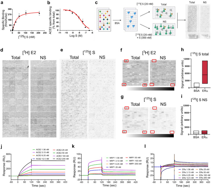
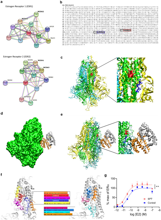
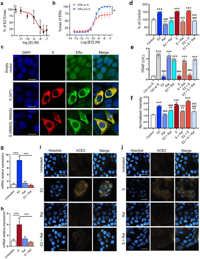
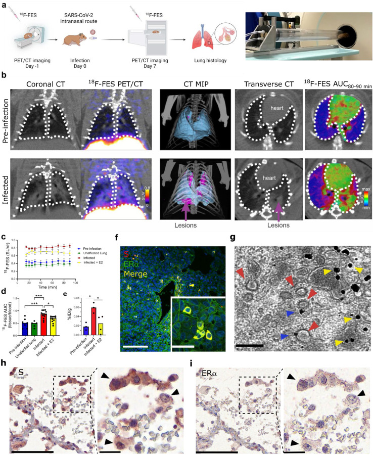
The severe acute respiratory syndrome coronavirus 2 (SARS-CoV-2) spike (S) protein binds angiotensin-converting enzyme 2 (ACE2) at the cell surface, which constitutes the primary mechanism driving SARS-CoV-2 infection. Molecular interactions between the transduced S and endogenous proteins likely occur post-infection, but such interactions are not well understood. We used an unbiased primary screen to profile the binding of full-length S against >9,000 human proteins and found significant S-host protein interactions, including one between S and human estrogen receptor alpha (ERα). After confirming this interaction in a secondary assay, we used bioinformatics, supercomputing, and experimental assays to identify a highly conserved and functional nuclear receptor coregulator (NRC) LXD-like motif on the S2 subunit and an S-ERα binding mode. In cultured cells, S DNA transfection increased ERα cytoplasmic accumulation, and S treatment induced ER-dependent biological effects and ACE2 expression. Noninvasive multimodal PET/CT imaging in SARS-CoV-2-infected hamsters using [ 18 F]fluoroestradiol (FES) localized lung pathology with increased ERα lung levels. Postmortem experiments in lung tissues from SARS-CoV-2-infected hamsters and humans confirmed an increase in cytoplasmic ERα expression and its colocalization with S protein in alveolar macrophages. These findings describe the discovery and characterization of a novel S-ERα interaction, imply a role for S as an NRC, and are poised to advance knowledge of SARS-CoV-2 biology, COVID-19 pathology, and mechanisms of sex differences in the pathology of infectious disease.


Figures

Figure 1.
Figure 1.. S binds ERα with high affinity.
(a) [125I]S saturation and (b) competition binding to recombinant ACE2. (c) Schematic of ProtoArray® experimental design. (d) Positive control ProtoArray® autoradiograms showing total and non-specific (NS) binding of [3H]estradiol (E2). (e) ProtoArray® autoradiograms showing total and NS binding of [125I]S. (f, g) Representative array blocks showing total and NS [3H]E2 and [125I]S binding. Red rectangles show location of ERα proteins. (h, i) Quantification of total and NS [125I]S binding at ERα and BSA (control). Data are representative of three independent experiments. (j-l) Representative SPR sensorgrams showing kinetic and equilibrium binding analyses of immobilized S exposed to increasing concentrations of ACE2, NRP1 and ERα protein (Kon=2.03 ×10⁵, Koff= 1.96 × 10⁻³, KD= 9.7 nM). In a-b, data are represented as mean ± SEM. In h, i, data are presented as median ± min and max limits.
Figure 2.
Figure 2.. S and ER interact at conserved LXD nuclear receptor coregulatory (NRC) motifs.
(a) ER interaction network showing known and predicted protein associations. (b) LXD-like patterns in the S sequence. The LXXLL motif and a homologous region are highlighted in blue and red boxes, respectively, with dark grey background. −1 and −2 positions are reported in italic and light grey background. (c) The LPPLL and IEDLL residues of the two motifs are shown in the 3D X-ray S structure (pdb id 6VYB) with blue and red colors, respectively. The three S chains are shown in yellow, cyan and green. (d) S-ER motif-oriented docking. The best 3D docking hypothesis is shown. The ER dimer is in orange and gray, S is green. (e) Alignment between the best-pose and the 3OLL model tied with NCOA1. The region occupied by S’s alpha-helix interacts in the area where the NCOA fragment was crystallized. (f) S protein peptides and their location with respect to the S 3D structure. (g) The SP7 peptide containing the LPPLL motif significantly increased ERα activation (F (1,48) = 30.38; **P<0.01; two-way ANOVA, peptide treatment main effect). Data are mean ± SEM.
Figure 3.
Figure 3.. S modulates ER-dependent biological functions.
S inhibits (a) E2-induced ERα DNA binding in MCF-7 nuclear extracts and (b) transcriptional activation in an ERα reporter cell line (F (1, 28) = 21.73, *P=0.01; two-way ANOVA, S treatment × E2 concentration interaction effect). (c) Immunofluorescent staining of S and endogenous ERα in MCF-7 cells transfected with empty vector, wild-type (WT) or the furin cleavage site mutant S(R682S, R685S). Scale bar = 16 μm. (d) S increases MCF-7 cell proliferation in an ER-dependent manner (**P<0.01, ***P<0.001 versus control; ###P<0.001 versus E2; &&&P<0.001 versus S; one-way ANOVA with post hoc Tukey test). (e) S decreases osteoclast differentiation in an ER-dependent manner (***p<0.001 versus control w/o RANKL; ###P<0.001 versus control w/ RANKL; one-way ANOVA with post hoc Tukey test). (f) S and E2 increase ACE2 protein levels in MCF-7 cells in an ER-dependent manner (***P<0.001 versus control; ###P<0.001 versus E2; &&&P<0.001 versus S, one-way ANOVA with post hoc Tukey test). (g, h) E2 and S increase ACE2 mRNA (***P<0.001, one-way ANOVA with post hoc Tukey test) and (i, j) protein in the Calu-3 lung cell line in an ER-dependent manner. Scale bar = 30 μm. All data shown as mean ± SEM.
Figure 4.
Figure 4.. SARS-CoV-2 infection increases cytoplasmic ERα accumulation and S-ERα colocalization in pulmonary macrophages.
(a) Schematic showing experimental design of SARS-CoV-2 hamster studies and BSL-3 imaging compartment. (b) CT, [18F]FES PET and AUC heatmap overlay images from hamsters at pre-infection (Day −1) and infection (Day 7) (MIP; maximum intensity projection, SUV; standard uptake value; AUC, area under the curve). (c) Time activity curves showing SUV ratio (SUVr; tissue/blood [18F]FES content) in each experimental group. n = 45/group. ***P<0.001 versus pre-infection (d) [18F]FES uptake expressed as area under the curve ratio (AUCr; tissue/blood [18F]FES content). F (3, 32) = 12.15; ***P<0.001, *P<0.05 (e) [18F]FES uptake expressed as % injected dose (ID)/g body weight in postmortem hamster lung (harvested 110 min post-injection; n = 3–4 /group). F (2, 7) = 7.161; *P<0.05. (f) Hamster lung immunohistochemistry showing colocalization of S and ERα immunoreactivity. (g) Immunogold EM showing SARS-CoV-2 particles (red arrowheads) and ERα-bound gold nanoparticles (blue arrowheads) in a hamster alveolar macrophage (scale bar = 200 nm). Yellow arrowheads correspond to cytoplasmic ERα accumulation. (h) S and (i) ERα immunostaining in SARS-CoV-2-infected human lung showing S-ERα colocalization in macrophages (black arrowheads). Scale bar (low mag = 100 nm; high mag = 25 nm). All data shown as mean ± SEM.

References

    1. Zhou X. et al. Incidence and impact of disseminated intravascular coagulation in COVID-19 a systematic review and meta-analysis. Thromb. Res. 201, 23–29 (2021). - PMC - PubMed
    1. Mitrovic M. et al. Rotational thromboelastometry (ROTEM) profiling of COVID-19 patients. Platelets 32, 690–696 (2021). - PubMed
    1. La Vignera S. et al. Sex-Specific SARS-CoV-2 Mortality: Among Hormone-Modulated ACE2 Expression, Risk of Venous Thromboembolism and Hypovitaminosis D. Int. J. Mol. Sci. 21, 2948 (2020). - PMC - PubMed
    1. Montopoli M. et al. Clinical outcome of SARS-CoV-2 infection in breast and ovarian cancer patients who underwent antiestrogenic therapy. Ann. Oncol. 32, 676–677 (2021). - PMC - PubMed
    1. Chen Y., Liu Q. & Guo D. Emerging coronaviruses: genome structure, replication, and pathogenesis. J. Med. Virol. 92, 418–423 (2020). - PMC - PubMed

Publication types