This is a preprint.
A robust, highly multiplexed mass spectrometry assay to identify SARS-CoV-2 variants
- PMID: 35665019
- PMCID: PMC9164449
- DOI: 10.1101/2022.05.28.22275691
A robust, highly multiplexed mass spectrometry assay to identify SARS-CoV-2 variants
Update in
-
A Robust, Highly Multiplexed Mass Spectrometry Assay to Identify SARS-CoV-2 Variants.Microbiol Spectr. 2022 Oct 26;10(5):e0173622. doi: 10.1128/spectrum.01736-22. Epub 2022 Sep 7. Microbiol Spectr. 2022. PMID: 36069609 Free PMC article.
Abstract
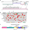
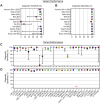
Severe acute respiratory syndrome coronavirus 2 (SARS-CoV-2) variants are characterized by differences in transmissibility and response to therapeutics. Therefore, discriminating among them is vital for surveillance, infection prevention, and patient care. While whole viral genome sequencing (WGS) is the "gold standard" for variant identification, molecular variant panels have become increasingly available. Most, however, are based on limited targets and have not undergone comprehensive evaluation. We assessed the diagnostic performance of the highly multiplexed Agena MassARRAY ® SARS-CoV-2 Variant Panel v3 to identify variants in a diverse set of 391 SARS-CoV-2 clinical RNA specimens collected across our health systems in New York City, USA as well as in Bogotá, Colombia (September 2, 2020 - March 2, 2022). We demonstrate almost perfect levels of interrater agreement between this assay and WGS for 9 of 11 variant calls (κ ≥ 0.856) and 25 of 30 targets (κ ≥ 0.820) tested on the panel. The assay had a high diagnostic sensitivity (≥93.67%) for contemporary variants (e.g., Iota, Alpha, Delta, Omicron [BA.1 sublineage]) and a high diagnostic specificity for all 11 variants (≥96.15%) and all 30 targets (≥94.34%) tested. Moreover, we highlight distinct target patterns that can be utilized to identify variants not yet defined on the panel including the Omicron BA.2 and other sublineages. These findings exemplify the power of highly multiplexed diagnostic panels to accurately call variants and the potential for target result signatures to elucidate new ones.
Importance: The continued circulation of SARS-CoV-2 amidst limited surveillance efforts and inconsistent vaccination of populations has resulted in emergence of variants that uniquely impact public health systems. Thus, in conjunction with functional and clinical studies, continuous detection and identification are quintessential to inform diagnostic and public health measures. Furthermore, until WGS becomes more accessible in the clinical microbiology laboratory, the ideal assay for identifying variants must be robust, provide high resolution, and be adaptable to the evolving nature of viruses like SARS-CoV-2. Here, we highlight the diagnostic capabilities of a highly multiplexed commercial assay to identify diverse SARS-CoV-2 lineages that circulated at over September 2, 2020 - March 2, 2022 among patients seeking care at our health systems. This assay demonstrates variant-specific signatures of nucleotide/amino acid polymorphisms and underscores its utility for detection of contemporary and emerging SARS-CoV-2 variants of concern.
Conflict of interest statement
Competing Interests
Robert Sebra is VP of Technology Development and a stockholder at Sema4, a Mount Sinai Venture. This work, however, was conducted solely at Icahn School of Medicine at Mount Sinai. Otherwise, the authors declare no competing interests.
Figures


References
-
- Kalia K, Saberwal G, Sharma G. 2021. The lag in SARS-CoV-2 genome submissions to GISAID. Nat Biotechnol 39:1058–1060. - PubMed
-
- Deng X, Gu W, Federman S, du Plessis L, Pybus OG, Faria N, Wang C, Yu G, Bushnell B, Pan C-Y, Guevara H, Sotomayor-Gonzalez A, Zorn K, Gopez A, Servellita V, Hsu E, Miller S, Bedford T, Greninger AL, Roychoudhury P, Starita LM, Famulare M, Chu HY, Shendure J, Jerome KR, Anderson C, Gangavarapu K, Zeller M, Spencer E, Andersen KG, MacCannell D, Paden CR, Li Y, Zhang J, Tong S, Armstrong G, Morrow S, Willis M, Matyas BT, Mase S, Kasirye O, Park M, Masinde G, Chan C, Yu AT, Chai SJ, Villarino E, Bonin B, Wadford DA, Chiu CY. 2020. Genomic surveillance reveals multiple introductions of SARS-CoV-2 into Northern California. Science 10.1126/science.abb9263. - DOI - PMC - PubMed
-
- Hernandez MM, Gonzalez-Reiche AS, Alshammary H, Fabre S, Khan Z, van De Guchte A, Obla A, Ellis E, Sullivan MJ, Tan J, Alburquerque B, Soto J, Wang C-Y, Sridhar SH, Wang Y-C, Smith M, Sebra R, Paniz-Mondolfi AE, Gitman MR, Nowak MD, Cordon-Cardo C, Luksza M, Krammer F, van Bakel H, Simon V, Sordillo EM. 2021. Molecular evidence of SARS-CoV-2 in New York before the first pandemic wave. Nat Commun 12:3463. - PMC - PubMed
Publication types
Grants and funding
LinkOut - more resources
Full Text Sources
Miscellaneous
