Dynamic Changes in the Aorta During the Cardiac Cycle Analyzed by ECG-Gated Computed Tomography
- PMID: 35665265
- PMCID: PMC9160308
- DOI: 10.3389/fcvm.2022.793722
Dynamic Changes in the Aorta During the Cardiac Cycle Analyzed by ECG-Gated Computed Tomography
Abstract
Background: To characterize the difference in aortic dimensions during the cardiac cycle with electrocardiogram (ECG)-gated computed tomography angiography (CTA) and to determine whether other parameters in comparison to diameter could potentially provide a more accurate size reference for stent selection at the aortic arch and the proximal thoracic descending aorta.
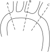
Methods: The CTA imaging of 90 patients during the cardiac cycle was reviewed. Three anatomic locations were selected for analysis (level A: 1 cm proximal to the innominate artery; level B: 1 cm distal to the left common carotid artery; and level C: 1 cm distal to the left subclavian artery). We measured the maximum diameter, the minimum diameter, the lumen area, the lumen perimeter, and the diameter derived from the lumen area, and the changes of each parameter at each level during the cardiac cycle were compared.
Results: The mean age was 60.9 ± 12.4 years (range, 16-78 years). There was a significant difference in the aortic dimensions during the cardiac cycle (p < 0.001). The diameter derived from the lumen area at all three levels was changed least over time when compared to the area, perimeter, and the maximum aortic diameter (all p < 0.01).
Conclusion: The aortic dimensional differences during the cardiac cycle are significant. The aortic diameter derived from the lumen area over other parameters may provide a better evaluation for selecting the size of the stent at the aortic arch and the proximal thoracic descending aorta. A prospective study comparing these different measurement parameters regarding the outcomes is still needed to evaluate the clinical implications.
Keywords: aorta; cardiac cycle; diameter; dynamic changes; oversizing.
Copyright © 2022 Zhu, Wang, Chen, Liu, Zhou, Shi, Huang, Yang, Li and Xiong.
Conflict of interest statement
The authors declare that the research was conducted in the absence of any commercial or financial relationships that could be construed as a potential conflict of interest.
Figures


 ) with the location of level C (1 cm distal to the left subclavian artery). The system calculated the maximum and minimum aortic diameter (D: 22.9/24.4 mm), area (area: 437.5 mm2), perimeter (perim: 74.3 mm), and diameter derived from the lumen area [Deff (area): 23.6 mm] automatically.

Similar articles
-
Thoracic Aorta Dimension Changes During Systole and Diastole: Evaluation with ECG-Gated Computed Tomography.Ann Vasc Surg. 2016 Aug;35:168-73. doi: 10.1016/j.avsg.2016.01.050. Epub 2016 Jun 3. Ann Vasc Surg. 2016. PMID: 27263817
-
Dynamic aortic changes in patients with thoracic aortic aneurysms evaluated with electrocardiography-triggered computed tomographic angiography before and after thoracic endovascular aneurysm repair: preliminary results.Ann Vasc Surg. 2009 May-Jun;23(3):291-7. doi: 10.1016/j.avsg.2008.08.007. Epub 2008 Sep 21. Ann Vasc Surg. 2009. PMID: 18809281
-
Comparison of dynamic changes in aortic diameter during the cardiac cycle measured by computed tomography angiography and transthoracic echocardiography.J Vasc Surg. 2019 May;69(5):1538-1544. doi: 10.1016/j.jvs.2018.07.083. J Vasc Surg. 2019. PMID: 31010518
-
Endovascular stent grafting and open surgical replacement for chronic thoracic aortic aneurysms: a systematic review and prospective cohort study.Health Technol Assess. 2022 Jan;26(6):1-166. doi: 10.3310/ABUT7744. Health Technol Assess. 2022. PMID: 35094747
-
[Dimensions of the proximal thoracic aorta from childhood to adult age: reference values for two-dimensional echocardiography. Ligurian Group of SIEC (Italian Society of Echocardiography)].G Ital Cardiol. 1997 Jul;27(7):686-96. G Ital Cardiol. 1997. PMID: 9303859 Review. Italian.
Cited by
-
Hypertension-Induced Biomechanical Modifications in the Aortic Wall and Their Role in Stanford Type B Aortic Dissection.Biomedicines. 2024 Oct 2;12(10):2246. doi: 10.3390/biomedicines12102246. Biomedicines. 2024. PMID: 39457560 Free PMC article.
References
LinkOut - more resources
Full Text Sources

