Case Reports
. 2022 May 31;2022(5):rjac221.
doi: 10.1093/jscr/rjac221. eCollection 2022 May.

Spinal myxomas: review of a rare entity

Affiliations
Case Reports

Spinal myxomas: review of a rare entity

Sabina Patel et al. J Surg Case Rep. .

Abstract

Intramuscular myxomas are rare, benign mesenchymal tumours, occurring predominantly in large skeletal muscles as large, slow-growing and painless masses. Spinal occurrence is rare, and may present incidentally, or diagnosed via localized symptoms secondary to local infiltration of surrounding structures. Differential diagnosis based on imaging includes sarcomas, meningiomas and lipomas. We discuss two contrasting cases presenting with well-circumscribed cystic paraspinal lesions indicative of an infiltrative tumour and discuss the radiological and histological differences that distinguish myxomas from similar tumours. Surgical resection of the tumour was performed in both cases, however one patient required surgical fixation due to bony erosion secondary to tumour infiltration. Immuno-histopathological analysis confirmed the diagnosis of a cellular myxoma. Follow up imaging at 6 months confirmed no symptomatic or tumour recurrence in both cases. Histological analysis is the definitive means for diagnosis to differentiate myxomas from other tumours. Recurrence is rare if full resection is achieved.


Figures

Figure 1
Figure 1
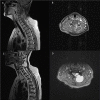
Pre-operative T1 and T2 weighted MRI: left-sided extra-axial mass at C4/5 hypointense on T1 and hyperintense on T2.
Figure 2
Figure 2
Histological staining of intraoperative tumour smears: (A) intraoperative smear Toluidine blue stained slide. (B): H&E ×10. (C): H&E ×10. (D): vimentin IHC ×10.
Figure 3
Figure 3
Pre-operative T1 post Gadolinium and T2 weighted MRI: left-sided extra-axial mass at C5/6 with heterogenous enhancement on T1 and hyperintense on T2. Moderate compression of the spinal cord and invasion into surrounding soft tissue.
Figure 4
Figure 4
Tumour specimen resected en bloc. Solitary, relatively well-delineated nodule covered by a thick capsule measuring 30 × 25 × 20 mm.
Figure 5
Figure 5
PRISMA flow of literature search.

References

    1. Manoharan SR, Shaw AB, Arnold CA, Farhadi HF. Infiltrative intramuscular myxoma of the cervical spine: a case report. Spine J 2015;15:e1–4. - PubMed
    1. Falavigna A, Righesso O, Volquind D, Teles AR. Intramuscular myxoma of the cervical paraspinal muscle. Eur Spine J 2009;18:245–9. - PMC - PubMed
    1. Rachidi S, Sood AJ, Rumboldt T, Day TA. Intramuscular myxoma of the paraspinal muscles: a case report and systematic review of the literature. Oncol Lett 2016;11:466–70. - PMC - PubMed
    1. Bell WO, Gill A, Babiak T, Patterson RH. Epidural myxoma causing compression of the cauda equina: a case report. Neurosurgery 1983;12:325–6. - PubMed
    1. Choi DY, Kim JT, Kim J, Lee HJ. Atypical intramuscular myxoma of the lumbosacral paraspinal muscle: the first case report in Asian. J Korean Neurosurg Soc 2015;58:566–70. - PMC - PubMed

Publication types