

Modern imaging in Cushing's disease
- PMID: 35666391
- PMCID: PMC9587975
- DOI: 10.1007/s11102-022-01236-w
Modern imaging in Cushing's disease
Abstract
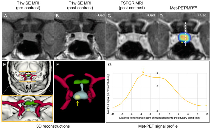
Management of Cushing's disease is informed by dedicated imaging of the sella and parasellar regions. Although magnetic resonance imaging (MRI) remains the investigation of choice, a significant proportion (30-50%) of corticotroph tumours are so small as to render MRI indeterminate or negative when using standard clinical sequences. In this context, alternative MR protocols [e.g. 3D gradient (recalled) echo, with acquisition of volumetric data] may allow detection of tumors that have not been previously visualized. The use of hybrid molecular imaging (e.g. 11C-methionine positron emission tomography coregistered with volumetric MRI) has also been proposed as an additional modality for localizing microadenomas.
Keywords: MRI; Molecular / functional imaging; PET; Pituitary Cushing's.
© 2022. The Author(s).
Conflict of interest statement
The authors declare no competing interests.
Figures
References
-
- Erickson D, Erickson B, Watson R, Patton A, Atkinson J, Meyer F, et al. 3 Tesla magnetic resonance imaging with and without corticotropin releasing hormone stimulation for the detection of microadenomas in Cushing’s syndrome. Clin Endocrinol (Oxf) 2010;72:793–799. doi: 10.1111/j.1365-2265.2009.03723.x. - DOI - PubMed
-
- Kasaliwal R, Sankhe SS, Lila AR, Budyal SR, Jagtap VS, Sarathi V, et al. Volume interpolated 3D-spoiled gradient echo sequence is better than dynamic contrast spin echo sequence for MRI detection of corticotropin secreting pituitary microadenomas. Clin Endocrinol (Oxf) 2013;78:825–830. doi: 10.1111/cen.12069. - DOI - PubMed
Publication types
MeSH terms
Substances
Grants and funding
LinkOut - more resources
Full Text Sources
Medical
