. 2022 Jun 6;18(6):e1010227.
doi: 10.1371/journal.pgen.1010227. eCollection 2022 Jun.

Widespread phages of endosymbionts: Phage WO genomics and the proposed taxonomic classification of Symbioviridae

Affiliations

Widespread phages of endosymbionts: Phage WO genomics and the proposed taxonomic classification of Symbioviridae

Sarah R Bordenstein et al. PLoS Genet. .

Abstract

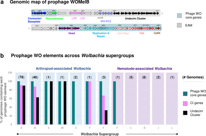
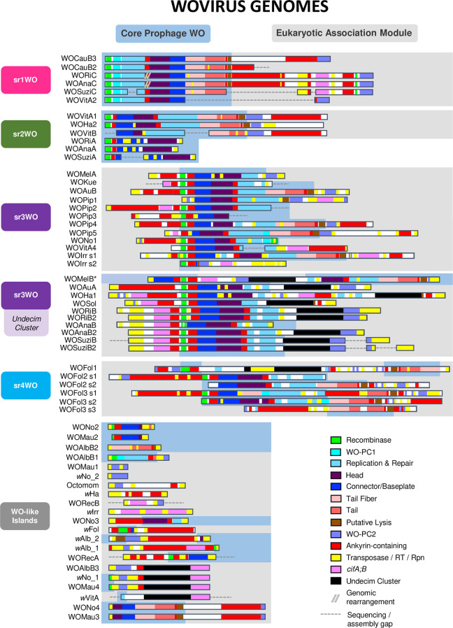
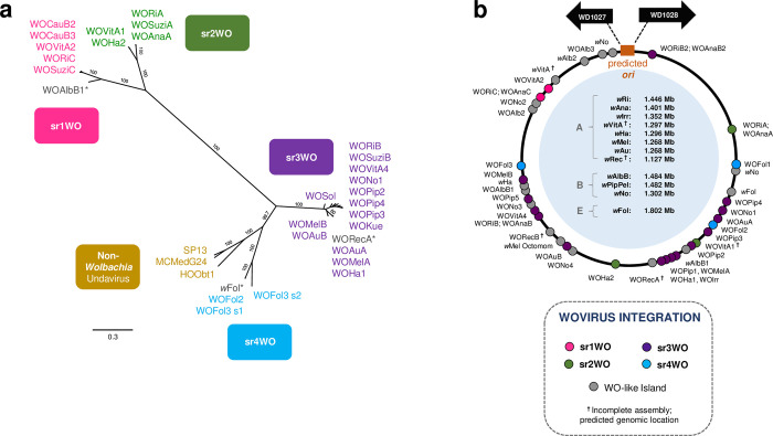
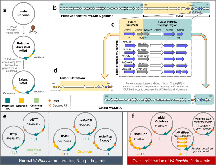
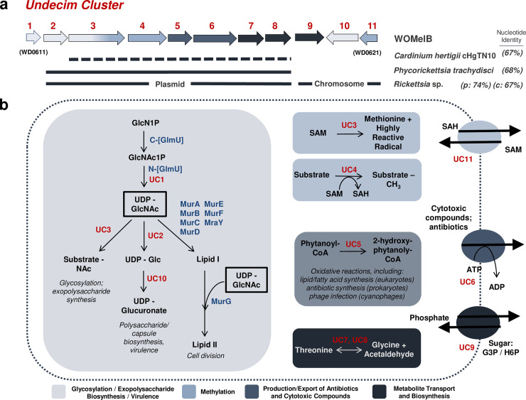
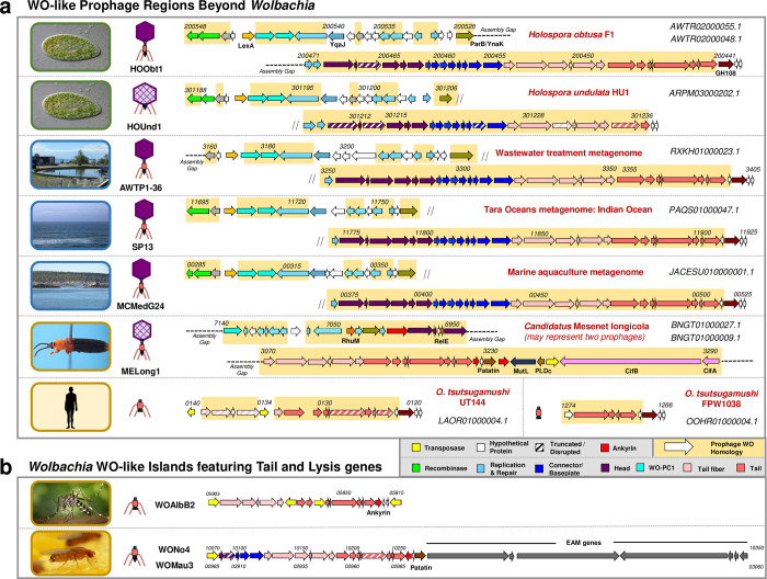
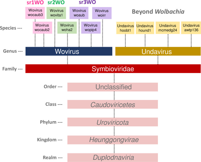
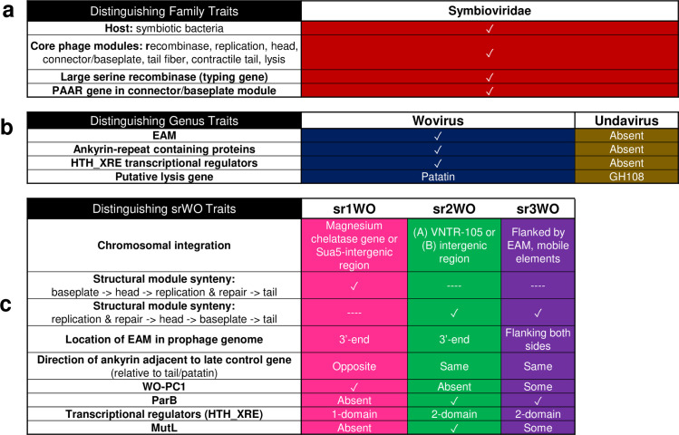
Wolbachia are the most common obligate, intracellular bacteria in animals. They exist worldwide in arthropod and nematode hosts in which they commonly act as reproductive parasites or mutualists, respectively. Bacteriophage WO, the largest of Wolbachia's mobile elements, includes reproductive parasitism genes, serves as a hotspot for genetic divergence and genomic rearrangement of the bacterial chromosome, and uniquely encodes a Eukaryotic Association Module with eukaryotic-like genes and an ensemble of putative host interaction genes. Despite WO's relevance to genome evolution, selfish genetics, and symbiotic applications, relatively little is known about its origin, host range, diversification, and taxonomic classification. Here we analyze the most comprehensive set of 150 Wolbachia and phage WO assemblies to provide a framework for discretely organizing and naming integrated phage WO genomes. We demonstrate that WO is principally in arthropod Wolbachia with relatives in diverse endosymbionts and metagenomes, organized into four variants related by gene synteny, often oriented opposite the putative origin of replication in the Wolbachia chromosome, and the large serine recombinase is an ideal typing tool to distinguish the four variants. We identify a novel, putative lytic cassette and WO's association with a conserved eleven gene island, termed Undecim Cluster, that is enriched with virulence-like genes. Finally, we evaluate WO-like Islands in the Wolbachia genome and discuss a new model in which Octomom, a notable WO-like Island, arose from a split with WO. Together, these findings establish the first comprehensive Linnaean taxonomic classification of endosymbiont phages, including non-Wolbachia phages from aquatic environments, that includes a new family and two new genera to capture the collective relatedness of these viruses.


Conflict of interest statement

The authors have declared that no competing interests exist.

Figures

Fig 1
Fig 1. Prophage WO is modular in structure and associates with all arthropod-infecting Wolbachia.
(a) A genomic map of prophage WOMelB from the D. melanogaster wMel Wolbachia strain highlights phage WO core genes in blue and EAM genes in gray. Genes are illustrated as arrows, and direction correlates with forward/reverse strand. The phage WO core consists of recombinase (green), connector/baseplate (royal blue), head (purple), replication and repair (light blue), tail fiber (light pink), tail (salmon), and lysis (brown). The WOMelB EAM encodes cifA and cifB (pink), WO-PC2 containing HTH_XRE transcriptional regulators (lavender), and a conserved set of genes termed the Undecim Cluster (black). (b) At least one phage WO core gene (teal) is associated with all sequenced arthropod-Wolbachia Supergroups and Supergroup F, which infects both arthropods (blue) and nematodes (purple). The Undecim Cluster (black) is found in the majority of sequenced Supergroup A, B, E, and M Wolbachia genomes, and CI genes (pink) are encoded by the majority of sequenced Supergroup A, B, T, and F genomes. Phage WO elements are absent from all strictly-nematode Wolbachia Supergroups. The number of genomes analyzed is listed in parentheses above each Supergroup. Each bar indicates the % of genomes containing each phage WO element. Source data is provided in S1 Table.
Fig 2
Fig 2. Integrated Wovirus genomes feature distinguishable module synteny.
Prophage WO variants are organized by genome content and synteny of their structural modules. Sr1WO and sr2WO feature a 5’-core prophage WO region (blue) and a 3’-EAM (gray). Sr3WO features an internal core prophage WO region that is flanked by EAM genes and mobile elements (yellow). Sr4WO is only present in wFol and features three genomic regions with multiple prophage segments. WO-like Islands feature small clusters of prophage WO-like genes; they are comprised of singular structural modules and/or subsets of EAM genes. All modules are color coded: green = recombinase; turquoise = WO-PC1; light blue = replication; purple = head; blue = connector/baseplate; light pink = tail fiber; salmon = tail; brown = putative lysis; lavender = WO-PC2; and black = Undecim Cluster. In addition, ankyrins are shown in red; transposable elements are shown in yellow; and cifA;cifB are shown in pink. Dotted lines represent breaks in the assembly; module organization is estimated based on closely related variants. Sr1WO is highlighted in hot pink; sr2WO is highlighted in green; sr3WO is highlighted in purple; sr4WO is highlighted in blue; WO-like Islands are highlighted in gray. * The WOMelB genome is rearranged relative to similar variants. Rather than 5’- and 3’-flanking EAM regions, module synteny reflects that of active phage particles whereby the EAM is internally oriented [39].
Fig 3
Fig 3. Phylogeny of Wovirus large serine recombinase correlates with module synteny and genomic integration.
(a) A phylogenetic tree of the proposed Wovirus recombinase sequence illustrates the utility of this gene as a WO-typing tool to distinguish prophage WO variants. Four distinct clades correlate with sr1WO-sr4WO genome organization shown in Fig 2. Non-Wolbachia sequences represent similar prophages (undaviruses, discussed below) from other bacterial hosts, such as the prophage HOObt1 of Holospora obtusa, an endonuclear symbiont of Paramecium. The tree was generated by Bayesian analysis of 283 amino acids using the JTT-IG model of evolution. Consensus support values are indicated for each branch. (*) indicates that the prophage regions are highly degraded; while they likely originated from the corresponding prophage group, they are now classified as WO-like Islands (S7 Fig). (b) Wovirus integration loci are concentrated opposite the putative origin of replication, ori. All Wolbachia genomes have been standardized where each dot represents % nucleotide distance calculated by: (nucleotide distance between 5’-WO and ori / genome size) * 100. () indicates the genome is not closed/circularized; genomic locations are estimated based on alignment of contigs to a reference genome (obtained from authors in [51,52]) and may not reflect true orientation.
Fig 4
Fig 4. Comparative genomics supports a WO:Octomom origin model for Wolbachia proliferation in wMelPop.
(a) A new model for Octomom origin predicts the initial infection of wMel with a WOMelA phage. After integration, Octomom splits from the WOMelA core prophage region to form a WO-like Island. (b) A genome map of the putative, intact, ancestral WOMelA where Octomom is highlighted in yellow and the extant WOMelA genome in teal illustrates placement of Octomom in the WO EAM. (c-d) An alignment of the WO-PC2 region with closely related prophages shows that half of the conserved module (WD0507-WD0508) is now associated with Octomom and the other half (WD0257-WD0254) remained with WOMelA prophage region. DUF2466 is split across the genomic regions and, when concatenated, shares homology to intact DUF2466 genes of WO-PC2 (see S11 Fig). An IS5 insertion (d) is associated with single-copy Octomom stability in the wMel chromosome. In wMelCS-like genomes, where the flanking RTs are intact (see S10 Fig), Octomom varies in copy number. (e) When Octomom (orange-yellow) and Octomom-like (green, defined by homology to WD0512, WD0513 and WO-PC2; illustrated in S10 Fig) regions exist in a single copy, either within or outside the corresponding prophage region, Wolbachia proliferation is normal, and it is non-pathogenic. (f) If the WO-like Island occurs in multiple copies or is absent from the genome, Wolbachia over-proliferate and are pathogenic. (*) Restoring the 1:1 (WO:Octomom) ratio returns the wMelPop phenotype back to normal levels. The association of Octomom with pathogenicity (i.e., correlation vs. causation) is still to be determined [–68]. NCBI accession numbers are listed for each genome; (†) indicates circular genomes are unavailable and genomic locations are putative.
Fig 5
Fig 5. The Undecim Cluster contributes a wide range of cellular processes associated with host-symbiont interactions.
(a) A genome map illustrates prophage WO’s Undecim Cluster. Gene labels UC1—UC11 correlate with wMel locus tags WD0611-WD0621. Lines under the genes indicate lateral gene transfer events of this region between Cardinium hertigii cHgTN10, Phycorickettsia trachydisci, and multiple strains of Rickettsia, including the Rickettsia endosymbiont of Ixodes scapularis (REIS) and its plasmid (pREIS2). Nucleotide identity is listed to the right. Dashed lines indicate that the region is not contiguous in the genome. UC1 shares partial homology with a core Wolbachia gene, glmU (WD0133) and was either not involved in the transfer event or has since been lost from non-Wolbachia genomes. (b) A cellular model illustrates the putative functions associated with this region. Cellular reactions are highlighted in boxes and membrane transporters are drawn as ovals. Wolbachia genes are labeled in blue; Undecim Cluster genes are labeled in red. UC3 (WD0613) is a fusion protein with an N-terminal glycosyltransferase and C-terminal radical SAM domain; therefore, it is listed twice. Reactions in light gray (UC1, UC2, UC3, and UC10) are likely precursors to multiple pathways in glycosylation, exopolysaccharide biosynthesis, cell division, and/or virulence. Light blue (UC3, UC4, and UC11) is associated with methylation; dark gray (UC5 and UC6) is associated with the production and export of antibiotics and cytotoxic compounds; and dark blue (UC7, UC8, and UC9) is associated with metabolite transport and biosynthesis. The above functions are predicted based on annotation and homology to other systems. Given the contiguous conservation of the Undecim Cluster throughout prophage WO, all functions, including those not captured in this model, are likely interrelated and influence host-symbiont dynamics.
Fig 6
Fig 6. WO-like prophage regions are found in endonuclear Paramecium endosymbionts, aquatic environments, and other animal-associated bacteria.
(a) Genome maps of non-Wolbachia prophage regions illustrate similar gene content and synteny to prophage WO. Locus tags are listed in italics above the genes; NCBI contig accession numbers are shown in the right-hand corner of each genome. Dashed lines represent breaks in the assembly whereas small diagonal lines represent a continuation of the genome onto the next line. Genes with nucleotide homology to prophage WO are highlighted in yellow and genes of similar function are similarly color-coded according to the figure legend. Candidatus Mesenet longicola is the only genome to feature EAM genes, including cifA and cifB. Arrows with diagonal stripes represent genes that may be pseudogenized relative to homologs in other prophage genomes. Genome maps for H. elegans and H. curviuscula prophages are not shown. (b) WO-like Islands featuring tail and lysis genes share homology with the Orientia regions and may represent phage-derived bacteriocins. Predicted physical structures are illustrated to the left of each genome. Images illustrate the isolation source for each prophage: green borders represent protozoa; blue borders represent aquatic environments; gold borders represent animals. All images are available under creative commons or public domain; attribution information is provided in S8 Table.
Fig 7
Fig 7. Comparative genomics support a new family-level designation for prophage WO classification.
Symbioviridae is proposed as a new taxonomic family of tailed phages within the class Caudoviricetes. It contains viruses that primarily infect Wolbachia (proposed genus Wovirus) and other symbionts including Holospora and metagenome-assembled genomes (MAGs) from aquatic environments (proposed genus Undavirus).
Fig 8
Fig 8. Linnaean classification of prophage WO-like viruses is supported by taxonomic traits at the family and genus level.
(a) Proposed family Symbioviridae encompasses viruses that infect symbiotic bacteria, contain a large serine recombinase for integration and a Proline-Alanine-Alanine-aRginine repeat (PAAR) gene in the connector/baseplate module, and feature a conserved set of core phage modules. They share nucleotide homology to Wolbachia’s prophages. (b) Genera are distinguished by presence (Wovirus) or absence (Undavirus) of an EAM and ankyrin repeat containing proteins. Woviruses may utilize patatin for lysis whereas undaviruses encode a canonical GH108 endolysin. (c) Proposed Wovirus clades are further distinguished by multiple factors including structural module synteny, HTH_XRE domains, and genome composition.

References

    1. Newton IL, Bordenstein SR. Correlations between bacterial ecology and mobile DNA. Curr Microbiol. 2011;62(1):198–208. doi: 10.1007/s00284-010-9693-3 - DOI - PMC - PubMed
    1. Cerveau N, Leclercq S, Leroy E, Bouchon D, Cordaux R. Short- and long-term evolutionary dynamics of bacterial insertion sequences: insights from Wolbachia endosymbionts. Genome Biol Evol. 2011;3:1175–86. doi: 10.1093/gbe/evr096 - DOI - PMC - PubMed
    1. Masui S, Kamoda S, Sasaki T, Ishikawa H. Distribution and evolution of bacteriophage WO in Wolbachia, the endosymbiont causing sexual alterations in arthropods. J Mol Evol. 2000;51(5):491–7. doi: 10.1007/s002390010112 - DOI - PubMed
    1. Kent BN, Bordenstein SR. Phage WO of Wolbachia: lambda of the endosymbiont world. Trends Microbiol. 2010;18(4):173–81. doi: 10.1016/j.tim.2009.12.011 - DOI - PMC - PubMed
    1. Reveillaud J, Bordenstein SR, Cruaud C, Shaiber A, Esen OC, Weill M, et al. The Wolbachia mobilome in Culex pipiens includes a putative plasmid. Nat Commun. 2019;10(1):1051. doi: 10.1038/s41467-019-08973-w - DOI - PMC - PubMed

Publication types