. 2022 Jun 14;119(24):e2121804119.
doi: 10.1073/pnas.2121804119. Epub 2022 Jun 6.

Rituximab abrogates aquaporin-4-specific germinal center activity in patients with neuromyelitis optica spectrum disorders

Affiliations

Rituximab abrogates aquaporin-4-specific germinal center activity in patients with neuromyelitis optica spectrum disorders

Valentina Damato et al. Proc Natl Acad Sci U S A. .

Abstract

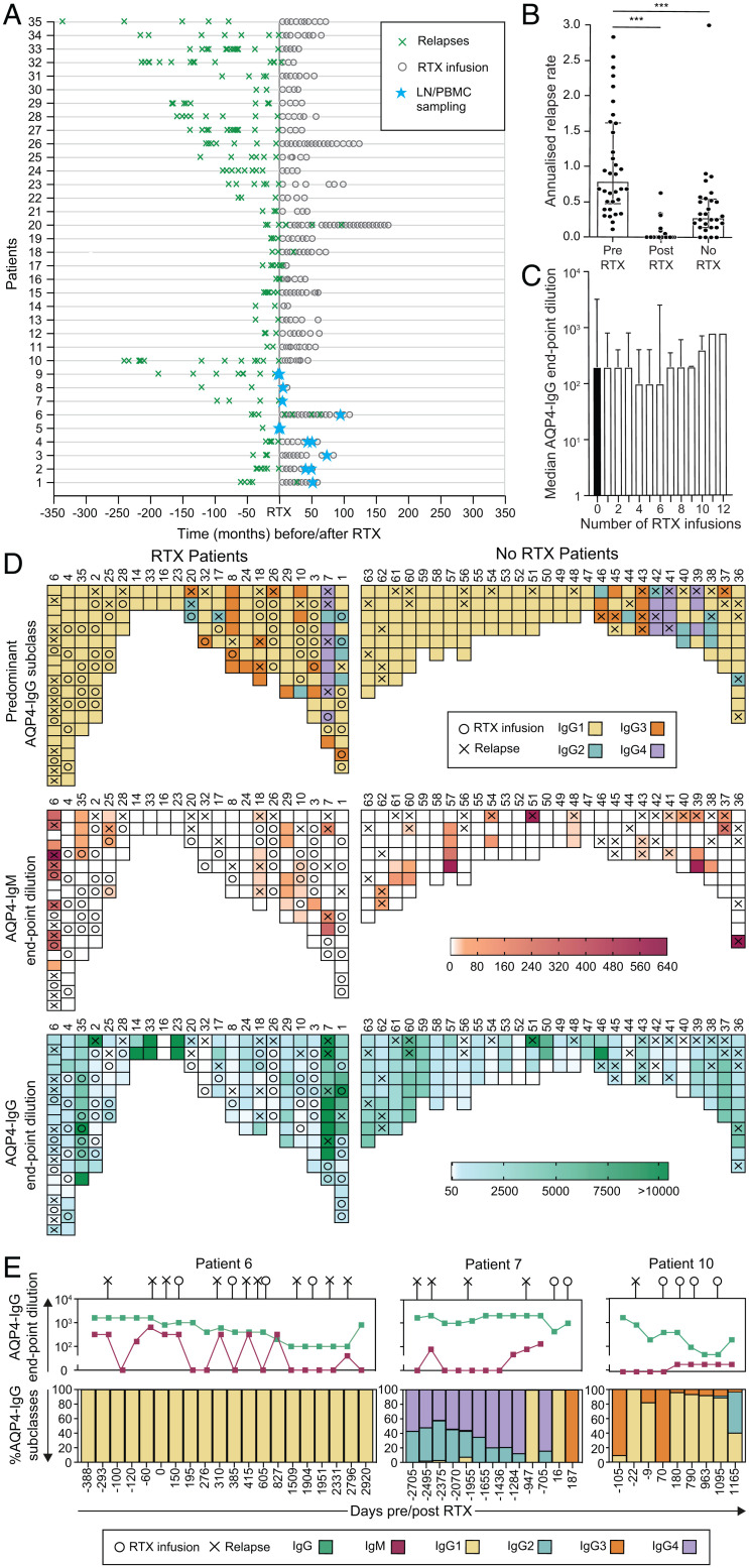
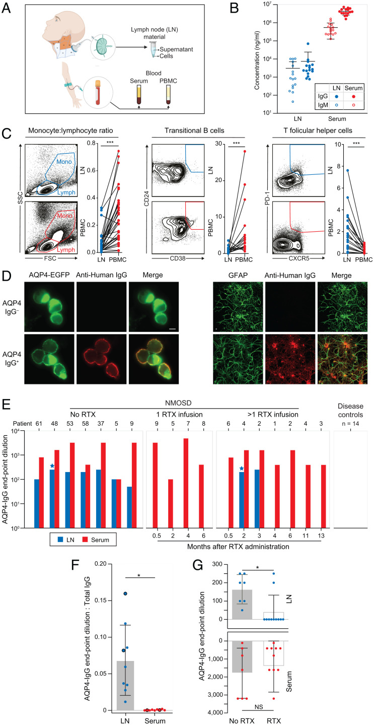
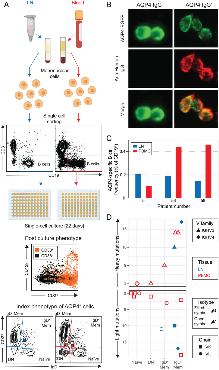
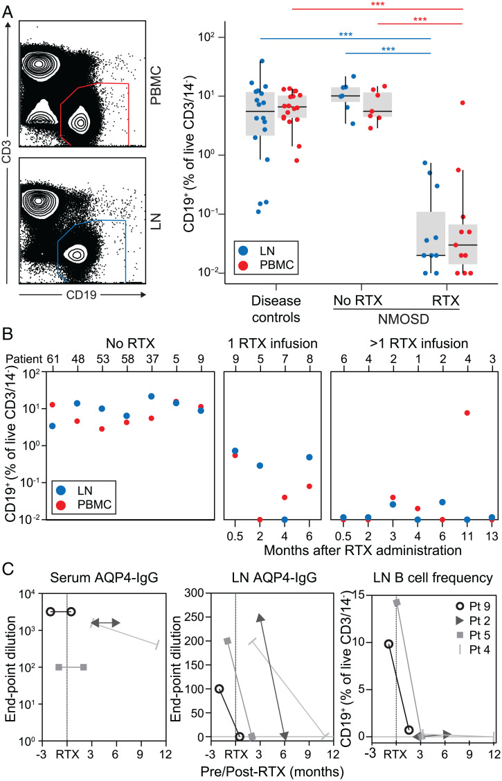
Neuromyelitis optica spectrum disorders (NMOSDs) are caused by immunoglobulin G (IgG) autoantibodies directed against the water channel aquaporin-4 (AQP4). In NMOSDs, discrete clinical relapses lead to disability and are robustly prevented by the anti-CD20 therapeutic rituximab; however, its mechanism of action in autoantibody-mediated disorders remains poorly understood. We hypothesized that AQP4-IgG production in germinal centers (GCs) was a core feature of NMOSDs and could be terminated by rituximab. To investigate this directly, deep cervical lymph node (dCLN) aspirates (n = 36) and blood (n = 406) were studied in a total of 63 NMOSD patients. Clinical relapses were associated with AQP4-IgM generation or shifts in AQP4-IgG subclasses (odds ratio = 6.0; range of 3.3 to 10.8; P < 0.0001), features consistent with GC activity. From seven dCLN aspirates of patients not administered rituximab, AQP4-IgGs were detected alongside specific intranodal synthesis of AQP4-IgG. AQP4-reactive B cells were isolated from unmutated naive and mutated memory populations in both blood and dCLNs. After rituximab administration, fewer clinical relapses (annual relapse rate of 0.79 to 0; P < 0.001) were accompanied by marked reductions in both AQP4-IgG (fourfold; P = 0.004) and intranodal B cells (430-fold; P < 0.0001) from 11 dCLNs. Our findings implicate ongoing GC activity as a rituximab-sensitive driver of AQP4 antibody production. They may explain rituximab’s clinical efficacy in several autoantibody-mediated diseases and highlight the potential value of direct GC measurements across autoimmune conditions.

Keywords: aquaporin; autoimmunity; cervical lymph nodes; neuromyelitis optica; rituximab.


Conflict of interest statement

Competing interest statement: S.R.I. is supported by the Medical Research Council (MR/V007173/1), Wellcome Trust (104079/Z/14/Z), BMA Research Grants - 2013 Vera Down grant, Epilepsy Research UK (P1201), and the Fulbright UK-US commission (MS-Research Society Award). This research was funded in whole, or in part, by the Wellcome Trust (grant number 104079/Z/14/Z). For the purpose of Open Access, the author has applied a CC BY public copyright license to any Author Accepted Manuscript version arising from this submission. J.T. is supported by the Swedish Wenner-Gren foundations. P.W. is supported by the UK NMO commissioning group. B.S. is supported by the Association of British Neurologists via the Patrick Berthoud Charitable Trust. The work is supported by the National Institute for Health Research (NIHR) Oxford Biomedical Research Centre. P.W. and S.R.I. are coapplicants and receive royalties on patent application WO/2010/046716 entitled “Neurological Autoimmune Disorders” (the patent has been licensed for the development of assays for LGI1 and other VGKC-complex antibodies) and have filed two other patents regarding autoantibody diagnostic algorithms.

Figures

Fig. 1.
Fig. 1.
Serological associations with relapses and RTX administration in patients with NMOSD. (A) The effect of RTX administration on relapses (green crosses) in 35 NMOSD patients administered RTX at time 0. The timing of paired LN and PBMC sampling is shown (blue stars). (B) Mean ARR at last follow-up compared between patients before RTX and after RTX (P < 0.001) and to those administered no RTX (P < 0.001, Mann–Whitney U tests). (C) AQP4-IgG serum endpoint dilutions (shown as reciprocal of 1:dilution) were not reduced after multiple infusions of RTX (P = 0.99, Kruskal–Wallis test). (D) Heat maps to represent associations between relapses (X) and the dominant AQP4-IgG subclass (>50% of total AQP4-IgG, Top; determined by flow cytometry), AQP4-IgM (Middle; levels in red; determined by end-point titrations using live cell–based assays), and total AQP4-IgG (Bottom; levels in green; by endpoint titrations using live cell–based assays) in patients either administered RTX (n = 22) or naive to RTX (n = 28). (E) Longitudinal data in individual NMOSD patients (6, 7, 10), with negative values indicating days before the first RTX infusion; ***P < 0.001.
Fig. 2.
Fig. 2.
CLN aspirations contain AQP4 antibodies, which are abrogated after RTX administration. (A) CLNs across anatomical levels I, II, III, and V (orange) were aspirated under ultrasound guidance. Paired blood was obtained in parallel, resulting in cellular and soluble fractions from both sampled sites. (B) Markedly different levels of total IgG (filled symbols) and IgM (open symbols) were measured in LN aspirates (blue; IgG diluted at 1:800; IgM diluted at 1:100) versus matched sera of NMOSD patients (red; IgG diluted at 1:100,000; IgM diluted at 1:6,400). (C) Differences between PBMCs and LN cell populations are highlighted by the ratio of monocytes to lymphocytes (SSC = side scatter; FSC = forward scatter) and frequencies of both transitional B cells and Tfh cells (all P < 0.001, Wilcoxon signed-rank test), confirming that ultrasound LN sampling resulted in limited blood contamination of the LN aspirates. (D) AQP4-IgG was detected in LN aspirates by binding (anti-human IgG, red) to the surface of live AQP4–EGFP-transfected HEK293T cells (green; Right) and to the surface of live rat astrocytes (identified with GFAP, green; Left). (Scale bar, 10 µm). (E) AQP4-IgG levels in serum (red) and LN aspirates (blue) were measured in seven patients naive to RTX (of whom patients 5 and 9 went onto receive RTX), in four patients between 0.5 and 6 mo after one RTX infusion, and in seven patients between 0.5 and 13 mo after more than one RTX infusion (including two sampled longitudinally: patients 2 and 4). Blue stars indicate AQP4-IgM detected in two LN samples. (F) The ratios of AQP4-IgG levels (endpoint dilutions) to total IgG levels were compared between LN aspirates and sera (P = 0.02, Wilcoxon signed-rank test). Large dots highlight the two RTX-treated patients with detectable AQP4-IgGs from LN aspirates. (G) AQP4-IgG levels from LN aspirates (P = 0.04, Mann–Whitney U test) and serum (nonsignificant) in patients who did versus did not receive RTX; NS, not significant; *P < 0.05; **P < 0.01; ***P < 0.001.
Fig. 3.
Fig. 3.
AQP4-specific B cells characterized from LNs and blood of patients with NMOSD. (A) Single B cells (CD3CD19+) from paired blood (red) and LN (blue) samples were labeled with detection antibodies, index sorted as single cells, and cultured. By day 22, ∼50% of cells proliferate and differentiate into antibody-secreting cells (CD19 cells gated to show CD27, CD38, and CD138 expression). Indexing revealed the original B cell subsets: naive, double-negative (DN), IgD, and IgD+ memory (Mem). (B) AQP4-reactive IgGs/IgMs in culture supernatants were identified by reactivity (red) directed against live HEK293T cells, which expressed surface AQP4–EGFP (green). (Scale bar, 10 µm.) (C) AQP4-specific B cell frequencies detected from three patients in LNs (blue) and PBMCs (red). (D) The heavy and light chains of these AQP4-specific BCRs (7 and 11 recovered, respectively) arose from all four studied B cell subsets and showed varied gene families (heavy variable IGHV3 as triangles and IGHV4 as diamonds) and light chain usage (kappa or lambda as squares or circles, respectively) from both LNs (blue) and PBMCs (red). The AQP4-reactive isotype detected in supernatants is shown using open (AQP4-IgM) or filled (AQP4-IgG) symbols; DN, double-negative cells; IGHV, immunoglobulin heavy chain variable region.
Fig. 4.
Fig. 4.
Intranodal B cells are rapidly depleted after RTX administration. (A) After RTX administration, B cells (CD19+ of CD14DAPICD3) were markedly depleted from both LNs (blue, P < 0.001, Mann–Whitney U test) and blood (red, P < 0.001, Mann–Whitney U test) in comparison to both disease controls and NMOSD patients that did not receive RTX. (B) Overall, the LN depletion was more pronounced in patients after more than one versus only one RTX infusion (P = 0.04, Mann–Whitney U test). (C) From two patients sampled before and after the first RTX infusion (patients 5 and 9) and two sampled at sequential timepoints after RTX infusion (patients 2 and 4), the unchanged serum AQP4-IgG levels (red) contrast with marked reductions in LN aspirates of both AQP4-IgG (blue) and LN B cell frequency (black); ***P < 0.001.

References

    1. Lennon V. A., et al. , A serum autoantibody marker of neuromyelitis optica: Distinction from multiple sclerosis. Lancet 364, 2106–2112 (2004). - PubMed
    1. Lennon V. A., Kryzer T. J., Pittock S. J., Verkman A. S., Hinson S. R., IgG marker of optic-spinal multiple sclerosis binds to the aquaporin-4 water channel. J. Exp. Med. 202, 473–477 (2005). - PMC - PubMed
    1. Hillebrand S., et al. , Circulating AQP4-specific auto-antibodies alone can induce neuromyelitis optica spectrum disorder in the rat. Acta Neuropathol. 137, 467–485 (2019). - PMC - PubMed
    1. Rossi A., Ratelade J., Papadopoulos M. C., Bennett J. L., Verkman A. S., Consequences of NMO-IgG binding to aquaporin-4 in neuromyelitis optica. Proc. Natl. Acad. Sci. U.S.A. 109, E1511; author reply E1512 (2012). - PMC - PubMed
    1. Soltys J., et al. , Membrane assembly of aquaporin-4 autoantibodies regulates classical complement activation in neuromyelitis optica. J. Clin. Invest. 129, 2000–2013 (2019). - PMC - PubMed

Publication types

MeSH terms

LinkOut - more resources