

Dacryops with dacryolithiasis in a dog
- PMID: 35667039
- PMCID: PMC9514479
- DOI: 10.1002/vms3.853
Dacryops with dacryolithiasis in a dog
Abstract
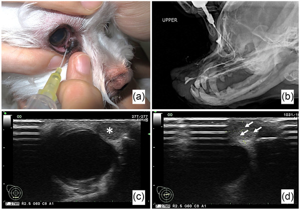
Background: A 10-year-old castrated male Maltese dog was presented with chronic swelling that had been present for at least 5 years in the medial canthus of the right eye (OD).
Objectives: To describe the treatment outcome of dacryops with dacryolithiasis.
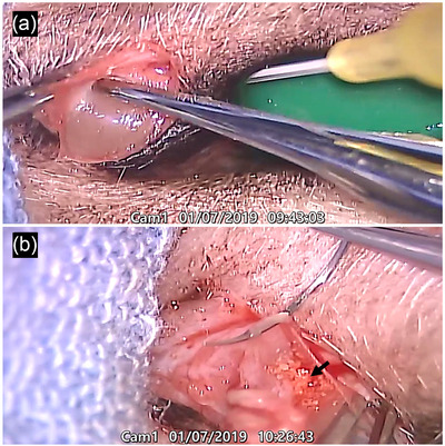
Methods: Bilateral patency of the nasolacrimal system was confirmed by flushing of both upper and lower puncta. Ocular ultrasonography revealed a well-defined, oval-shaped, heterogeneous mass with several hyperechoic foci. Dacryocystorhinography revealed no connection between the mass and lacrimal canaliculus. Gentle blunt dissection of the fibrous connective tissue around the cystic mass was performed. The mass was removed, which intraluminally contained multiple calculi.
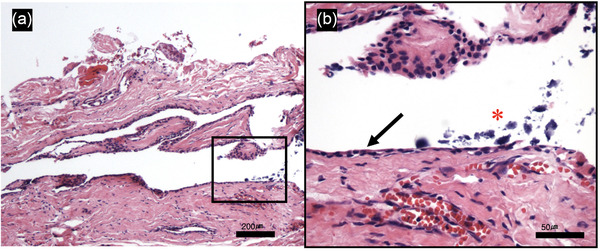
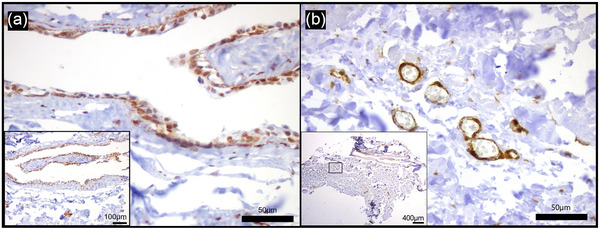
Results: Histopathologically, the cystic structure was lined by simple cuboidal epithelium and surrounded by smooth muscle actin positive myoepithelial cells consistent with dacryops derived from the lacrimal glandular ductal system. In addition, several spherical basophilic minerals were observed in the lumen, which were identified as dacryoliths.
Conclusion: Surgical removal of this dacryops with dacryolithiasis was curative without recurrence after four months.
Keywords: Maltese; dacryolithiasis; dacryops; ectopic lacrimal gland; lacrimal system; nasolacrimal cyst.
© 2022 The Authors. Veterinary Medicine and Science published by John Wiley & Sons Ltd.
Conflict of interest statement
None of the authors of this article has a financial or personal relationship with other people or organisations that could inappropriately influence or bias the content of the paper.
Figures
References
-
- Cassotis, N. J. , & Schiffman, P. (2006). Calcification associated with the nasolacrimal system of a horse: Case report and mineralogic composition. Veterinary Ophthalmology, 9, 187–190. - PubMed
-
- Delgado, E. (2013). Case report: Dacryops of the lacrimal gland in a dog. Veterinary Ophthalmology, 16, 153–158. - PubMed
-
- Giuliano, E. A. (2013). Diseases and surgery of the canine lacrimal secretory system. In Gellat K. N., Gilger B. C., & Kern (Eds.), T. J. Veterinary Ophthalmology (5th ed., pp. 912–944 ). Willey‐Blackwell.
-
- Iliadelis, E. , Karabatakis, V. , & Sofoniou, M. (1999). Dacryoliths in chronic dacryocystitis and their composition (spectrophotometric analysis). European Journal of Ophthalmology, 9, 266–268. - PubMed
Publication types
MeSH terms
Substances
LinkOut - more resources
Full Text Sources
Medical
