. 2022 Jun 6;14(6):a041238.
doi: 10.1101/cshperspect.a041238. Online ahead of print.

Fibroblast Heterogeneity in Healthy and Wounded Skin

Affiliations

Fibroblast Heterogeneity in Healthy and Wounded Skin

Clarisse Ganier et al. Cold Spring Harb Perspect Biol. .

Abstract

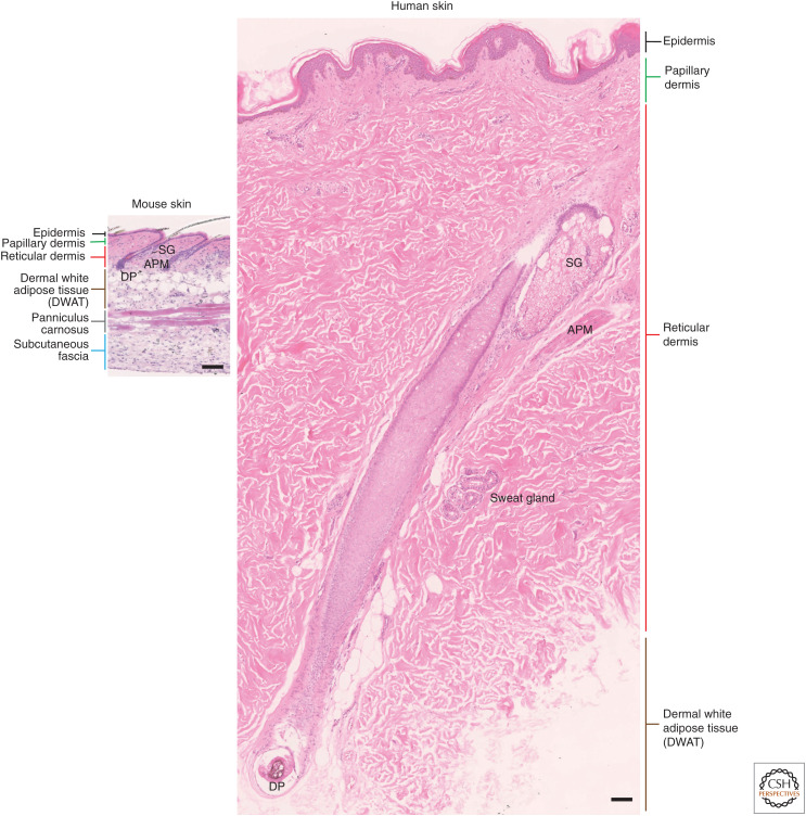
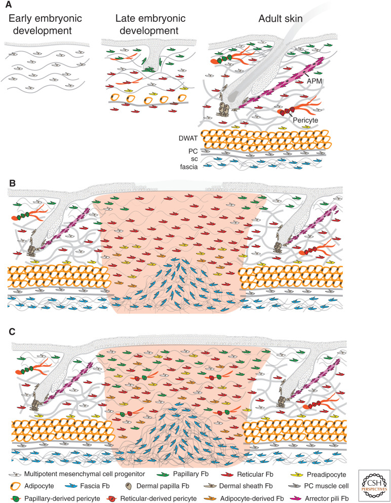
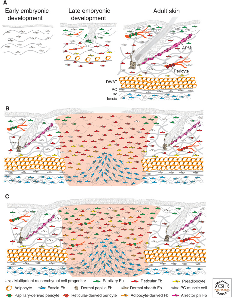
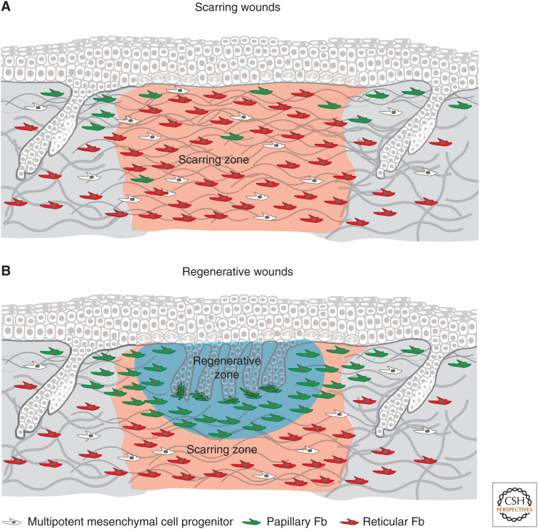
Fibroblasts are the main cell type in the dermis. They are responsible for the synthesis and deposition of structural proteins such as collagen and elastin, which are integrated into the extracellular matrix (ECM). Mouse and human studies using flow cytometry, cell culture, skin reconstitution, and lineage tracing experiments have shown the existence of different subpopulations of fibroblasts, including papillary fibroblasts, reticular fibroblasts, and fibroblasts comprising the dermal papilla at the base of the hair follicle. In recent years, the technological advances in single-cell sequencing have allowed researchers to study the repertoire of cells present in full-thickness skin including the dermis. Multiple groups have confirmed that distinct fibroblast populations can be identified in mouse and human dermis on the basis of differences in the transcriptional profile. Here, we discuss the current state of knowledge regarding dermal fibroblast heterogeneity in healthy mouse and human skin, highlighting the similarities and differences between mouse and human fibroblast subpopulations. We also discuss how fibroblast heterogeneity may provide insights into physiological wound healing and its dysfunction in pathological states such as hypertrophic and keloid scars.


Figures

Figure 1.
Figure 1.
Comparison of mouse and human body skin. The epidermis and dermis are thicker in human than in mouse skin. In most body sites, mouse body skin has a higher density of hair follicles compared to human body skin and the sweat glands are segregated to non-hair-bearing regions. In both mouse and human skin, the dermis is composed of a papillary and a reticular layer. Underneath the dermis, in mice, the subcutaneous fat is composed of a dermal with adipose tissue (DWAT), a panniculus carnosus (striated muscle), and the fascia beneath. In human, the dermis is separated from the underlying fascia by a large (many centimeters in the truck area) layer of subcutaneous fat (DWAT). Scale bars, 100 μm. (SG) Sebaceous gland, (APM) arrector pili muscle, (DP) dermal papilla.
Figure 2.
Figure 2.
Dermal cell organization during mouse skin development (A) and early (B) and advanced adult wound healing (C). In early embryonic development, there are multipotent mesenchymal cell progenitors present in the dermis. In late embryonic development, the mesenchymal progenitors have differentiated into papillary and reticular fibroblasts (Fb). The papillary fibroblasts form the dermal papillae of developing hair follicles, whereas the reticular fibroblasts gave rise to preadipocytes and mature adipocytes. In mouse adult skin, many hair follicles project into the dermis. There are specific fibroblasts in the dermal papilla at the base of each follicle, in the dermal sheath around each follicle, and in the APM that connects to the hair follicle bulge. The papillary dermis is composed of multipotent mesenchymal cell progenitors, papillary fibroblasts, and papillary derived pericytes. The reticular dermis is composed of multipotent mesenchymal cell progenitors, reticular fibroblasts, preadipocytes, and reticular derived pericytes. The subcutaneous fat is mainly composed by mature adipocytes in the dermal white adipose tissue (DWAT), panniculus carnosus (PC), muscle cells in the panniculus muscle layer, and fascia fibroblasts in the subcutaneous (sc) fascia. (A) In wounded adult skin, the initial wave of dermal repair is dominated by reticular fibroblasts; papillary fibroblasts from the upper dermis follow later. Fascia fibroblasts collectively migrate into the wound to contribute to the repair of deep full thickness wounds in the early phase. Also, preadipocytes and adipocytes in the lower dermis start to be recruited. At this stage, fibroblasts are highly proliferative and start to secrete collagen fibers. Reepithelialization is not complete. (B) In the advanced phase of wound healing, reepithelialization is complete and reticular fibroblasts, papillary fibroblasts, preadipocytes, adipocytes, and multipotent mesenchymal cell progenitors have migrated into the wound and are randomly distributed. More fascia fibroblasts from the subcutaneous fascia may have migrated and expanded into the lower area of deep full thickness wounds. At this stage, collagen fibers are secreted and remodeled by fibroblasts.
Figure 2.
Figure 2.
Dermal cell organization during mouse skin development (A) and early (B) and advanced adult wound healing (C). In early embryonic development, there are multipotent mesenchymal cell progenitors present in the dermis. In late embryonic development, the mesenchymal progenitors have differentiated into papillary and reticular fibroblasts (Fb). The papillary fibroblasts form the dermal papillae of developing hair follicles, whereas the reticular fibroblasts gave rise to preadipocytes and mature adipocytes. In mouse adult skin, many hair follicles project into the dermis. There are specific fibroblasts in the dermal papilla at the base of each follicle, in the dermal sheath around each follicle, and in the APM that connects to the hair follicle bulge. The papillary dermis is composed of multipotent mesenchymal cell progenitors, papillary fibroblasts, and papillary derived pericytes. The reticular dermis is composed of multipotent mesenchymal cell progenitors, reticular fibroblasts, preadipocytes, and reticular derived pericytes. The subcutaneous fat is mainly composed by mature adipocytes in the dermal white adipose tissue (DWAT), panniculus carnosus (PC), muscle cells in the panniculus muscle layer, and fascia fibroblasts in the subcutaneous (sc) fascia. (A) In wounded adult skin, the initial wave of dermal repair is dominated by reticular fibroblasts; papillary fibroblasts from the upper dermis follow later. Fascia fibroblasts collectively migrate into the wound to contribute to the repair of deep full thickness wounds in the early phase. Also, preadipocytes and adipocytes in the lower dermis start to be recruited. At this stage, fibroblasts are highly proliferative and start to secrete collagen fibers. Reepithelialization is not complete. (B) In the advanced phase of wound healing, reepithelialization is complete and reticular fibroblasts, papillary fibroblasts, preadipocytes, adipocytes, and multipotent mesenchymal cell progenitors have migrated into the wound and are randomly distributed. More fascia fibroblasts from the subcutaneous fascia may have migrated and expanded into the lower area of deep full thickness wounds. At this stage, collagen fibers are secreted and remodeled by fibroblasts.
Figure 3.
Figure 3.
Dermal wound healing zones in scarring and regenerative wounds. In mouse skin, the wound healing process differs depending on the size of the wound. Both large (>1 cm2) and small (<1 cm2) wounds heal by a combination of contraction and reepithelialization, which is followed by the formation of new connective tissue leading to scar formation (A). However, large wounds also contain a regenerative zone that is rich in fibroblasts in which hair follicle neogenesis occurs (B). Conversely, a small wound (<1 cm2) repairs without de novo hair follicle formation, resulting in scarring (A).

References

    1. Abbasi S, Sinha S, Labit E, Rosin NL, Yoon G, Rahmani W, Jaffer A, Sharma N, Hagner A, Shah P, et al. 2020. Distinct regulatory programs control the latent regenerative potential of dermal fibroblasts during wound healing. Cell Stem Cell 27: 396–412.e6. 10.1016/j.stem.2020.07.008 - DOI - PubMed
    1. Armulik A, Genové G, Betsholtz C. 2011. Pericytes: developmental, physiological, and pathological perspectives, problems, and promises. Dev Cell 21: 193–215. 10.1016/j.devcel.2011.07.001 - DOI - PubMed
    1. Ascensión AM, Fuertes-Álvarez S, Ibañez-Solé O, Izeta A, Araúzo-Bravo MJ. 2021. Human dermal fibroblast subpopulations are conserved across single-cell RNA sequencing studies. J Invest Dermatol 141: 1735–1744.e35. 10.1016/j.jid.2020.11.028 - DOI - PubMed
    1. Berry R, Rodeheffer MS. 2013. Characterization of the adipocyte cellular lineage in vivo. Nat Cell Biol 15: 302–308. 10.1038/ncb2696 - DOI - PMC - PubMed
    1. Birbrair A, Zhang T, Wang ZM, Messi ML, Enikolopov GN, Mintz A, Delbono O. 2013. Role of pericytes in skeletal muscle regeneration and fat accumulation. Stem Cells Dev 22: 2298–2314. 10.1089/scd.2012.0647 - DOI - PMC - PubMed

LinkOut - more resources