. 2022 Jun 6;13(6):529.
doi: 10.1038/s41419-022-04978-4.

Syntaxin 18 regulates the DNA damage response and epithelial-to-mesenchymal transition to promote radiation resistance of lung cancer

Affiliations

Syntaxin 18 regulates the DNA damage response and epithelial-to-mesenchymal transition to promote radiation resistance of lung cancer

Clotilde Thumser-Henner et al. Cell Death Dis. .

Abstract

Radiotherapy is an important modality in lung cancer treatment. Despite advances in treatment planning and dose delivery, patient benefit is still limited by in-field relapse and metastatic recurrence. Simultaneous application of cisplatinum-based chemotherapy leads to moderately improved outcomes, thus providing proof-of-concept for radiosensitization strategies in lung cancer. In an unbiased functional genetic screen for radiosensitization targets in lung cancer, we identified syntaxin 18, a protein involved in retrograde vesicular transport between the Golgi apparatus and endoplasmic reticulum, as mediator of radioresistance. Downregulation of endogenous syntaxin 18 specifically reduced clonogenic survival of radioresistant and radiosensitive lung cancer cells following X-radiation. Gene expression programs regulating DNA repair, mitotic checkpoints and mitosis were altered in isogenic cells with reduced syntaxin 18 expression. Functionally, this translated into impaired DNA damage-induced cell cycle checkpoints leading to cell death by mitotic catastrophe. Interestingly, downregulation of syntaxin 18 in lung cancer cells also impaired expression of markers of epithelial-mesenchymal-transition, and reduced migration and invasion capacity. These findings suggest that syntaxin 18 is a key player regulating genes responsible for controlling the growth of the primary tumor as well as metastases upon radiotherapy of lung cancer. They provide a promising lead for biologically rational radiosensitization strategies impacting on radiation-induced cell death as well as metastasis.


Conflict of interest statement

The authors declare no competing interests.

Figures
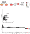
Fig. 1
Fig. 1. An in vitro shRNA screen identifies STX18 as a modulator of radiosensitivity in A549 cells.
a A549 cells were transduced with the DECIPHER shRNA library Human Module 1, comprising 27500 pooled shRNA with a transduction efficiency of 30% and cells were selected with puromycin. After 5 days, cells were pooled and either frozen, irradiated with 10 Gy or not irradiated. DNA was then isolated and sequenced. Analysis distinguished barcodes that were enriched or reduced after irradiation. n = 2. b Differential representation of individual shRNA with highest reduction after irradiation in both repetitions of the screen. Data were normalized to shRNA representation in the initial cell population. c Normalized differential expression of the most enriched and reduced shRNA after irradiation. Data were normalized to shRNA representation in the initial cell population.
Fig. 2
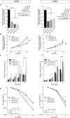
Fig. 2. STX18 knockdown sensitizes radioresistant A549 and radiosensitive H460 cell lines to irradiation.
a A549 and H460 cells were transduced with a shRNA targeting STX18 and its expression was quantified by RT-qPCR. n = 3–4. The insert shows respective immunoblotting analyses of STX18 expression. Here, quantification was achieved after normalization to the loading control (Actin) and the shScr sample. n = 3. b Proliferation of unirradiated cells was measured by proliferation/cell viability assay. n = 3. c Quantification of sub-G1 fraction by flow cytometry after irradiation. Cells were irradiated and cell cycle distribution was quantified by PI staining after 72 h. n = 3–5. d Colony formation ability was assessed after irradiation. Cells were irradiated and colonies were counted after 10 days. n = 3.
Fig. 3
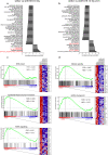
Fig. 3. RNA profiling of A549 cells identifies differentially expressed gene sets after STX18 knockdown and irradiation.
Differentially represented gene sets in shScr vs shSTX18 at basal level (a) and 24 h after irradiation with 10 Gy (b). Only the gene sets with an FDR < 0.25 are represented. For representation of A549-shSTX18 cells, counts from A549-shSTX18 K2 and K3 were pooled. c Enrichment plots of the gene sets “DNA repair”, “epithelial mesenchymal transition” and “TGFβ signaling” enriched in A549-shScr vs A549-shSTX18 K2 and K3. Biological replicates are indicated with I, II, and III. The 30 most enriched genes for each gene sets are represented. d Enrichment plots of the gene sets “mitotic spindle” and “G2/M checkpoint” enriched in A549-shScr vs A549-shSTX18 K2 and K3 after irradiation. Biological replicates are indicated with I, II, and III. The 30 most enriched genes for each gene sets are represented. n = 3.
Fig. 4
Fig. 4. STX18 knockdown leads to a defect in G2/M checkpoint in line with a premature entry into mitosis in A549 cells.
For the representation of STX18 knockdown, A549-shSTX18 K3 cells were used. a Immunoblotting analysis of proteins involved in cell cycle checkpoints after 10 Gy. Vinculin was used as control. After normalization to the loading control, the samples were compared to the shScr-0.5 h sample. b Detection by flow cytometry of Annexin V positive cells following irradiation with 10 Gy and/or pre-treatment with 1 µM berzosertib or Chir-124. Cells were incubated for 72 h then stained with Annexin V. c Quantification of mitotic index after irradiation with 2 Gy by quantification of cells positive for phosphorylation of Histone H3 by flow cytometry. d Quantification of fragmented nuclei and nuclei with abnormal shape in the cell population 72 h after irradiation. e Cell cycle analysis by flow cytometry after irradiation. Cells were irradiated and cell cycle distribution was analyzed by PI staining after 72 h. n = 3 for all experiments.
Fig. 5
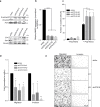
Fig. 5. Syntaxin 18 knockdown leads to an epithelial phenotype and decreased migration and invasion capacities in A549 cells.
a Immunoblotting analysis of E-cadherin, vimentin, ZO-1 and Zeb1 expression. Vinculin and actin were used as controls. After normalization to the loading control, the samples were compared to the shScr sample (set to 1). b RT-qPCR analysis of MMP9 mRNA expression. c Detection by flow cytometry of Annexin V positive cells. Cells were incubated for 72 h on poly(2-hydroxyethyl methacrylate)-coated (Poly-Hema) plates and stained with Annexin V. d Boyden chamber assay for A549 migration and invasion. Cells were seeded on the transwell plate after overnight serum starvation and incubated for 24 h. Migrated and invaded cells were then stained with crystal violet. e Representative pictures from Boyden chamber assay results shown in (d). Scale bar, 500 µm. n = 3 for all experiments.

Similar articles

Cited by

References

    1. Auperin A, Le Pechoux C, Rolland E, Curran WJ, Furuse K, Fournel P, et al. Meta-analysis of concomitant versus sequential radiochemotherapy in locally advanced non-small-cell lung cancer. J Clin Oncol: Off J Am Soc Clin Oncol. 2010;28:2181–90. doi: 10.1200/JCO.2009.26.2543. - DOI - PubMed
    1. Wu YL, Tsuboi M, He J, John T, Grohe C, Majem M, et al. Osimertinib in resected EGFR-mutated non-small-cell lung cancer. N Engl J Med. 2020;383:1711–23. doi: 10.1056/NEJMoa2027071. - DOI - PubMed
    1. Uramoto H, Tanaka F. Recurrence after surgery in patients with NSCLC. Transl Lung Cancer Res. 2014;3:242–9. - PMC - PubMed
    1. Durant ST, Zheng L, Wang Y, Chen K, Zhang L, Zhang T, et al. The brain-penetrant clinical ATM inhibitor AZD1390 radiosensitizes and improves survival of preclinical brain tumor models. Sci Adv. 2018;4:eaat1719. doi: 10.1126/sciadv.aat1719. - DOI - PMC - PubMed
    1. Ferreira S, Dutreix M. DNA repair inhibitors to enhance radiotherapy: Progresses and limitations. Cancer Radiother. 2019;23:883–90. doi: 10.1016/j.canrad.2019.08.008. - DOI - PubMed

Publication types

Substances