Review
. 2022 Aug;21(8):578-600.
doi: 10.1038/s41573-022-00477-5. Epub 2022 Jun 6.

Thinking outside the box: non-canonical targets in multiple sclerosis

Affiliations
Review

Thinking outside the box: non-canonical targets in multiple sclerosis

Laura Bierhansl et al. Nat Rev Drug Discov. 2022 Aug.

Abstract

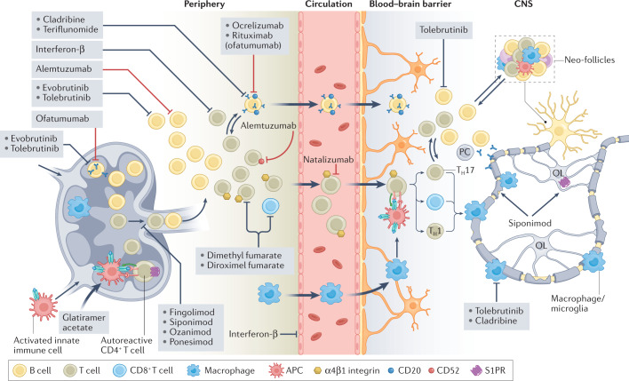
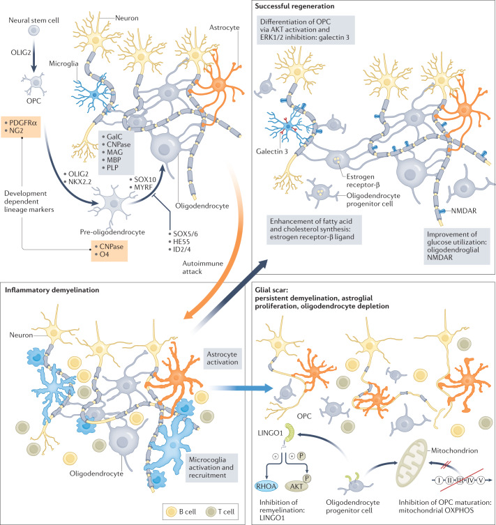
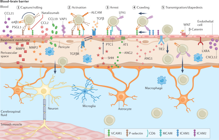
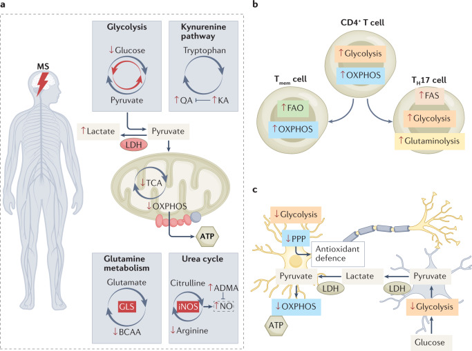
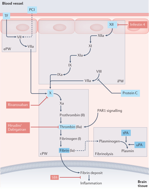
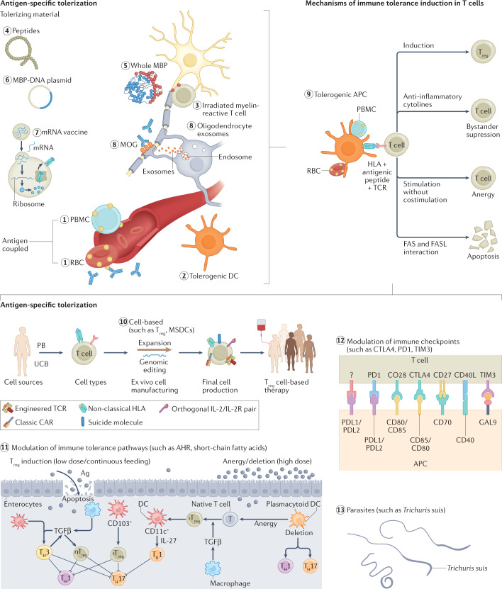
Multiple sclerosis (MS) is an immune-mediated disease of the central nervous system that causes demyelination, axonal degeneration and astrogliosis, resulting in progressive neurological disability. Fuelled by an evolving understanding of MS immunopathogenesis, the range of available immunotherapies for clinical use has expanded over the past two decades. However, MS remains an incurable disease and even targeted immunotherapies often fail to control insidious disease progression, indicating the need for new and exceptional therapeutic options beyond the established immunological landscape. In this Review, we highlight such non-canonical targets in preclinical MS research with a focus on five highly promising areas: oligodendrocytes; the blood-brain barrier; metabolites and cellular metabolism; the coagulation system; and tolerance induction. Recent findings in these areas may guide the field towards novel targets for future therapeutic approaches in MS.


Conflict of interest statement

The authors declare no competing interests.

Figures

Fig. 1
Fig. 1. Targets of current disease-modifying therapies in MS.
Overview of the immunopathogenesis and targets of available disease-modifying therapies in multiple sclerosis (MS). The established therapeutic approaches have diverse mechanisms of action (pleiotropic effects, immune cell depletion, reduction of proliferation and blockade of migration) that modify or inhibit the different steps of the inflammatory process in MS within the peripheral immune system, blood–brain barrier or within the central nervous system (CNS). The therapies depicted are subdivided into monoclonal antibodies (red lines) and pharmacological agents (black lines). APC, antigen-presenting cell; OL, oligodendrocyte; S1PR, sphingosine 1-phosphate receptor; TH cell, T helper cell.
Fig. 2
Fig. 2. Role of oligodendrocytes in MS.
Oligodendrocytes differentiate from neuronal stem cells to oligodendrocyte progenitor cells (OPCs) and pre-oligodendrocytes, a process orchestrated by several pathways and factors (oligodendrocyte transcription factor 2 (OLIG2), homeobox protein NKX2.2, SRY-box transcription factor 10 (SOX10), myelin regulatory factor (MYRF), SOX5/6, HES family bHLH transcription factor 5 (HES5), inhibitor of DNA binding 2 (ID2) and ID4). The different development stages of oligodendrocytes are typified by various oligodendroglial lineage markers (OPC: platelet-derived growth factor receptor-α (PDGFRα) and neuron-glial antigen 2 (NG2); pre-oligodendrocytes: 2′,3′-cyclic-nucleotide 3′-phosphodiesterase (CNPase) and oligodendrocyte marker 4 (O4); oligodendrocytes: galactocerebroside (GalC), CNPase, myelin-associated glycoprotein (MAG), myelin basic protein (MBP) and proteolipid protein (PLP)). In multiple sclerosis (MS), the autoimmune attack (orange arrow) drives inflammatory demyelination, which is characterized by astrocyte activation and microglia recruitment/activation. This leads to demyelination and axonal damage and is associated with astroglial proliferation and oligodendrocyte depletion, resulting in a glial scar (blue arrow). Strategies to modulate OPC differentiation to enhance remyelination (leucine-rich repeat and immunoglobin-like domain-containing protein 1 (LINGO1) and oxidative phosphorylation (OXPHOS)) are shown. To force successful regeneration (grey arrow), potential pathways are metabolic support (enhancement of glucose utilization or fatty acid and cholesterol synthesis) or promotion of OPC differentiation to oligodendrocyte (galectin 3).
Fig. 3
Fig. 3. Blood–brain barrier disruption in MS.
In physiological conditions, blood–brain barrier integrity is tightly regulated. In multiple sclerosis (MS), the tight barrier can be dysfunctional, leading to increased leukocyte recruitment and transmigration. In addition, autoreactive T cells can enter the CNS through peripheral activation of cellular locomotion molecules, together with chemokine and adhesion receptors. T cell trans-endothelial migration proceeds in several steps (steps 1 to 5) that are controlled by various adhesion molecules (selectins, lymphocyte function-associated antigen 1 (LFA1) and α4β1 integrin) and their receptors (selectin ligands, intercellular adhesion molecule 1 (ICAM1) and vascular cell adhesion protein 1 (VCAM1)) expressed by endothelial cells (ECs). Furthermore, tissue-resident macrophages can secrete factors (cytokines, chemokines and growth factors) that lead to EC activation, characterized by increased expression of selectin and adhesion molecules. ALCAM, activated leukocyte cell adhesion molecule; ANG, angiopoietin; CCL2, CC-chemokine ligand 2; CCL21, CC-chemokine ligand 21; CXCL12, CXC-chemokine ligand 12; LXRα, liver X receptor-α; MCAM, melanoma cell adhesion molecule; MMP2/9, matrix metalloproteinase 2/9; PSGL1, P-selectin glycoprotein ligand 1; TGFβ, transforming growth factor-β; TIE2, tyrosine-protein kinase receptor TIE2; VAP1, vascular adhesion protein 1.
Fig. 4
Fig. 4. Overview of metabolic adaptations in MS.
a | A variety of metabolic changes indicated by the different levels of metabolites in multiple sclerosis (MS) are known to occur. It is assumed that the MS brain metabolism has increased glycolysis and therefore a reduction of the flux through the tricarboxylic acid cycle (TCA) and oxidative phosphorylation (OXPHOS). Furthermore, other pathways such as the urea cycle, the kynurenine pathway and glutamine metabolism are also affected in MS. Red upwards and downwards arrows indicate changes in the metabolite level or metabolic pathways (grey circular arrows, changes in red) in MS. b | T cell subtypes show different metabolic properties. In MS, T cells seem to increase their metabolic fluxes (indicated by red upwards and downwards arrows); however, after differentiation, they display a different metabolic profile. T memory (Tmem) cells rely on fatty acid oxidation (FAO) and OXPHOS, whereas T helper 17 (TH17) cells display increased fatty acid synthesis (FAS), glycolysis and glutaminolysis. c | Metabolic active pathways of neurons (yellow) and astrocytes (blue). Neurons rely on the astrocyte–neuron lactate shuttle for their energy supply. Astrocytes take up glucose and metabolize it to pyruvate using glycolysis, and, finally, lactate is generated through lactate dehydrogenase (LDH). Lactate can exit the cell and the extracellular lactate can be shuttled into neurons to either fuel neuronal ATP synthesis or to generate reducing agents such as NADPH to maintain redox homeostasis. In MS, this mechanism seems to be diminished (indicated by red upwards and downwards arrows). ADMA, asymmetric dimethylarginine; BCAA, branched-chain amino acid; GLS, glutamine synthetase; iNOS, inducible nitric oxide synthetase; KA, kynurenic acid; NO, nitric oxide; PPP, pentose phosphate pathway; QA, quinolinic acid.
Fig. 5
Fig. 5. Alteration of the coagulation system in MS.
Several components of the coagulation pathway are involved in the neuroinflammatory process in multiple sclerosis (MS) and could serve as potential therapeutic targets. This overview demonstrates the classical coagulation pathway, including the extrinsic pathway (ePW), the intrinsic pathway (iPW), the common pathway (cPW) and fibrinolysis. In MS lesions the coagulation factors protein C inhibitor (PCI) and tissue factor (TF) are expressed, and the interaction of TF and factor VII initiates the ePW. Protein C levels are increased in MS patients, and recombinant activated protein C can reduce severity in autoimmune encephalomyelitis (EAE). The exposure of collagen and other intracellular molecules following cell damage can activate the iPW via factor XII. Both the ePW and iPW converge in the common pathway, which includes the cascade from factor X to thrombin and finally fibrin strands. Fibrin deposits accumulate within the brain tissue and can thereby activate inflammatory pathways. Tissue plasminogen activator (tPa) and urokinase plasminogen activator (uPa) activate plasminogen to plasmin, which can degrade the fibrin strands. The highlighted components are known to be altered in MS patients. Several known inhibitors (coloured red), including approved drugs (rivaroxaban, dabigatran and hirudin) and preclinical components (infestin 4 and monoclonal antibody 5B8), are known to inhibit the cascade at certain steps, which has potentially protective effects in in vitro models of MS.
Fig. 6
Fig. 6. Tolerance induction in MS.
Immune tolerance induction aims to treat multiple sclerosis (MS) at the initial stages of pathogenesis. Various approaches are of interest to induce immune tolerance and can be separated into antigen-specific (labelled 1 to 8) and unspecific tolerization strategies (labelled 10 to 13). The mechanisms (labelled 9) of immune tolerance are driven by tolerogenic antigen-presenting cells (APCs). Application of the different potential auto-pathogen factors can lead to the development of those cells. APC interaction with T cells leads to several changes in the immune response: the induction of regulatory T (Treg) cells; reduction of effector T cells; an increase in exhausted T cells and Treg cells with strong bystander immunosuppression capacity; apoptosis of autoreactive T cells; and anergy in T cells. Ag, antigen; AHR, aryl hydrocarbon receptor; CAR, chimeric antigen receptor; CTLA4, cytotoxic T lymphocyte antigen 4; DC, dendritic cell; GAL9, galectin 9; MBP, myelin basic protein; MOG, myelin oligodendrocyte glycoprotein; PB, peripheral blood; PBMC, peripheral blood mononuclear cell; PD1, programmed cell death 1; PDL1, programmed cell death 1 ligand; RBC, red blood cell; TH, T helper; TCR, T cell receptor; TGFβ, transforming growth factor-β; TIM3, T cell immunoglobulin mucin receptor 3; UCB, umbilical cord blood.

Similar articles

Cited by

References

    1. Reich DS, Lucchinetti CF, Calabresi PA. Multiple sclerosis. N. Engl. J. Med. 2018;378:169–180. doi: 10.1056/NEJMra1401483. - DOI - PMC - PubMed
    1. Baecher-Allan C, Kaskow BJ, Weiner HL. Multiple sclerosis: mechanisms and immunotherapy. Neuron. 2018;97:742–768. doi: 10.1016/j.neuron.2018.01.021. - DOI - PubMed
    1. Walton C, et al. Rising prevalence of multiple sclerosis worldwide: insights from the Atlas of MS, third edition. Mult. Scler. 2020;26:1816–1821. doi: 10.1177/1352458520970841. - DOI - PMC - PubMed
    1. Sorensen PS, et al. The apparently milder course of multiple sclerosis: changes in the diagnostic criteria, therapy and natural history. Brain. 2020;143:2637–2652. doi: 10.1093/brain/awaa145. - DOI - PubMed
    1. Cree BAC, et al. Secondary progressive multiple sclerosis: new insights. Neurology. 2021;97:378–388. doi: 10.1212/WNL.0000000000012323. - DOI - PMC - PubMed