. 2022 Jun 6;15(1):192.
doi: 10.1186/s13071-022-05291-x.

Plasmodium infection suppresses colon cancer growth by inhibiting proliferation and promoting apoptosis associated with disrupting mitochondrial biogenesis and mitophagy in mice

Affiliations

Plasmodium infection suppresses colon cancer growth by inhibiting proliferation and promoting apoptosis associated with disrupting mitochondrial biogenesis and mitophagy in mice

Xin Yao et al. Parasit Vectors. .

Abstract

Background: Colon cancer is a common gastrointestinal tumor with a poor prognosis, and thus new therapeutic strategies are urgently needed. The antitumor effect of Plasmodium infection has been reported in some murine models, but it is not clear whether it has an anti-colon cancer effect. In this study, we investigated the anti-colon cancer effect of Plasmodium infection and its related mechanisms using a mouse model of colon cancer.

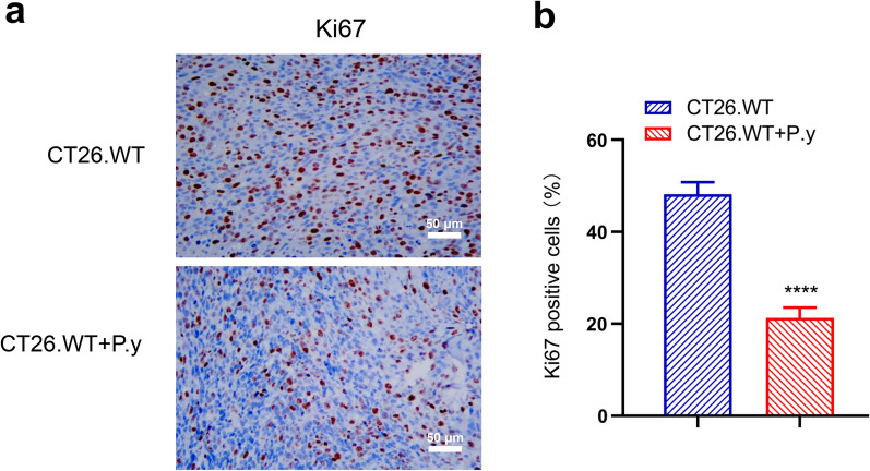
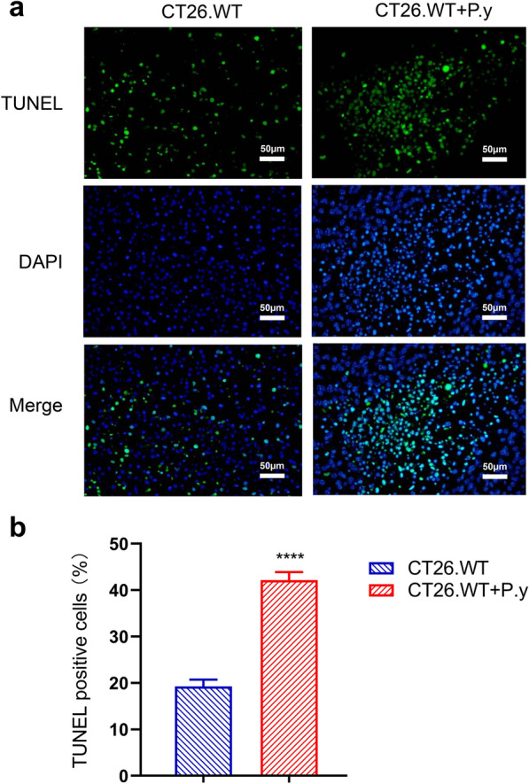
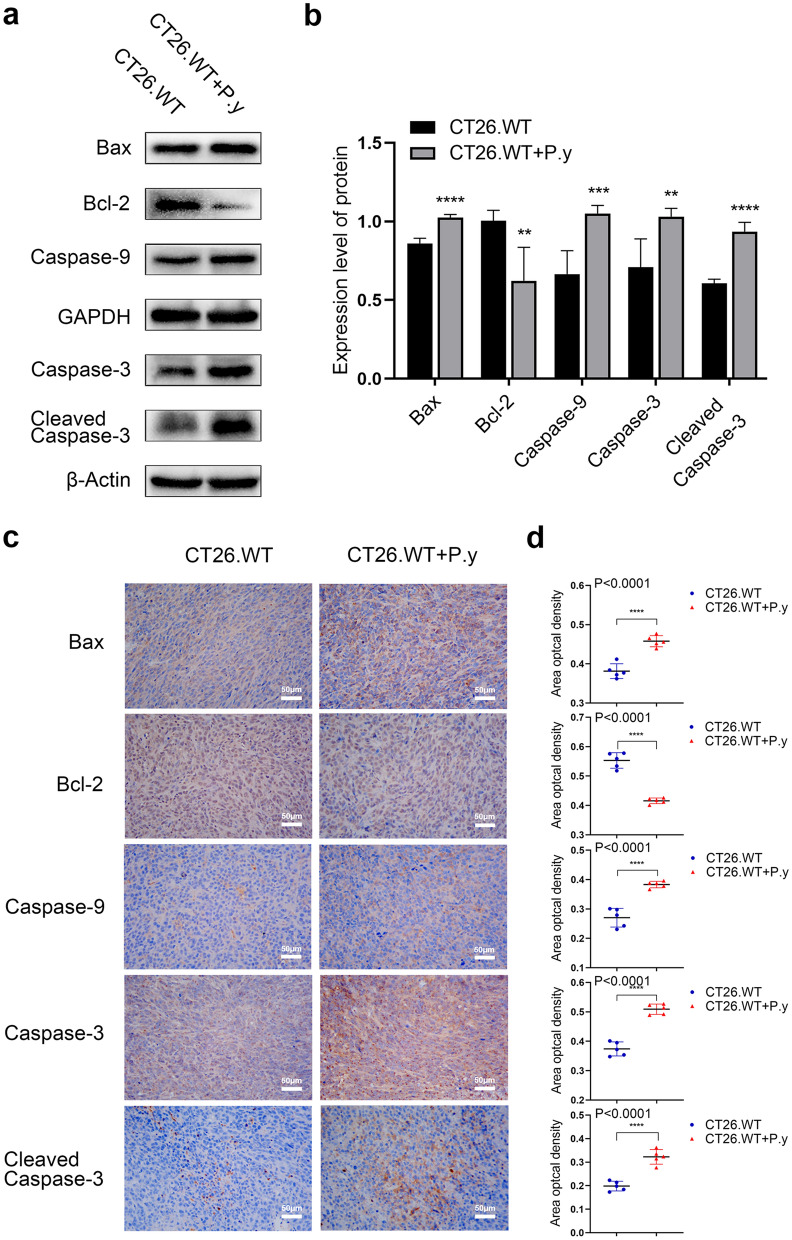
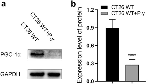
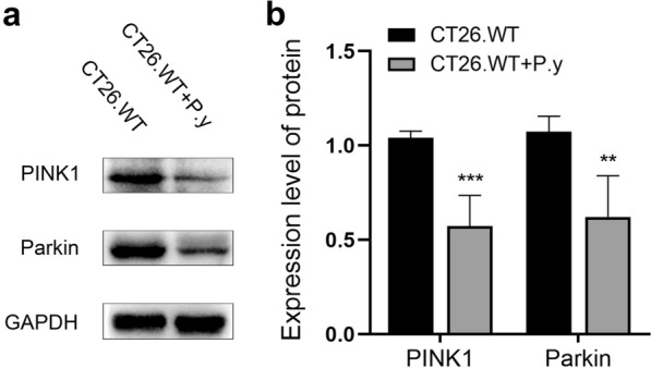
Methods: An experimental model was established by intraperitoneal injection of Plasmodium yoelii 17XNL-infected erythrocytes into mice with colon cancer. The size of tumors was observed dynamically in mice, and the expression of Ki67 detected by immunohistochemistry was used to analyze tumor cell proliferation. Apoptosis was assessed by terminal deoxynucleotidyl transferase (TdT) dUTP nick-end labeling (TUNEL) staining, and the expression of apoptosis-related proteins including Bax, Bcl-2, caspase-9, and cleaved caspase-3 was detected by western blot and immunohistochemistry, respectively. Transmission electron microscopy (TEM) was used to observe the ultrastructural change in colon cancer cells, and the expression of mitochondrial biogenesis correlative central protein, PGC-1α, and mitophagy relevant crucial proteins, PINK1/Parkin, were detected by western blot.

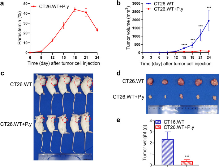
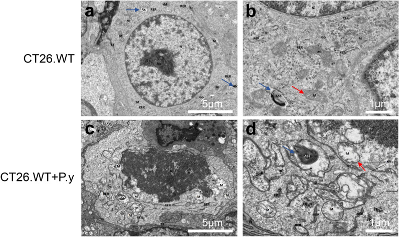
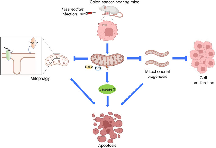
Results: We found that Plasmodium infection reduced the weight and size of tumors and decreased the expression of Ki67 in colon cancer-bearing mice. Furthermore, Plasmodium infection promoted mitochondria-mediated apoptosis in colon cancer cells, as evidenced by the increased proportion of TUNEL-positive cells, the upregulated expression of Bax, caspase-9, and cleaved caspase-3 proteins, and the downregulated expression of Bcl-2 protein. In colon cancer cells, we found destroyed cell nuclei, swollen mitochondria, missing cristae, and a decreased number of autolysosomes. In addition, Plasmodium infection disturbed mitochondrial biogenesis and mitophagy through the reduced expression of PGC-1α, PINK1, and Parkin proteins in colon cancer cells.

Conclusions: Plasmodium infection can play an anti-colon cancer role in mice by inhibiting proliferation and promoting mitochondria-mediated apoptosis in colon cancer cells, which may relate to mitochondrial biogenesis and mitophagy.

Keywords: Colon cancer; Mitochondrial apoptosis; Mitochondrial biogenesis; Mitophagy; Plasmodium.


Conflict of interest statement

The authors declare that they have no competing interests.

Figures

Fig. 1
Fig. 1
Plasmodium yoelii infection suppressed tumor growth in a murine colon cancer model. BALB/c mice were subcutaneously injected with CT26.WT cells under the right forelimb. On the sixth day, the tumor-bearing mice were intraperitoneally injected with either P. yoelii-infected erythrocytes or normal erythrocytes. a Parasitemia of the CT26.WT + P.y group. b Tumor volume was measured over time from the day of tumor cell inoculation. Day 15 (t-test, t(8) = 10.01, P < 0.0001); day 18 (t-test, t(8) = 6.515, P = 0.0002); day 21 (t-test, t(8) = 6.290, P = 0.0002); day 24 (t-test, t(8) = 6.730, P = 0.0001). c, d The mice were euthanized on day 24, and tumors were harvested for weighing, photographing, and further analysis. e Weight of the tumors (t-test, t(8) = 6.618, P = 0.0002). CT26.WT denotes the control group and CT26.WT + P.y denotes the P. yoelii-infected group. The results are shown as mean ± SEM (n = 5). ***P < 0.001, ****P < 0.0001
Fig. 2
Fig. 2
Plasmodium infection inhibited the proliferation of tumor cells in colon cancer tissues. a Immunohistochemical staining demonstrates that the Ki67-positive cells were decreased in the CT26.WT + P.y group (×400). b The percentage of Ki67 expression in tumor tissues of the two groups (t-test, t(8) = 17.51, P < 0.0001). Brown areas represent positive expression. Scale bar = 50 µm. The results are shown as mean ± SEM (n = 5). ****P < 0.0001
Fig. 3
Fig. 3
Apoptosis in colon cancer induction by Plasmodium infection. a Apoptosis was observed under a fluorescence microscope (×400). b Quantitative estimation of the proportion of apoptotic cells in each group (t-test, t(8) = 22.31, P < 0.0001). Green fluorescence indicates the TUNEL-positive nuclei, blue represents DAPI stained nuclei, and a blend indicates apoptotic cells. The results are shown as mean ± SEM (n = 5). Scale bar = 50 µm. **** P < 0.0001
Fig. 4
Fig. 4
The mitochondrial pathway regulation by Plasmodium infection in colon cancer-bearing mice. a The expression of mitochondria-mediated apoptosis proteins (Bax, Bcl-2, caspase-9, caspase-3 and cleaved caspase-3) was analyzed by western blot. b Quantification of relative intensity of western blot signals: Bax (t-test, t(8) = 9.438, P < 0.0001); Bcl-2 (t-test, t(8) = 3.822, P = 0.0051); caspase-9 (t-test, t(8) = 5.462, P = 0.0006); caspase-3 (t-test, t(8) = 3.836, P = 0.0050); cleaved caspase-3 (t-test, t(8) = 11.36, P < 0.0001). c, d Immunohistochemical staining and quantification of apoptosis protein expression in the mitochondrial pathway. Bax (t-test, t(8) = 7.222, P < 0.0001); Bcl-2 (t-test, t(8) = 10.92, P < 0.0001); caspase-9 (t-test, t(8) = 7.465, P < 0.0001); caspase-3 (t-test, t(8) = 10.23, P < 0.0001); cleaved caspase-3 (t-test, t(8) = 7.458, P < 0.0001). The results are presented as mean ± SEM (n = 5). Scale bar = 50 µm. ** P < 0.01, *** P < 0.001, **** P < 0.0001
Fig. 5
Fig. 5
Changes in tumor cell ultrastructure after Plasmodium infection. The ultrastructural changes in tumor cells in the two groups were observed by TEM. The damaged cell nuclei and mitochondria were observed in the CT26.WT + P.y group. The mitochondria (M, red arrow) were swollen and the cristae disappeared and vacuolated, with chromatin condensation and nuclear disintegration (N: nucleus). The number of autolysosomes (ASS, blue arrow) in the CT26.WT + P.y group was more than that in the CT26.WT group. a, c The magnification is ×2000, scale bar = 5 µm. b, d The magnification is ×6000, scale bar = 1 µm
Fig. 6
Fig. 6
Plasmodium infection inhibited mitochondrial biogenesis in colon cancer. a Western blot analysis showed that the expression of the PGC-1α protein was decreased in the CT26.WT + P.y group; GAPDH was used as a loading control. b The density ratio of PGC-1α/GAPDH was decreased in the CT26.WT + P.y group compared to the control group (t-test, t(8) = 8.356, P < 0.0001). The results are shown as the density mean ± SEM (n = 5). **** P < 0.0001
Fig. 7
Fig. 7
Plasmodium infection inhibited mitophagy in the murine colon cancer model. a The western blot results showed that the expression of PINK1 and Parkin were both decreased in the CT26.WT + P.y group. b Quantification of protein expression was normalized to GAPDH: PINK1 (t-test, t(8) = 6.558, P < 0.0002); Parkin (t-test, t(8) = 4.758, P = 0.0014). The results are shown as the density mean ± SEM (n = 5). **P < 0.01, ***P < 0.001
Fig. 8
Fig. 8
The anti-colon cancer mechanisms of Plasmodium infection in vivo. In the murine colon cancer model, Plasmodium infection inhibits the proliferation of tumor cells and induces mitochondrial apoptosis. Furthermore, Plasmodium infection disturbs mitochondrial biogenesis, leading to the inhibition of proliferation and the promotion of apoptosis. Mitophagy is inhibited by Plasmodium infection, contributing to mitochondrial dysfunction. Plasmodium infection inhibits the growth of tumors to exert the tumor suppression function

References

    1. Sung H, Ferlay J, Siegel RL, Laversanne M, Soerjomataram I, Jemal A, et al. Global cancer statistics 2020: GLOBOCAN estimates of incidence and mortality worldwide for 36 cancers in 185 countries. CA Cancer J Clin. 2021;71:209–249. doi: 10.3322/caac.21660. - DOI - PubMed
    1. Taieb J, André T, Auclin E. Refining adjuvant therapy for non-metastatic colon cancer, new standards and perspectives. Cancer Treat Rev. 2019;75:1–11. doi: 10.1016/j.ctrv.2019.02.002. - DOI - PubMed
    1. Vogel G. Malaria as lifesaving therapy. Science. 2013;342:686. doi: 10.1126/science.342.6159.686. - DOI - PubMed
    1. Qin L, Chen C, Chen L, Xue R, Ou-Yang M, Zhou C, et al. Worldwide malaria incidence and cancer mortality are inversely associated. Infect Agent Cancer. 2017;12:14. doi: 10.1186/s13027-017-0117-x. - DOI - PMC - PubMed
    1. Chen L, He Z, Qin L, Li Q, Shi X, Zhao S, et al. Antitumor effect of malaria parasite infection in a murine Lewis lung cancer model through induction of innate and adaptive immunity. PLoS ONE. 2011;6:e24407. doi: 10.1371/journal.pone.0024407. - DOI - PMC - PubMed

MeSH terms