. 2022 Jun 6;41(1):195.
doi: 10.1186/s13046-022-02402-5.

Linear DNA amplicons as a novel cancer vaccine strategy

Affiliations

Linear DNA amplicons as a novel cancer vaccine strategy

Antonella Conforti et al. J Exp Clin Cancer Res. .

Abstract

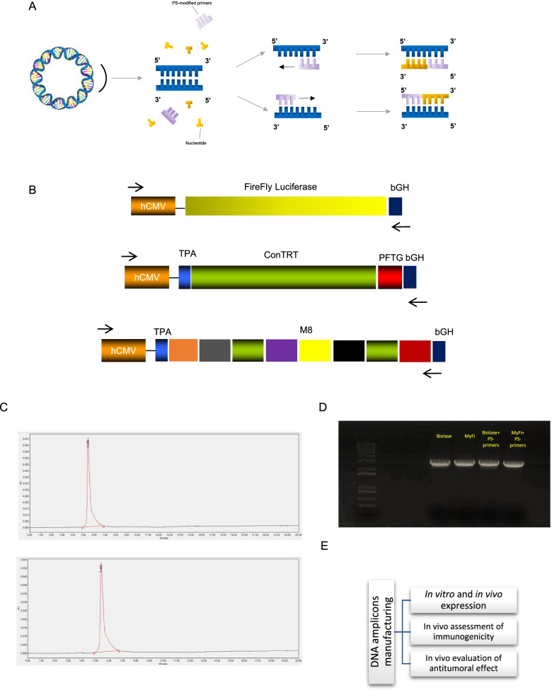
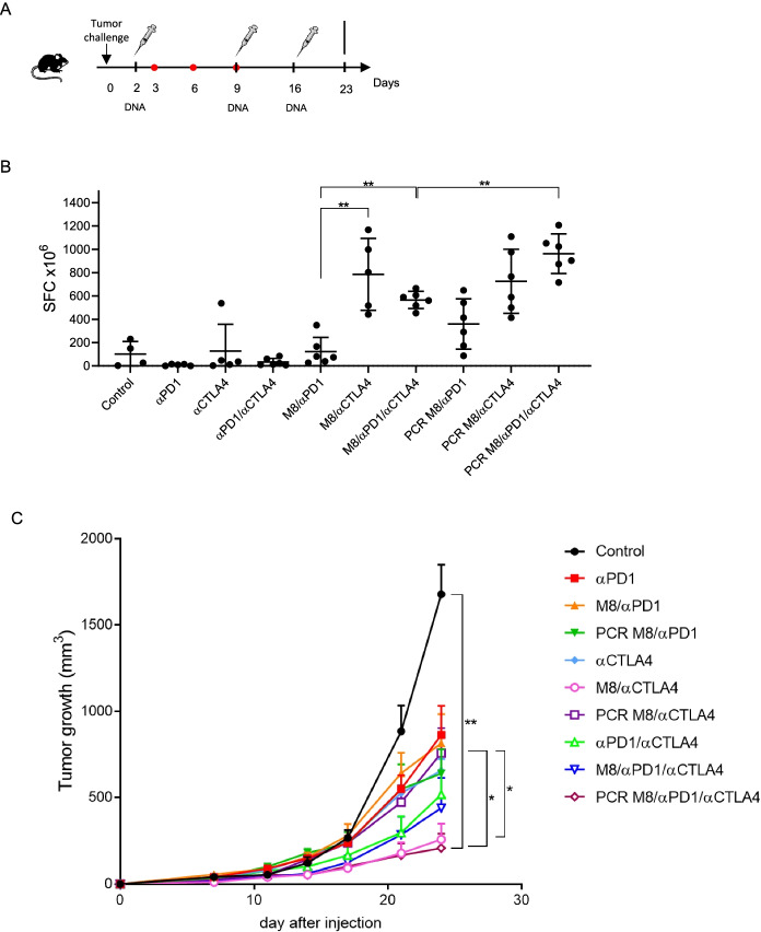
Background: DNA-based vaccines represent a simple, safe and promising strategy for harnessing the immune system to fight infectious diseases as well as various forms of cancer and thus are considered an important tool in the cancer immunotherapy toolbox. Nonetheless, the manufacture of plasmid DNA vaccines has several drawbacks, including long lead times and the need to remove impurities from bacterial cultures. Here we report the development of polymerase chain reaction (PCR)-produced amplicon expression vectors as DNA vaccines and their in vivo application to elicit antigen-specific immune responses in animal cancer models.

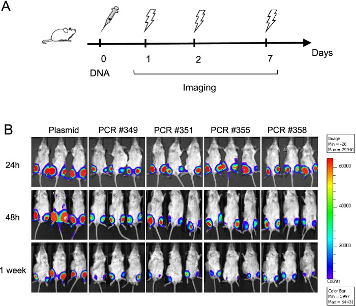
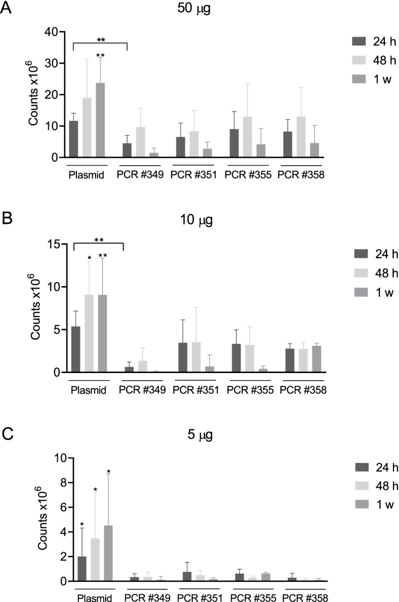
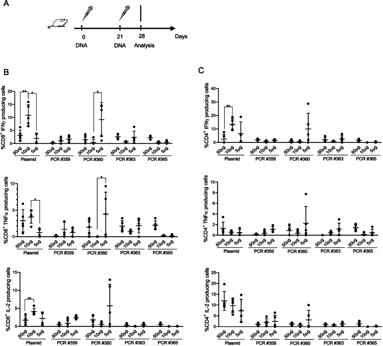
Methods: Plasmid DNA and amplicon expression was assessed both in vitro, by Hela cells transfection, and in vivo, by evaluating luciferase expression in wild-type mice through optical imaging. Immunogenicity induced by DNA amplicons was assessed by vaccinating wild-type mice against a tumor-associated antigen, whereas the antitumoral effect of DNA amplicons was evaluated in a murine cancer model in combination with immune-checkpoint inhibitors (ICIs).

Results: Amplicons encoding tumor-associated-antigens, such as telomerase reverse transcriptase or neoantigens expressed by murine tumor cell lines, were able to elicit antigen-specific immune responses and proved to significantly impact tumor growth when administered in combination with ICIs.

Conclusions: These results strongly support the further exploration of the use of PCR-based amplicons as an innovative immunotherapeutic approach to cancer treatment.

Keywords: Amplicons; Cancer immunotherapy; DNA vaccine; PCR.


Conflict of interest statement

Evvivax, Takis and NeoMatrix are currently developing proprietary nucleic-acid vaccines based on DNA-EP. Applied DNA is commercializing LinearDNA™, its proprietary, large-scale PCR-based manufacturing platform that allows for the large-scale production of specific DNA sequences. The Company’s common stock is listed on NASDAQ under ticker symbol ‘APDN,’ and its publicly traded warrants are listed on OTC under ticker symbol ‘APPDW.’

Figures

Fig. 1
Fig. 1
Manufacturing process of DNA amplicons. A A schematic representation showing the assembly of termini PS-modified amplicon expression vectors via a PCR device is shown. The antigen-encoding sequence, taken from a plasmid DNA, comprises a promotor, one or more ORFs and a terminator. Amplicon expression vector, encoding the selected sequence, is syntethized and then amplified through PS-modified PCR primers in order to produce a plurality of amplicon expression vectors. B A schematic representation of gene expression cassettes is shown for luciferase, conTRT and M8 constructs. C HPLC chromatogram of two representative DNA amplicons, one synthetized by means of Biolase™ Taq (top) and one synthetized by means of MyFi™ Taq (bottom). D Electrophoresis gel of four DNA amplicons, all encoding luciferase gene (synthetized by means of Biolase™ or MyFi.™ Taq polymerase, with or w/o PS-modified primers). E A schematic representation of experimental study passages is shown
Fig. 2
Fig. 2
In vivo expression of DNA amplicons. A Schematic representation of the experimental setup. BALB/c mice (3 mice/group) were injected i.m. by DNA-EP, with equimolar concentrations of plasmid DNA or DNA amplicons encoding for luciferase (50–10-5 μg/mouse). B After 1–2-7 days post DNA injection, luciferase signal was measured by performing optical imaging at IVIS. Although a significant decreased expression was detected in DNA amplicons injected mice after 7 days, no difference was measured between DNA amplicons synthetized by means of different Taq polymerase
Fig. 3
Fig. 3
Assessment of in vivo DNA amplicon expression. By means of optical imaging at IVIS, in vivo plasmid DNA and amplicon expression was assessed after 24 h, 48 h and one week post DNA injection at different DNA doses, 50 μg (A) – 10 μg (B) – 5 μg (C). In A) at 24 h post DNA injection, the only statistically significant difference was measured between DNA plasmid and PCR #349; at 48 h no significant difference was assessed between samples; after 1 week, DNA plasmid expression is significantly higher than DNA amplicons expression. In B) at 24 h post DNA injection, DNA plasmid expression is significantly higher only than PCR #349 expression; at 48 h and 1 week post injection, plasmid expression is significantly higher than each PCR. In C) DNA plasmid expression is significantly higher than each PCR at any time point. Significance was determined using Mann–Whitney test, *p < 0,05 **p < 0.01
Fig. 4
Fig. 4
In vivo immunogenicity of DNA amplicons. A Schematic representation of experimental setup. BALB/c mice (6 mice/group) were injected, by means of i.m. DNA-EP, with three doses (50–10-5 μg/mouse) of a DNA plasmid, and equimolar doses of DNA amplicons, both encoding for conTRT. All mice were vaccinated following a prime-boost regimen (days 0 and 21) and blood analysis was performed at day 28 by intracellular cytokine staining. Assessment of Th1 (B) and Th2 (C) immune response was done on PBMCs isolated at day 28. Significance was determined using Mann–Whitney test, *p < 0,05 **p < 0.01
Fig. 5
Fig. 5
In vivo antitumoral effect of DNA amplicons. A Schematic representation of experimental setup in a neoantigen cancer murine model. After tumor challenge, C57Bl/6 mice (6 mice/group) received a combination of DNA (plasmid or amplicon, administered at days 2,9 and 16) and ICIs (αCTLA-4 or αPD-1, administered at days 3,6 and 9). B Neoantigen-specific T cell response was assessed by IFNγ ELISpot performed on splenocytes collected at day 23 and stimulated with neoantigen peptide pools. C Panel depicts tumor growth curve up to sacrifice at day 23. The combination of PCR M8 with both ICIs is significantly higher than the combination of PCR M8 with the only αCTLA-4 or αPD1, whereas M8/αCTLA-4 is significantly more effective than PCR M8/αCTLA-4. Significance was determined using Mann–Whitney test, *p < 0,05 **p < 0.01

References

    1. Liu MA. DNA vaccines: an historical perspective and view to the future. Immunol Rev. 2011;239:62–84. doi: 10.1111/j.1600-065X.2010.00980.x. - DOI - PubMed
    1. Buonaguro L, Petrizzo A, Tornesello ML, Buonaguro FM. Translating tumor antigens into cancer vaccines. Clin Vaccine Immunol. 2010;18:23–34. doi: 10.1128/CVI.00286-10. - DOI - PMC - PubMed
    1. Aurisicchio L, Pallocca M, Ciliberto G, Palombo F. The perfect personalized cancer therapy: cancer vaccines against neoantigens. J Exp Clin Cancer Res. 2018;37:86. doi: 10.1186/s13046-018-0751-1. - DOI - PMC - PubMed
    1. Conforti A, Palombo F, Aurisicchio L. DNA/RNA based vaccines. In: Buonaguro L, Van der Burg S, editors. Cancer vaccines as Immunotherapy of Cance. Elsevier. 2022. In press.
    1. Salvatori E, Lione L, Compagnone M, Pinto E, Conforti A, Ciliberto G, et al. Neoantigen cancer vaccine augments anti-CTLA-4 efficacy. NPJ Vaccines. 2022;7:15. doi: 10.1038/s41541-022-00433-9. - DOI - PMC - PubMed