. 2023 Apr 12;227(7):838-849.
doi: 10.1093/infdis/jiac229.

People With Human Immunodeficiency Virus Receiving Suppressive Antiretroviral Therapy Show Typical Antibody Durability After Dual Coronavirus Disease 2019 Vaccination and Strong Third Dose Responses

Affiliations

People With Human Immunodeficiency Virus Receiving Suppressive Antiretroviral Therapy Show Typical Antibody Durability After Dual Coronavirus Disease 2019 Vaccination and Strong Third Dose Responses

Hope R Lapointe et al. J Infect Dis. .

Abstract

Background: Longer-term humoral responses to 2-dose coronavirus disease 2019 (COVID-19) vaccines remain incompletely characterized in people living with human immunodeficiency virus (HIV) (PLWH), as do initial responses to a third dose.

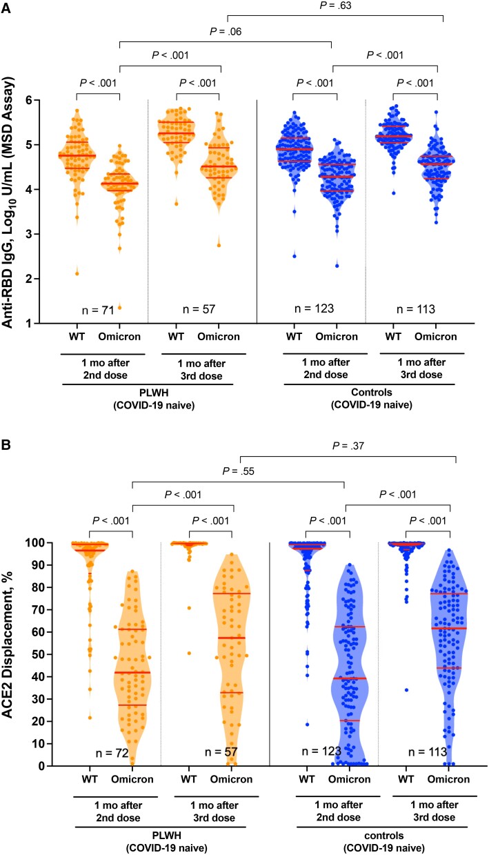
Methods: We measured antibodies against the severe acute respiratory syndrome coronavirus 2 (SARS-CoV-2) spike protein receptor-binding domain, angiotensin-converting enzyme 2 (ACE2) displacement, and viral neutralization against wild-type and Omicron strains up to 6 months after 2-dose vaccination, and 1 month after the third dose, in 99 PLWH receiving suppressive antiretroviral therapy and 152 controls.

Results: Although humoral responses naturally decline after 2-dose vaccination, we found no evidence of lower antibody concentrations or faster rates of antibody decline in PLWH compared with controls after accounting for sociodemographic, health, and vaccine-related factors. We also found no evidence of poorer viral neutralization in PLWH after 2 doses, nor evidence that a low nadir CD4+ T-cell count compromised responses. Post-third-dose humoral responses substantially exceeded post-second-dose levels, though Omicron-specific responses were consistently weaker than responses against wild-type virus. Nevertheless, post-third-dose responses in PLWH were comparable to or higher than controls. An mRNA-1273 third dose was the strongest consistent correlate of higher post-third-dose responses.

Conclusion: PLWH receiving suppressive antiretroviral therapy mount strong antibody responses after 2- and 3-dose COVID-19 vaccination. Results underscore the immune benefits of third doses in light of Omicron.

Keywords: COVID-19; HIV; antibodies; humoral; immune response; neutralization; third dose; vaccines.


Conflict of interest statement

Potential conflicts of interest. All authors: no reported conflicts. All authors have submitted the ICMJE Form for Disclosure of Potential Conflicts of Interest. Conflicts that the editors consider relevant to the content of the manuscript have been disclosed.

Figures

Figure 1.
Figure 1.
Concentrations of total binding antibodies in serum to spike receptor-binding domain (RBD) after 2 and 3 coronavirus disease 2019 (COVID-19) vaccine doses. A, Binding antibody responses to the severe acute respiratory syndrome coronavirus 2 spike RBD in serum samples 1, 3, and 6 months after the second vaccine dose and 1 month after the third dose, in people living with human immunodeficiency virus (PLWH) (orange) and controls (blue) who were COVID-19 naive at the studied time point, as well as in individuals who had recovered from COVID-19 at the studied time point (COVID group [black]). Participants who experienced a postvaccination infection were relocated from their original group into the COVID group at their first postinfection study visit, where they are denoted by red symbols. Numbers of participants are shown at the bottom of the plot. The thick horizontal red bar represents the median; thinner horizontal red bars, the interquartile range (IQR). P values were computed using the Mann-Whitney U test (for comparisons between groups) or the Wilcoxon matched-pairs test (for comparisons across time points within a group) and are uncorrected for multiple comparisons. Abbreviations: LLOD, lower limit of detection; ULOQ, upper limit of quantification. B, Temporal declines in serum binding antibody responses to spike RBD after 2 vaccine doses in individual PLWH (light orange) and controls (light blue) who remained COVID-19 naive during this period. Thick lines in corresponding colors denote averages for each group. Only participants with a complete longitudinal data series with no values above the ULOQ are shown. C, Binding antibody half-lives after 2 COVID-19 vaccine doses, in PLWH (orange) and controls (blue) who remained COVID-19 naive during this period. Values were calculated by fitting an exponential curve to the data shown in B. The 2 outliers are individuals whose antibody levels did not decay or decayed exceedingly slowly, producing half-lives >500 days. For visualization purposes, their half-lives are shown as 500 days. Numbers are indicated at the bottom of the plot. Red bars and whiskers represent medians and interquartile ranges, and P values were computed using the Mann-Whitney U test.
Figure 2.
Figure 2.
Live virus neutralization activities after 2 and 3 coronavirus disease 2019 (COVID-19) vaccine doses. Viral neutralization activity in plasma at 1, 3, and 6 months after the second dose, and 1 month after the third vaccine dose, in people living with human immunodeficiency virus (PLWH) (orange) and controls (blue) who were COVID-19 naive at the studied time point, as well as individuals who had recovered from COVID-19 at the studied time point (COVID group [black]). Plasma neutralization was defined as the reciprocal of the highest plasma dilution at which vial cytopathic effect was prevented in all triplicate assay wells. Plasma samples showing neutralization in <3 wells at the lowest plasma dilution of 1:20 were coded as having a reciprocal dilution of 10, corresponding to the lower limit of quantification (LLOQ) in this assay. The highest dilution tested was 1:2560, which corresponds to the upper limit of quantification (ULOQ). Participants who experienced a postvaccination infection were relocated from their original group into the COVID-19 group at their first postinfection study visit, where they are denoted by a red symbol. Numbers of participants are shown at the bottom of the plot. Thick horizontal red bars represent medians; thinner horizontal red bars, interquartile ranges. P values were computed using the Mann-Whitney U test (for comparisons between groups) or the Wilcoxon matched-pairs test (for comparisons across time points within a group) and are uncorrected for multiple comparisons.
Figure 3.
Figure 3.
Omicron-specific immunoglobulin G (IgG) binding and angiotensin-converting enzyme 2 (ACE2) displacement activities 1 month after the second and third coronavirus disease 2019 (COVID-19) vaccine doses. A, Binding IgG responses in plasma to the wild-type (WT) and Omicron spike receptor binding domain (RBD), measured using the Meso Scale Diagnostics (MSD) V-Plex assay, in people living with human immunodeficiency virus (PLWH) (orange) and controls (blue) who remained COVID-19 naive throughout the study. Numbers of participants are shown at the bottom of the plot. Thick horizontal red bars represent medians; thinner horizontal red bars, interquartile ranges. P values were computed using the Wilcoxon matched-pairs test (for all within-group comparisons) or the Mann-Whitney U test (for between-group comparisons) and are uncorrected for multiple comparisons. B, Same as A but for ACE2 displacement activity, measured using the V-plex severe acute respiratory syndrome coronavirus 2 ACE2 assay, with results reported as the percentage of ACE2 displacement.
Figure 4.
Figure 4.
Omicron-specific neutralization activities 1 month after the second and third coronavirus disease 2019 (COVID-19) vaccine doses. Neutralization activities, reported as the reciprocal of the highest plasma dilution at which neutralization was observed in all triplicate assay wells, against the wild-type (WT) and Omicron virus isolates in a subset of people living with human immunodeficiency virus (PLWH) (orange) and controls (blue) who remained COVID-19 naive throughout the study. Numbers of participants are shown at the bottom of the plot. Thick horizontal red bars represent medians; thinner horizontal red bars, interquartile ranges. P values were computed using the Wilcoxon matched-pairs test (for within-group comparisons) or the Mann-Whitney U test (for between-group comparisons) and are uncorrected for multiple comparisons. Abbreviations: LLOQ, lower limit of quantification; ULOQ, upper limit of quantification.

Update of

Comment in

References

    1. Geretti AM, Stockdale AJ, Kelly SH, et al. Outcomes of COVID-19 related hospitalization among people with HIV in the ISARIC WHO Clinical Characterization Protocol (UK): a prospective observational study. Clin Infect Dis 2021; 73:e2095–e2106. - PMC - PubMed
    1. Boulle A, Davies MA, Hussey H, et al. Risk factors for coronavirus disease 2019 (COVID-19) death in a population cohort study from the Western Cape Province, South Africa. Clin Infect Dis 2021; 73:e2005–e2015. - PMC - PubMed
    1. Tesoriero JM, Swain CE, Pierce JL, et al. COVID-19 outcomes among persons living with or without diagnosed HIV infection in New York State. JAMA Network Open 2021; 4:e2037069. - PMC - PubMed
    1. Bhaskaran K, Rentsch CT, MacKenna B, et al. HIV infection and COVID-19 death: a population-based cohort analysis of UK primary care data and linked national death registrations within the OpenSAFELY platform. Lancet HIV 2021; 8:e24–e32. - PMC - PubMed
    1. Poland GA, Ovsyannikova IG, Kennedy RB. SARS-CoV-2 immunity: review and applications to phase 3 vaccine candidates. Lancet 2020; 396:1595–606. - PMC - PubMed

Publication types