. 2022 May 20:13:904862.
doi: 10.3389/fimmu.2022.904862. eCollection 2022.

A Versatile Hemolin With Pattern Recognitional Contributions to the Humoral Immune Responses of the Chinese Oak Silkworm Antheraea pernyi

Affiliations

A Versatile Hemolin With Pattern Recognitional Contributions to the Humoral Immune Responses of the Chinese Oak Silkworm Antheraea pernyi

Xueshan He et al. Front Immunol. .

Abstract

Hemolin is a distinctive immunoglobulin superfamily member involved in invertebrate immune events. Although it is believed that hemolin regulates hemocyte phagocytosis and microbial agglutination in insects, little is known about its contribution to the humoral immune system. In the present study, we focused on hemolin in Antheraea pernyi (Ap-hemolin) by studying its pattern recognition property and humoral immune functions. Tissue distribution analysis demonstrated the mRNA level of Ap-hemolin was extremely immune-inducible in different tissues. The results of western blotting and biolayer interferometry showed recombinant Ap-hemolin bound to various microbes and pathogen-associated molecular patterns. In further immune functional studies, it was detected that knockdown of hemolin regulated the expression level of antimicrobial peptide genes and decreased prophenoloxidase activation in the A. pernyi hemolymph stimulated by microbial invaders. Together, these data suggest that hemolin is a multifunctional pattern recognition receptor that plays critical roles in the humoral immune responses of A. pernyi.

Keywords: antimicrobial peptide synthesis; hemolin; humoral immunity; pattern recognition receptor; prophenoloxidase activating system.


Conflict of interest statement

Author YL is employed by Liaoning Applos Biotechnology Co. Ltd. The remaining authors declare that the research was conducted in the absence of any commercial or financial relationships that could be construed as a potential conflict of interest.

Figures

Figure 1
Figure 1
Identification of His6-Ap-hemolin and its binding to insoluble pathogens. (A) SDS–PAGE and immunoblot analysis of Ap-hemolin. Lane 1: purified recombinant His6-Ap-hemolin (5 μg, Coomassie Brilliant Blue staining); Lane 2: natural hemolin in A. pernyi hemolymph (5 mg/ml); Lane 3: His6-Ap-hemolin (40 μg/ml); both Lanes 2 and 3 (30 μl volume per lane) were detected by western blotting assay using the anti-Ap-hemolin antibody. (B) Binding of His6-Ap-hemolin to microorganisms and insoluble PAMPs was detected by western blotting. A total of 10 μg of His6-Ap-hemolin was incubated with microbial cells or insoluble PAMPs. Supernatant and washing fractions were combined as the unbound sample (U). The bound sample (B) was eluted with 1 × SDS sample buffer. His6-Ap-hemolin was used as a positive control (PC). Three biological replicates were used.
Figure 2
Figure 2
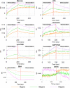
Expression profiles of hemolin in A. pernyi larvae. Tissue distribution analysis of Ap-hemolin mRNA in response to immune challenge was detected at different immune challenge times (0, 3, 6, 9, 12, 18, 24 and 48 hr). Tissues involved in the experiment were epidermis (A), midgut (B), Malpighian tube (C), fat body (D) and hemocytes (E). Each tissue was collected at different times from the samples (five individuals per time point) injected with insect saline (Control) or the ones immune challenged with the mixture of S. aureus, E. coli and C. albicans (Induced). Each bar represents the mean ± SD (N=3). (F) Changes in hemolin protein levels in cell-free plasma upon microbe injection. Samples were collected before injection (NT) or 12 h after challenge with microbe injection. Lane 1: nontreated 5th instar larvae plasma (NT); Lane 2: larvae plasma stimulated by S. aureus; Lane 3: larvae plasma stimulated by C. albicans; Lane 4: larvae plasma stimulated by E. coli; Lane 5: recombinant His6-Ap-hemolin (2.25 μg, a marker to indicate the migration of endogenous hemolin in hemolymph). All lanes were detected by western blotting assay using anti-His6-Ap-hemolin antibody (total protein in lane 1-4: 125 μg). Three biological replicates were used.
Figure 3
Figure 3
Binding of His6-Ap-hemolin to soluble PAMPs. A biolayer interferometry binding assay was performed to detect the binding of His6-Ap-hemolin to six soluble microbe-derived PAMPs, including mannan, LPS, LTA, laminarin, Lys-PGN and DAP-PGN. The vertical and horizontal axes represent the light shift distance (nm) and association/dissociation time (s), respectively. As negative controls, the binding of His6-Ap-hemolin to chitin (C9752) and bovine serum albumin (V900933) purchased from Sigma–Aldrich (Poole, USA) was also examined.
Figure 4
Figure 4
Effect of hemolin on the induction of antibacterial peptide synthesis in (A) pernyi larvae by qPCR analysis. RNAi efficiency within 24 hr post RNAi-treatment was estimation by qPCR (A) and western blotting analysis (B). For qPCR, the whole body of larvae was collected at 12 and 24 hr post-injection with dshemolin (dsHEM) or dsEGFP (dsEG) double-stranded RNA. Larvae injected with buffer (Bu) were used as controls. For western blotting analysis, 10 μg of plasma samples from dshemolin-treated and control larvae were used. NT: native sample without treatment. The results represent 3 biological replicates and the gray-scale statistical was shown below. (C-E) Relative mRNA expression levels of antibacterial peptides and the representative factors in Toll or Imd signaling pathway in response to microbial stimulation. Micro: larvae collected 12 hr after injection with formaldehyde-killed microbial cells (105 cells per larva); dsEG/Micro: larvae were first injected with dsEGFP for 24 hr and then immune-challenged with microbial cells for another 12 hr; dsHEM/Micro: larvae were first injected with dshemolin for 24 hr and then immune-challenged with microbes for another 12 hr. Microbes: E. coli, S. aureus and C. albicans. The relative expression level of antibacterial peptides and signaling pathway related factors detected by qPCR: defensin, attacin, moricin, spätzle and Fadd. Each bar represents the mean ± SD (N=3). ****: p<0.0001, ns, no significant difference.
Figure 5
Figure 5
PO activity assay triggered by microbes. (A) The effects of endogenous hemolin on PPO cascade activation by microbes were investigated by monitoring the PO activity produced in hemolin knockdown plasma. Hemocyte-free plasma of 2nd instar larvae was collected 24 hr after dsRNA injection. Then, the PO activity triggered by microbes was examined. dsEG and dsHEM: plasma samples from dsEGFP- and dshemolin-injected 2nd instar larvae, respectively; NT: nontreated 2nd instar larvae plasma. (B) The effects of exogenous hemolin on PPO activation stimulated by microbes were investigated with recombinant hemolin and 5th instar native larvae plasma. HL: native cell-free hemolymph (total protein: 12 mg/ml); HEM: recombinant His6-Ap-hemolin (500 ng); Micro: E. coli, S. aureus and C. albicans (5 × 10² cells per sample). Each bar represents the mean ± SD (N=3). ****: p<0.0001, ns, no significant difference.
Figure 6
Figure 6
PO activity assay stimulated by soluble PAMPs. The effects of endogenous (A) and exogenous (B) hemolin on PPO cascade activation by soluble PAMPs were investigated by monitoring the PO activity produced in the A. pernyi larval plasma as described in Figure 5. PAMPs: laminarin (1 μg/ml), LPS (100 μg/ml), LTA (100 μg/ml), DAP-PGN (500 μg/ml), Lys-PGN (500 μg/ml) and mannan (1 mg/ml). dsEG and dsHEM: plasma samples from dsEGFP- and dshemolin-injected 2nd instar larvae, respectively; NT: nontreated 2nd instar larvae plasma. HL: native cell-free hemolymph (total protein: 12 μg/ml); HEM: recombinant His6-Ap-hemolin (500 ng). Each bar represents the mean ± SD (N=3). ***: p<0.001, ****: p<0.0001, ns, no significant difference.
Figure 7
Figure 7
Pathogen dependent PO activation in Ap-hemolin blocked plasma. Hemocyte-free plasma samples were pre-incubated with anti-His6-Ap-hemolin antibody and 20 mM Tris-HCl, pH7.0, respectively. PO activity of these samples triggered by microbes (A) and soluble PAMPs (B) was examined and compared. HL: native cell-free hemolymph (total protein: 12 mg/ml); Bu: 20 mM Tris-HCl, pH7.0; Ab: anti-His6-hemolin antibody (0.8 mg/ml). Each bar represents the mean ± SD (N=3). ****: p<0.0001, ns, no significant difference.

References

    1. White JM, Littman DR. Viral Receptors of the Immunoglobulin Superfamily. Cell (1989) 56(5):725–8. doi: 10.1016/0092-8674(89)90674-0 - DOI - PubMed
    1. Verschueren E, Husain B, Yuen K, Sun Y, Paduchuri S, Senbabaoglu Y, et al. . The Immunoglobulin Superfamily Receptome Defines Cancer-Relevant Networks Associated With Clinical Outcome. Cell (2020) 182(2):329–44.e19. doi: 10.1016/j.cell.2020.06.007 - DOI - PubMed
    1. Akira S, Uematsu S, Takeuchi O. Pathogen Recognition and Innate Immunity. Cell (2006) 124(4):783–801. doi: 10.1016/j.cell.2006.02.015 - DOI - PubMed
    1. Ishii KJ, Koyama S, Nakagawa A, Coban C, Akira S. Host Innate Immune Receptors and Beyond: Making Sense of Microbial Infections. Cell Host Microbe (2008) 3(6):352–63. doi: 10.1016/j.chom.2008.05.003 - DOI - PubMed
    1. Beck G, Habicht GS. Immunity and the Invertebrates. Sci Am (1996) 275(5):60–3,6. doi: 10.1038/scientificamerican1196-60 - DOI - PubMed

Publication types

LinkOut - more resources