

Constrictive pericarditis caused by pericardial metastasis from esophageal squamous cell carcinoma: a case report
- PMID: 35669905
- PMCID: PMC9163270
- DOI: 10.1007/s13691-022-00543-0
Constrictive pericarditis caused by pericardial metastasis from esophageal squamous cell carcinoma: a case report
Abstract
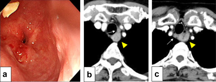
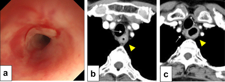
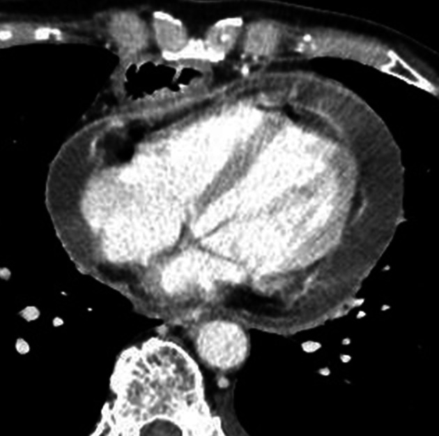
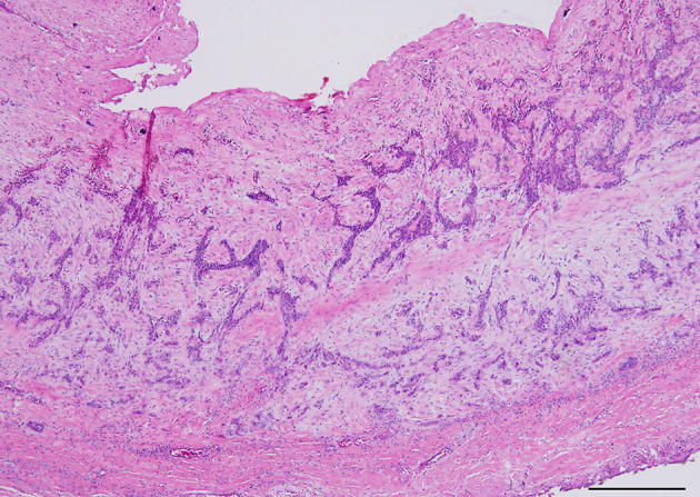
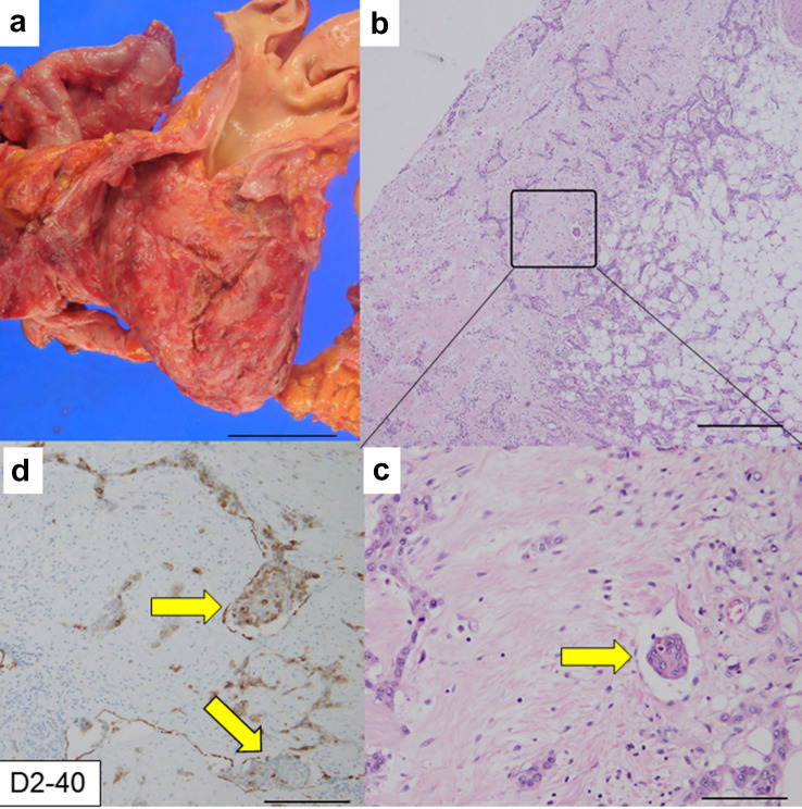
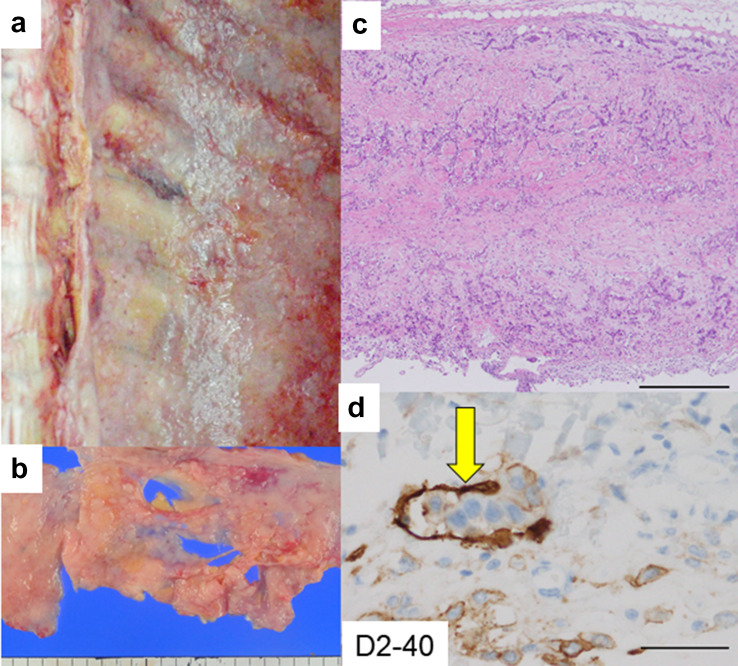
Constrictive pericarditis is a rare condition characterized by clinical signs of right heart failure subsequent to the loss of pericardial compliance. We report a case of constrictive pericarditis due to pericardial metastasis in a patient with a history of esophageal squamous cell carcinoma that had a pathological complete response (pCR) to preoperative chemoradiotherapy. A 66-year-old woman was referred to our division for the treatment of advanced esophageal cancer. Video-assisted thoracoscopic surgery esophagectomy (VATSE) with 3-field lymphadenectomy was performed after neoadjuvant chemoradiotherapy (NAC-CRT). Pathological examination revealed no residual tumor, lymph node metastasis, lymphatic invasion, or vessel invasion. The histological treatment effect of the chemoradiotherapy was pathological complete response (pCR). Five months after surgery, the patient was admitted to a nearby hospital for the treatment of acute pericarditis. However, a month after admission, acute pericarditis progressed to constrictive pericarditis, and she was referred to our hospital for further management. Subsequently, urgent pericardiectomy was performed through a lower half sternotomy incision. After surgery, heart failure improved for a while but worsened again. The patient died 7 days after the surgery. Pathological examination of the resected pericardium revealed evidence of metastasis from squamous cell carcinoma of the esophagus. An autopsy revealed the spread of esophageal cancer to the bilateral pleura, right lung, pericardium, diaphragm, soft tissue surrounding the tracheal bifurcation, and bilateral hilar lymph nodes. Similarly, tumor cells were found in the lymphatic vessels of the pericardium and pleura. Even if pCR is achieved with NAC-CRT, as in our case, esophageal cancer may metastasize and present as constrictive pericarditis within a short period; therefore, careful patient follow-up is essential.
Keywords: Constrictive pericarditis; Esophageal cancer; Pericardial tumor; Squamous cell carcinoma.
© The Author(s) under exclusive licence to The Japan Society of Clinical Oncology 2022.
Conflict of interest statement
Conflict of interestThe authors declare that they have no competing interests.
Figures
Similar articles
-
Constrictive pericarditis caused by a pericardial-occupying tumor due to esophageal cancer.Clin J Gastroenterol. 2014 Jun;7(3):243-6. doi: 10.1007/s12328-014-0489-z. Epub 2014 Apr 23. Clin J Gastroenterol. 2014. PMID: 26183744
-
Blunt trauma as a suspected cause of delayed constrictive pericarditis: a case report.J Med Case Rep. 2011 Feb 23;5:76. doi: 10.1186/1752-1947-5-76. J Med Case Rep. 2011. PMID: 21345214 Free PMC article.
-
Malignant pleural mesothelioma with constrictive pericarditis as the first manifestation: A case report.Clin Case Rep. 2023 Jun 20;11(6):e7555. doi: 10.1002/ccr3.7555. eCollection 2023 Jun. Clin Case Rep. 2023. PMID: 37351350 Free PMC article.
-
Constrictive Pericarditis 5 Months after Radiation Therapy in a 62-Year-Old Woman with Esophageal Cancer.Tex Heart Inst J. 2017 Dec 19;44(6):411-415. doi: 10.14503/THIJ-16-6054. eCollection 2017 Dec. Tex Heart Inst J. 2017. PMID: 29276442 Free PMC article. Review.
-
Radical Pericardiectomy for Pericardial Diseases.Curr Cardiol Rep. 2019 Feb 12;21(2):6. doi: 10.1007/s11886-019-1092-1. Curr Cardiol Rep. 2019. PMID: 30747309 Review.
Cited by
-
Myocardial metastasis from ZEB1- and TWIST-positive spindle cell carcinoma of the esophagus: A case report.World J Gastroenterol. 2024 Mar 21;30(11):1636-1643. doi: 10.3748/wjg.v30.i11.1636. World J Gastroenterol. 2024. PMID: 38617457 Free PMC article.
References
-
- Busch C, Penov K, Amorim PA, et al. Risk factors for mortality after pericardiectomy for chronic constrictive pericarditis in a large single-centre cohort. Eur J Cardio-thorac Surg. 2015;48:e110–e116. - PubMed
-
- Lam KY, Dickens P, Chan AC. Tumors of the heart. A 20-year experience with a review of 12,485 consecutive autopsies. Arch Pathol Lab Med. 1993;117:1027–1031. - PubMed
Publication types
LinkOut - more resources
Full Text Sources
Research Materials