. 2022 Jul 1;323(1):L69-L83.
doi: 10.1152/ajplung.00436.2021. Epub 2022 Jun 7.

PTPα promotes fibroproliferative responses after acute lung injury

Affiliations

PTPα promotes fibroproliferative responses after acute lung injury

Yael Aschner et al. Am J Physiol Lung Cell Mol Physiol. .

Abstract

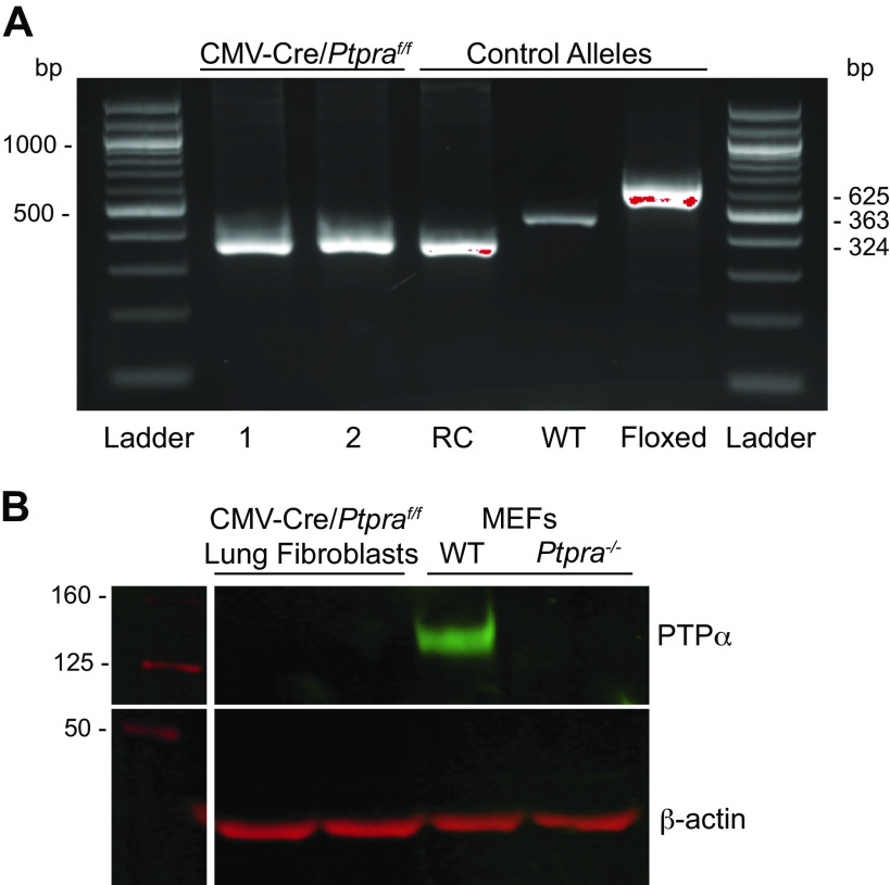
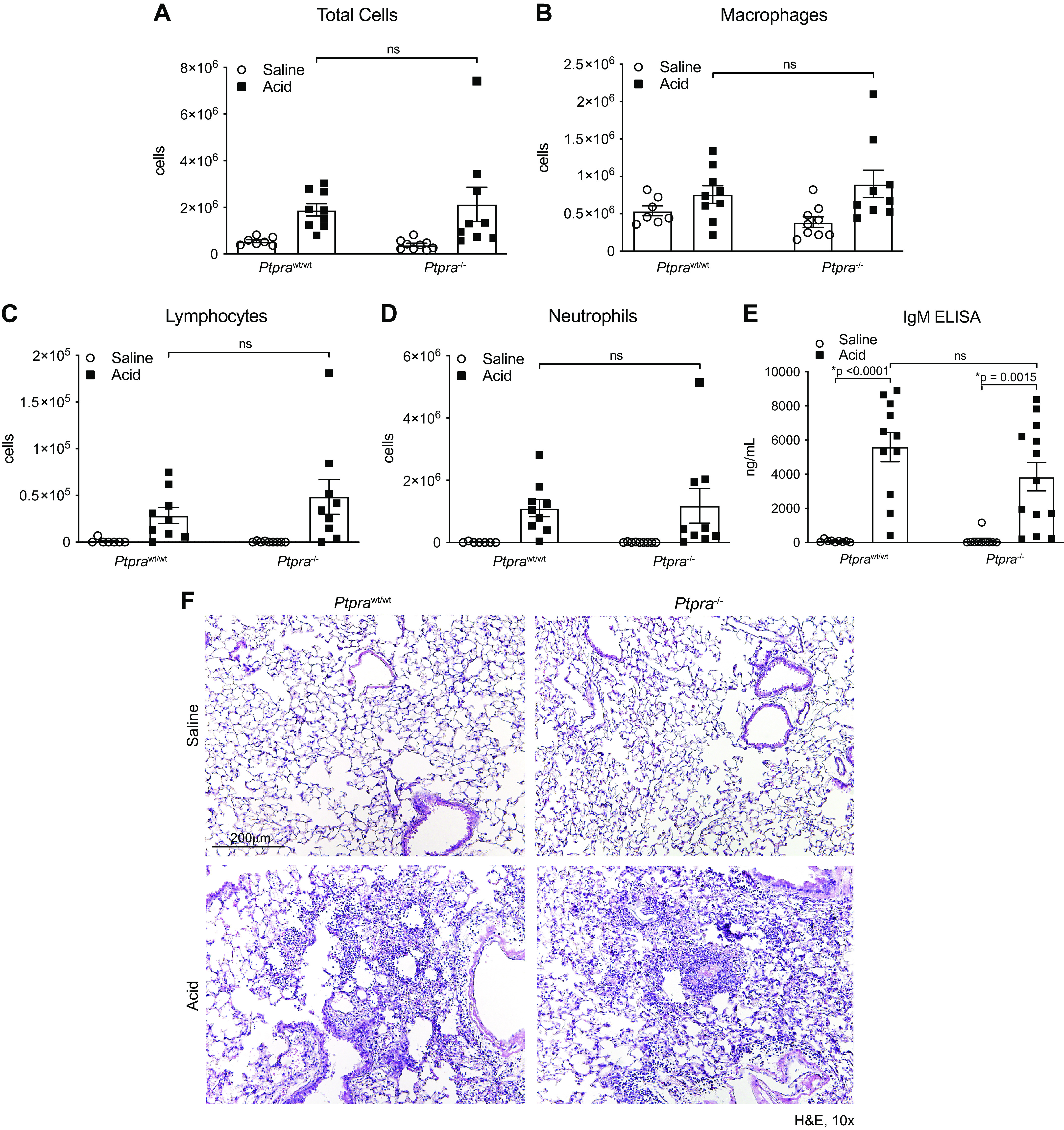
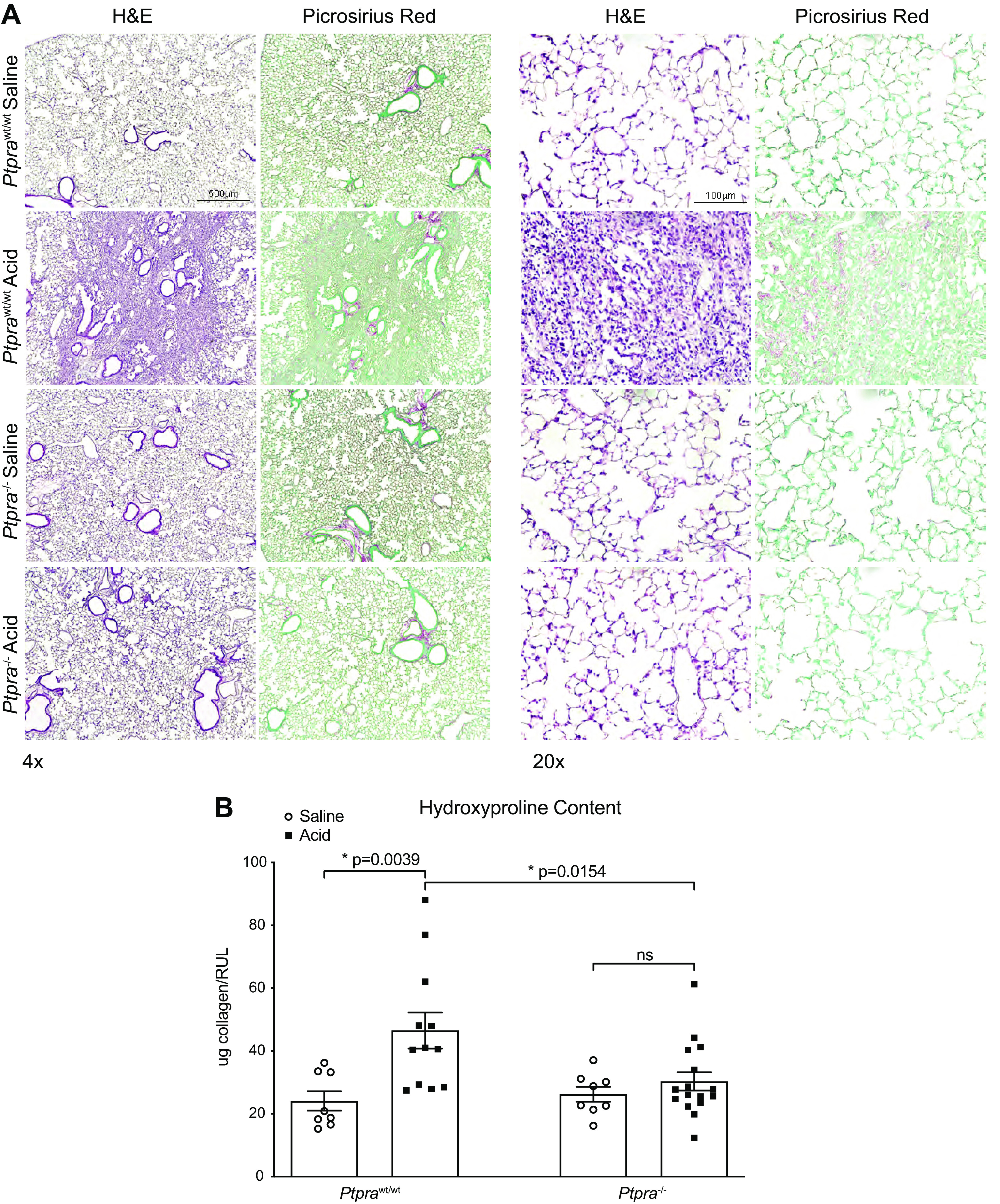
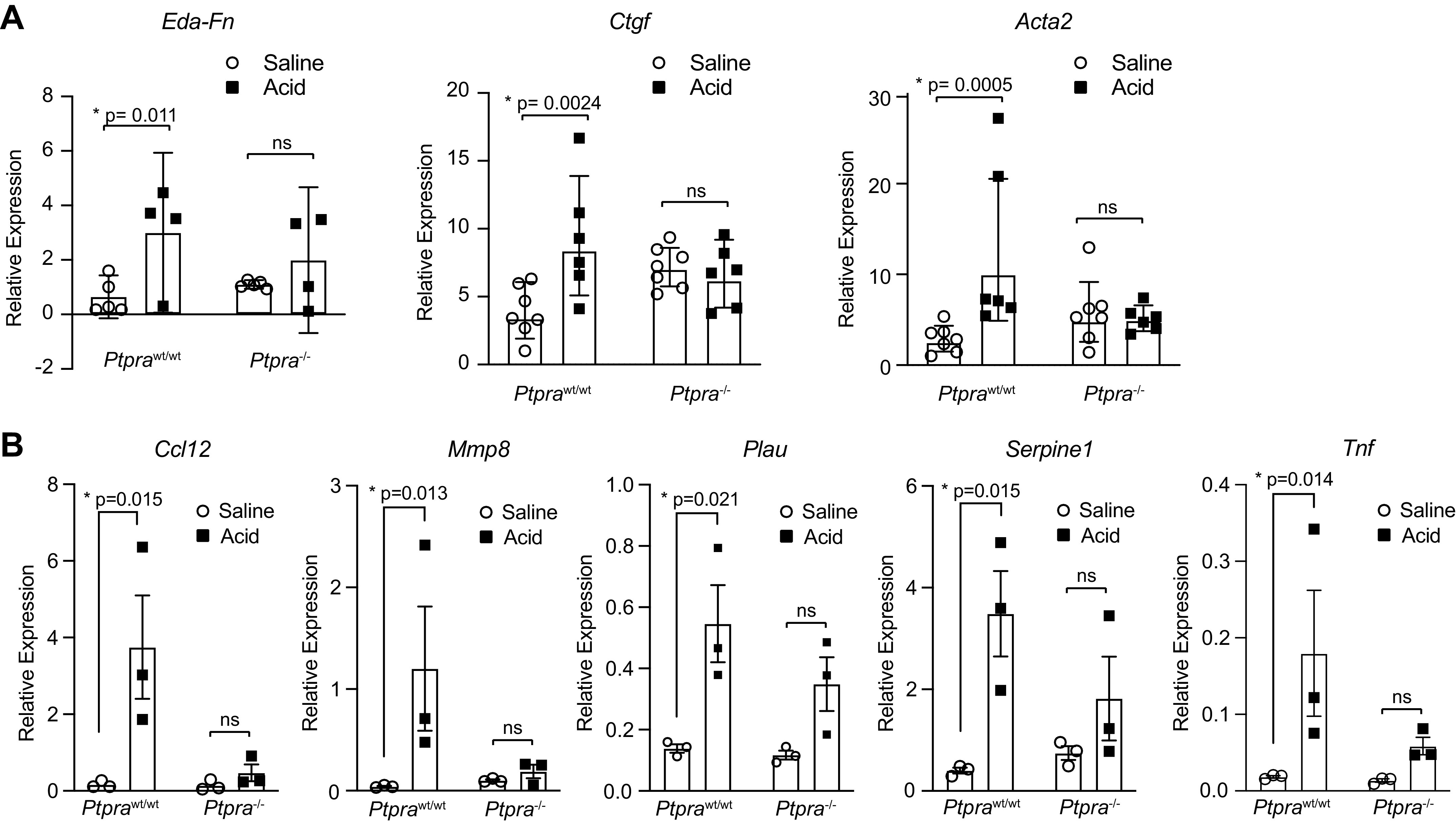
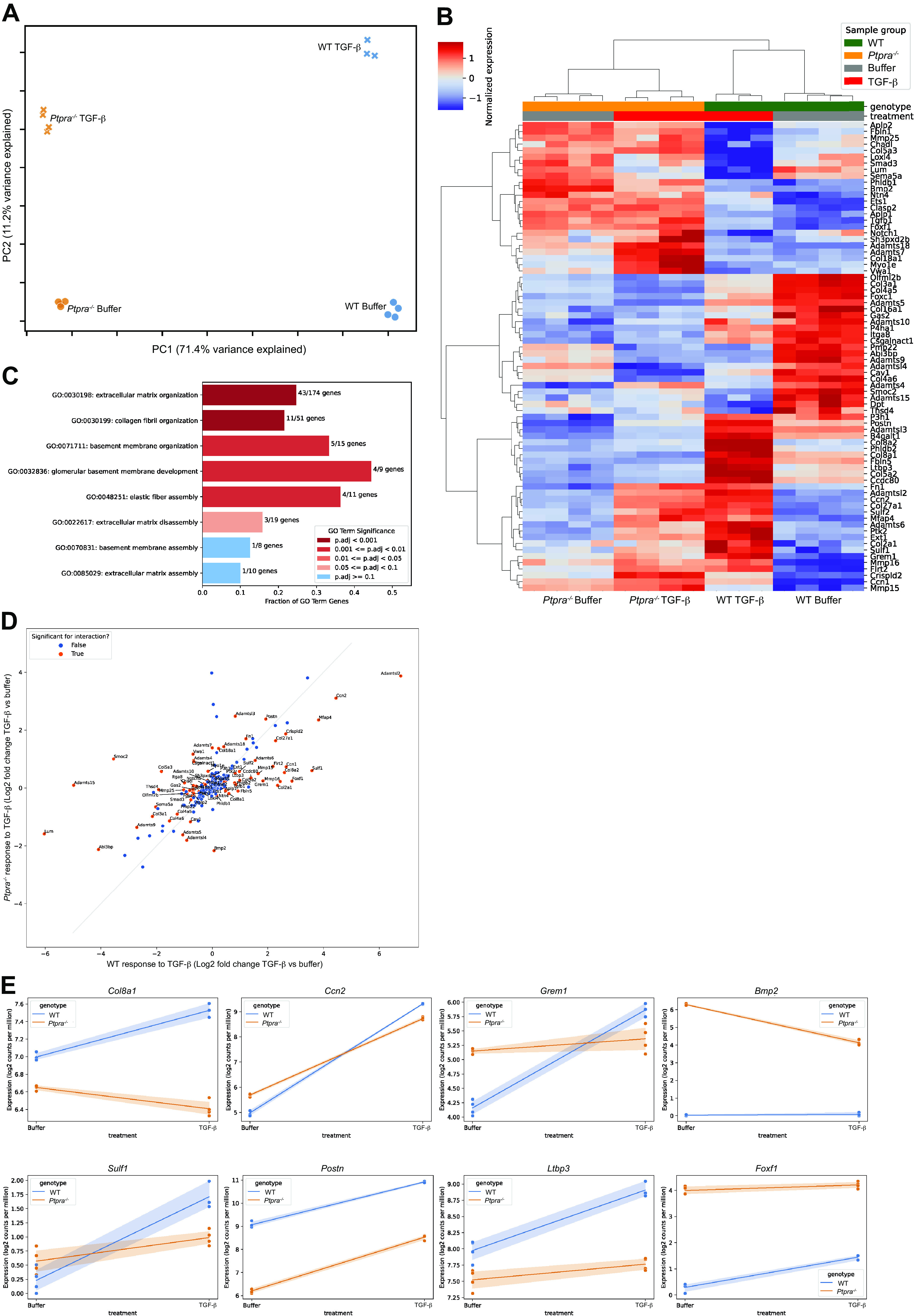
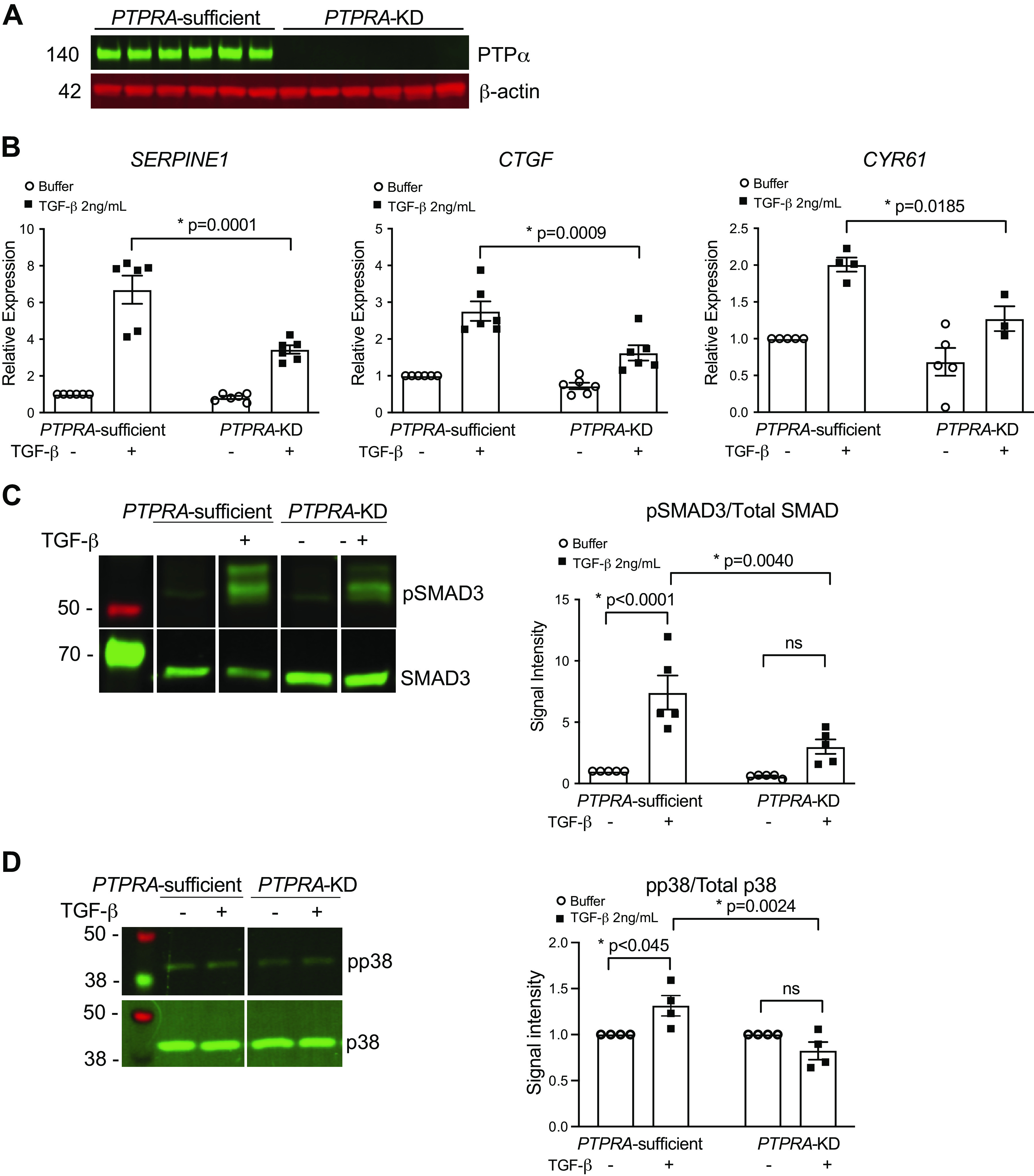
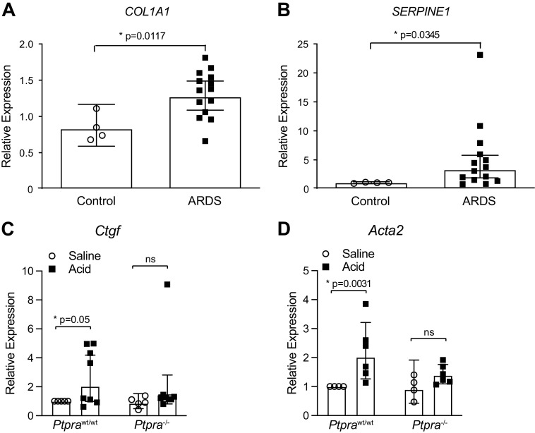
The acute respiratory distress syndrome (ARDS) is a major healthcare problem, accounting for significant mortality and long-term disability. Approximately 25% of patients with ARDS will develop an overexuberant fibrotic response, termed fibroproliferative ARDS (FP-ARDS) that portends a poor prognosis and increased mortality. The cellular pathological processes that drive FP-ARDS remain incompletely understood. We have previously shown that the transmembrane receptor-type tyrosine phosphatase protein tyrosine phosphatase-α (PTPα) promotes pulmonary fibrosis in preclinical murine models through regulation of transforming growth factor-β (TGF-β) signaling. In this study, we examine the role of PTPα in the pathogenesis of FP-ARDS in a preclinical murine model of acid (HCl)-induced acute lung injury. We demonstrate that although mice genetically deficient in PTPα (Ptpra-/-) are susceptible to early HCl-induced lung injury, they exhibit markedly attenuated fibroproliferative responses. In addition, early profibrotic gene expression is reduced in lung tissue after acute lung injury in Ptpra-/- mice, and stimulation of naïve lung fibroblasts with the BAL fluid from these mice results in attenuated fibrotic outcomes compared with wild-type littermate controls. Transcriptomic analyses demonstrate reduced extracellular matrix (ECM) deposition and remodeling in mice genetically deficient in PTPα. Importantly, human lung fibroblasts modified with a CRISPR-targeted deletion of PTPRA exhibit reduced expression of profibrotic genes in response to TGF-β stimulation, demonstrating the importance of PTPα in human lung fibroblasts. Together, these findings demonstrate that PTPα is a key regulator of fibroproliferative processes following acute lung injury and could serve as a therapeutic target for patients at risk for poor long-term outcomes in ARDS.

Keywords: ARDS; PTPα; acute lung injury; fibroproliferative ARDS.


Conflict of interest statement

No conflicts of interest, financial or otherwise, are declared by the authors.

Figures

Figure 1.
Figure 1.
CMV-cre/Ptpraf/f (Ptpra−/−) mice do not express protein tyrosine phosphatase-α (PTPα). A: tail clips from CMV-cre/Ptpraf/f (Ptpra−/−) mice were genotyped by endpoint PCR for the Ptpra allele. Genotyping results for two representative mice are shown on the left (1 and 2), with control alleles for the recombinant (RC), wild type (WT) and floxed alleles shown on the right. B: isolated lung fibroblasts from Ptpra−/− mice were lysed for Western blot analysis of expression of PTPα protein. WT and Ptpra−/− murine embryonic fibroblasts (MEFs) are shown as positive and negative controls.
Figure 2.
Figure 2.
Early lung injury is not altered by the presence or absence of Ptpra. Wild-type (Ptprawt/wt) and Ptpra−/− mice were treated with 0.19 N HCl or saline and harvested at 48 h. Bronchoalveolar lavage (BAL) cell counts were performed for total cells (A), macrophages (B), lymphocytes (C), and neutrophils (D). E: IgM ELISA was performed on BAL fluid collected at the 48-h time point. F: digital images of hematoxylin-eosin (H&E) staining at 48 h after treatment with HCl or saline. Magnification ×10. In AE, each symbol represents an individual mouse and horizontal lines represent means and SE. Values in each group represent results from pooled independent experiments with a total of 7–13 mice/group. *P < 0.05; one-way ANOVA.
Figure 3.
Figure 3.
Ptpra−/− mice are protected from the development of fibrosis. Wild-type and Ptpra/ mice were treated with 0.19 N HCl or saline and harvested at 14 days. A: digital images of hematoxylin-eosin (H&E) and Picrosirius Red staining for collagen deposition (red) were obtained. Magnification ×4 (left) and ×20 (right). B: effect of deletion of Ptpra as reflected by changes in lung hydroxyproline content. Each symbol represents an individual mouse and horizontal lines represent means and SE. Values in each group represent results from pooled independent experiments with a total of 8–16 mice/group. *P < 0.05; one-way ANOVA.
Figure 4.
Figure 4.
Profibrotic gene expression is reduced in the absence of Ptpra. Ptprawt/wt and Ptpra−/− mice were treated with 0.19 N HCl or saline and harvested at 48 h. A: mRNA expression of fibrotic markers Eda-fibronectin, Ctgf, and Acta2 was assessed in homogenized lung tissue using qPCR. Right-skewed variables were natural-log transformed prior to analysis for significance. Each symbol represents an individual mouse and horizontal lines represent geometric mean and 95% confidence interval (CI). Values in each group represent results from pooled independent experiments with a total of 4–7 mice/group. *P < 0.05; t test between specific treatment combinations. B: a fibrosis-focused qPCR array was performed on homogenized lung tissue. Data were normalized to the housekeeping gene GusB. qPCR array plates were performed in triplicate and data are represented as means ± SE. *P < 0.05.
Figure 5.
Figure 5.
Extracellular matrix responses to transforming growth factor-β (TGF-β) stimulation are altered in the absence of Ptpra. Wild type WT and Ptpra−/− murine embryonic fibroblasts (MEFs) were stimulated with 2 ng/mL of TGF-β for 24 h and harvested for bulk RNA sequencing. The “ECM Organization” GO term pathway was interrogated. A: principal component analysis (PCA) shows clear clustering of transcriptomic data. A single sample (WT, TGF-β, 24 h stimulation) was excluded due to poor quality RNA. B: heatmap of differentially expressed genes (DEGs) within the “ECM Organization” GO term pathway that were significant for the interaction comparison between WT and Ptpra−/− after stimulation with TGF-β at a false discovery rate (FDR) of 0.05. C: bar plot of the “parent” ECM Organization GO Term (GO:0030198) and its “children” ordered by P value significance on the y-axis, with x-axis showing the fraction of overlap between the GO term’s annotated genes and the significant genes in the analysis. D: scatterplot of genes within the ECM Organization GO term pathway. The x-axis shows log2-fold change for TGF-β over buffer in the WT fibroblasts, and the y-axis shows log2-fold change for TGF-β over buffer in the Ptpra−/− cells. Genes that reached significance are shown in orange. E: a subset of genes within the ECM Organization GO term that demonstrate differential responses to TGF-β based on genotype (WT vs. Ptpra−/−) are shown.
Figure 6.
Figure 6.
CRISPR-targeted knockdown of PTPRA results in attenuated responses of human fibroblasts to transforming growth factor-β (TGF-β) stimulation. A: CRISPR-targeted knockdown of PTPRA in hTERT-transformed immortalized human lung fibroblasts (IHLFs) results in efficient deletion of the protein tyrosine phosphatase-α (PTPα) protein by Western blotting. B: PTPRA-sufficient and PTPRA-KD fibroblasts were treated with 2 ng/mL of TGF-β, or buffer for 24 h and harvested to analyze the expression of SERPINE1, CTGF, and CYR61 by qPCR. C: PTPRA-sufficient and PTPRA-KD fibroblasts were treated with 2 ng/mL of TGF-β for 30 min and harvested, after which phosphorylation status of SMAD3 was assessed by Western blotting. Densitometry shows expression of phospho-SMAD3 over total SMAD3. D: PTPRA-sufficient and PTPRA-KD fibroblasts were treated with 2 ng/mL of TGF-β for 15 min and harvested, after which phosphorylation status of p38 was assessed by Western blotting. Densitometry shows expression of phospho-p38 over total p38. Data represent mean values ± SE from at least three independent experiments. *P < 0.05; one-way ANOVA.
Figure 7.
Figure 7.
Bronchoalveolar lavage (BAL) fluid from patients with acute respiratory distress syndrome (ARDS) and murine lung injury-BAL fluid induces expression of transforming growth factor-β (TGF-β)-dependent profibrotic genes in lung fibroblasts, which is attenuated in the absence of protein tyrosine phosphatase-α (PTPα). BAL fluid collected from patients with ARDS or normal human controls was used to stimulate cultured normal human lung fibroblasts (NHLFs). After 24 h, cells were harvested for qPCR expression of COL1A1 (A) and SERPINE1 (B). BAL fluid from Ptprawt/wt and Ptpra−/− mice treated for 48 h with HCl or saline was used to stimulate cultured NIH 3T3 fibroblasts. After 6 h, cells were harvested for qPCR expression of Ctgf (C) and Acta2 (D). Right-skewed variables were natural-log transformed prior to analysis for significance. Each symbol represents an individual patient (n = 14 ARDS patients, 4 controls) or experimental animal (n = 4–8 mice/group), and horizontal lines represent geometric mean and 95% confidence interval (CI). *P ≤ 0.05; t test between specific treatment combinations.

References

    1. Acute Respiratory Distress Syndrome N, Brower RG, Matthay MA, Morris A, Schoenfeld D, Thompson BT, Wheeler A. Ventilation with lower tidal volumes as compared with traditional tidal volumes for acute lung injury and the acute respiratory distress syndrome. N Engl J Med 342: 1301–1308, 2000. doi:10.1056/NEJM200005043421801. - DOI - PubMed
    1. Matthay MA, Zemans RL. The acute respiratory distress syndrome: pathogenesis and treatment. Annu Rev Pathol 6: 147–163, 2011. doi:10.1146/annurev-pathol-011110-130158. - DOI - PMC - PubMed
    1. National Heart L, Blood Institute Acute Respiratory Distress Syndrome Clinical Trials N, Wiedemann HP, Wheeler AP, Bernard GR, Thompson BT, Hayden D, deBoisblanc B, Connors AF Jr, Hite RD, Harabin AL. Comparison of two fluid-management strategies in acute lung injury. N Engl J Med 354: 2564–2575, 2006. doi:10.1056/NEJMoa062200. - DOI - PubMed
    1. Rubenfeld GD, Caldwell E, Peabody E, Weaver J, Martin DP, Neff M, Stern EJ, Hudson LD. Incidence and outcomes of acute lung injury. N Engl J Med 353: 1685–1693, 2005. doi:10.1056/NEJMoa050333. - DOI - PubMed
    1. Rubenfeld GD, Herridge MS. Epidemiology and outcomes of acute lung injury. Chest 131: 554–562, 2007. doi:10.1378/chest.06-1976. - DOI - PubMed

Publication types

MeSH terms

Substances