. 2022 Aug 1;323(2):L152-L164.
doi: 10.1152/ajplung.00503.2021. Epub 2022 Jun 7.

Impact of e-cigarette aerosol on primary human alveolar epithelial type 2 cells

Affiliations

Impact of e-cigarette aerosol on primary human alveolar epithelial type 2 cells

Katherine D Wick et al. Am J Physiol Lung Cell Mol Physiol. .

Abstract

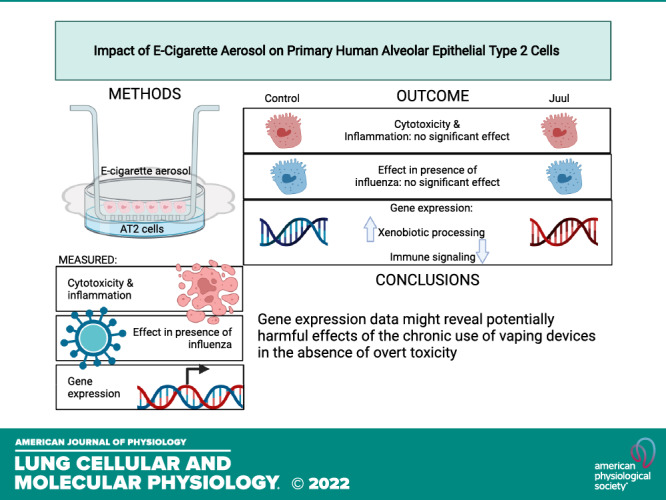
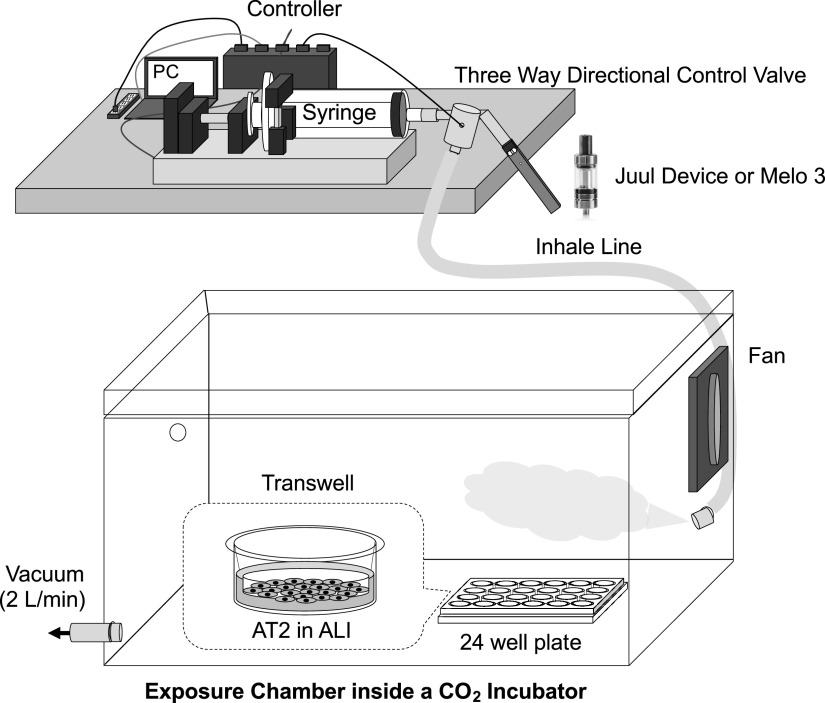
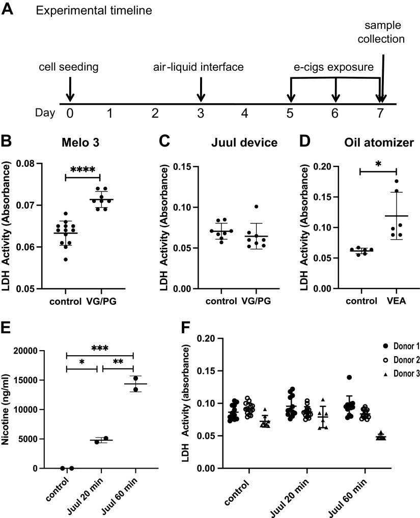
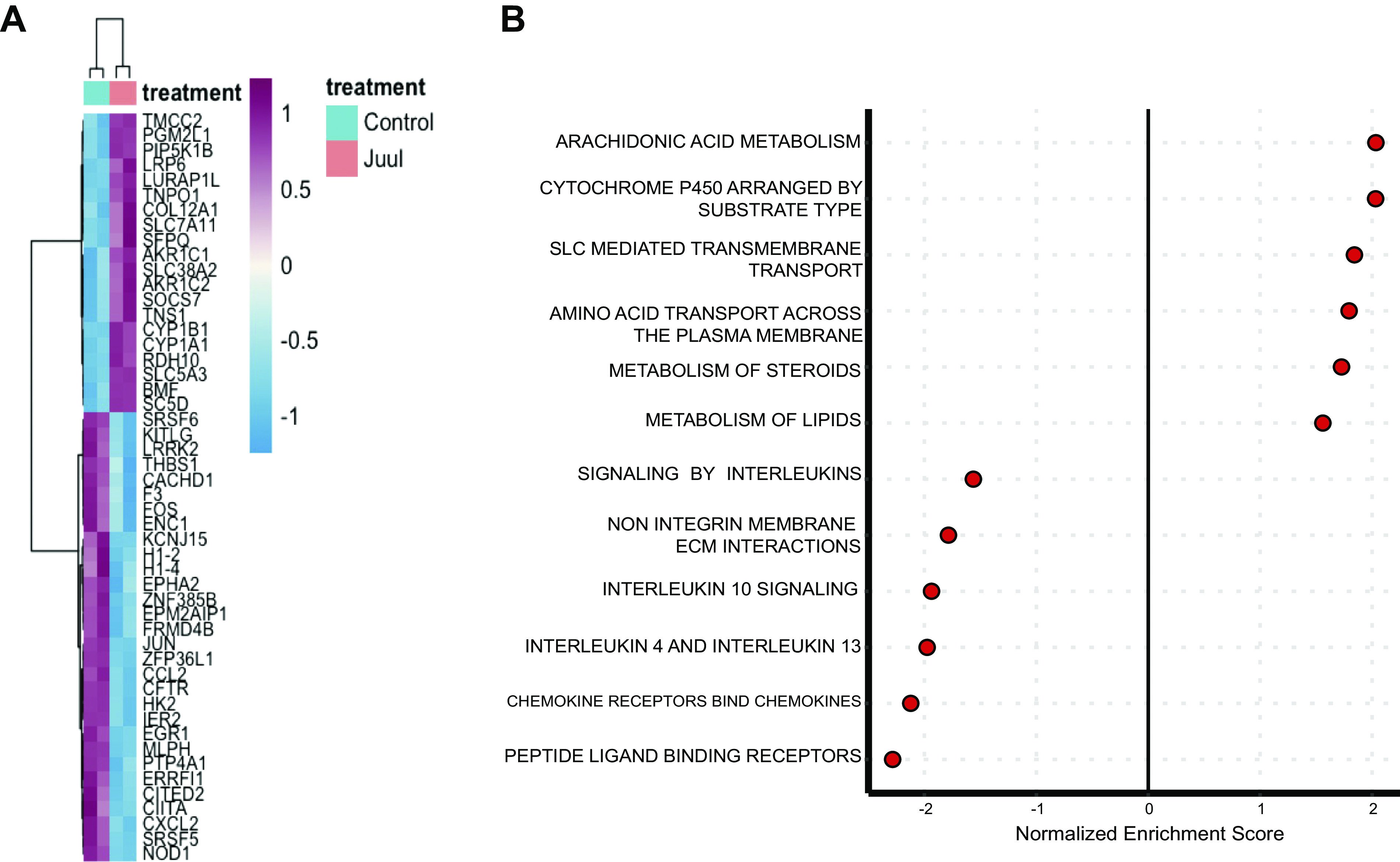
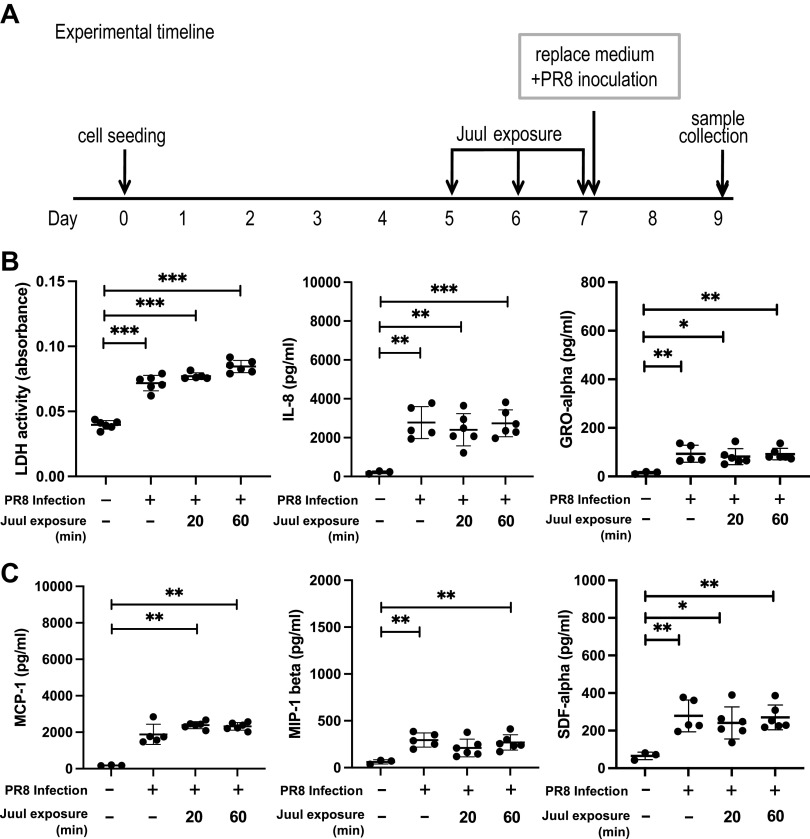
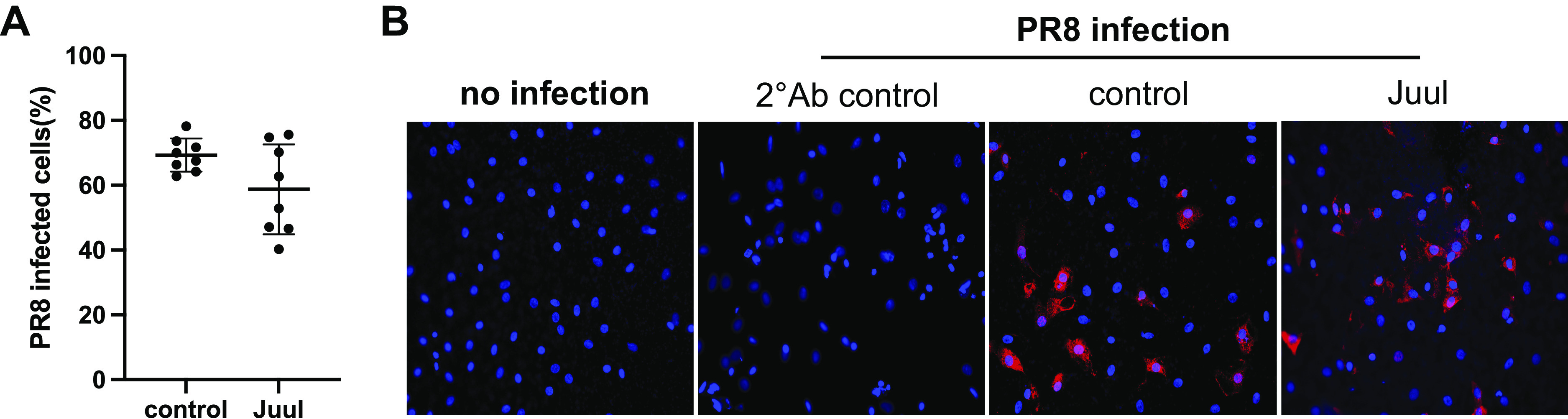
Electronic cigarettes (e-cigarettes) are designed to simulate combustible cigarette smoking and to aid in smoking cessation. Although the number of e-cigarette users has been increasing, the potential health impacts and biological effects of e-cigarettes are still not fully understood. Previous research has focused on the biological effects of e-cigarettes on lung cancer cell lines and distal airway epithelial cells; however, there have been few published studies on the effect of e-cigarettes on primary lung alveolar epithelial cells. The primary purpose of this study was to investigate the direct effect of e-cigarette aerosol on primary human lung alveolar epithelial type 2 (AT2) cells, both alone and in the presence of viral infection. The Melo-3 atomizer caused direct AT2 cell toxicity, whereas the more popular Juul pod's aerosol did not have a detectable cytotoxic effect on AT2 cells. Juul nicotine aerosol also did not increase short-term susceptibility to viral infection. However, 3 days of exposure upregulated genes central to the generation of reactive oxygen species, lipid peroxidation, and carcinogen metabolism and downregulated key innate immune system genes related to cytokine and chemokine signaling. These findings have implications for the potentially injurious impact of long-term use of popular low-power e-cigarette pods on the human alveolar epithelium. Gene expression data might be an important endpoint for evaluating the potential harmful effects of vaping devices that do not cause overt toxicity.

Keywords: EVALI; alveolar type II cells; e-cigarettes; inflammation; pulmonary edema.


Conflict of interest statement

C.S.C. has received grants and personal fees from Bayer and GlaxoSmithKline, personal fees from Boehringer Ingelheim, CSL Behring, Prometic, and Roche/Genentech. M.A.M. has received grants from Bayer Pharmaceuticals and GlaxoSmithKline, personal fees from Boehringer Ingelheim, Cerus Therapeutics, CSL Berhing, Quark Pharmaceuticals, Roche-Genentec, and Thesan Pharmaceuticals. None of the other authors has any conflicts of interest, financial or otherwise, to disclose.

Figures

None
Graphical abstract
Figure 1.
Figure 1.
Electronic nicotine aerosol delivery setup. A Gram Universal Vaping Machine controls a syringe pump and a three-way valve, enabling precise control over aerosol generation. The exposure chamber was placed inside a 37°C cell culture incubator containing 5% CO2. To create steady-state conditions, air was removed from the chamber at 2.0 L/min and fresh aerosol was injected during the entire exposure, with the volume inflow balance derived from the culture incubator, providing a source of fresh conditioned air and minimizing nonspecific stress on the cells.
Figure 2.
Figure 2.
Cytotoxic effect of VG/PG aerosol generated by Melo 3 atomizer and Juul device on cultured AT2 cells. Cells were cultured in serum-free medium during aerosol exposure for 20 or 60 min/day for 3 consecutive days. A: experimental timeline. B: LDH activity (absorbance) in the culture medium. AT2 cells exposed to VG/PG aerosols generated by Melo3 for 60 min. Experiments were carried out on AT2 cells from three human donor lungs. Figure shows representative data (experimental replicates) from one donor lung sample. n = 8–13 wells in each group. Data are represented as means ± SD. ****P < 0.0001 by one-way ANOVA. C: LDH activity (absorbance) in the culture medium. AT2 cells exposed to VG/PG aerosols generated by Juul Device for 60 min. Experiments were carried out on AT2 cells from three human donor lungs. Figure shows representative data (experimental replicates) from one donor lung sample. n = 8 wells in each group. Data are represented as means ± SD. D: LDH activity (absorbance) in the culture medium. AT2 cells exposed to VEA aerosol for 60 min. Experiments were carried out on AT2 cells from three human donor lungs. Figure shows representative data (experimental replicates) from one donor lung sample. n = 6 wells in each group. *P < 0.05 by one-way ANOVA. Difference remained significant after removing outliers. E: nicotine concentration measured in culture medium of AT2 cells exposed to Juul nicotine vapor for different durations. Experiments were carried out on AT2 cells from two human donor lungs. Figure shows representative data (experimental replicates) from one donor lung sample. n = 2 wells in each group. Data are represented as means ± SD. *P < 0.05, **P < 0.01, ***P < 0.001 by one-way ANOVA. F: LDH activity (absorbance) in the culture medium. Experiments were carried out on AT2 cells from three human donor lungs. n = 12 wells in each group for donor 1, n = 14 wells for donor 2, n = 6–8 wells for donor 3. Data are represented as means ± SD. P > 0.05 by grouped mixed-effects model followed by Tukey’s multiple comparison test. AT2, alveolar epithelial type 2; LDH, lactate dehydrogenase; PG, propylene glycol; VEA, vitamin E acetate; VG, vegetable glycerin.
Figure 3.
Figure 3.
Effect of nicotine aerosol on tight junctional protein expression of AT2 cells. A: tight junction organization rate (TiJOR) was measured by counting the number of tight junction intersections in five randomly selected fields from AT2 monolayers between Juul nicotine group and control. A: quantification of the tight junction organization with modified method of image processing using ImageJ software. TiJOR calculated in both control and Juul aerosol group. Experiments were done on cells from three human donor lungs. Figure shows representative data from cell preparation of one donor lung. Each dot represents one randomly selected field from each group. n = 5 fields. Data are represented as means ± SD. B: representative immunofluorescent staining in one field of tight junctional protein Claudin-18 on AT2 cell monolayers. No qualitative difference in Claudin-18 distribution in AT2 cell monolayers was seen. Images were taken with Leica SP5 laser scanning confocal microscope. Green: claudin-18; blue: DAPI. AT2, alveolar epithelial type 2.
Figure 4.
Figure 4.
Juul nicotine aerosol exposure effect on inflammatory cytokines in culture medium of AT2 cells. AT2 cells on Transwells were exposed to 20 or 60 min of Juul aerosol daily for 3 consecutive days. Inflammatory chemokines in the culture medium were measured with Luminex assay. Statistical differences between Juul and control groups were calculated with one-way ANOVA followed by Tukey’s multiple comparison test. Representative data (experimental replicates) from one of the experiments from three human donor lungs. n = 12 wells in each group. Data are represented as means ± SD. *P < 0.05. AT2, alveolar epithelial type 2.
Figure 5.
Figure 5.
RNA-Seq analysis for gene expression of AT2 cells exposed to Juul nicotine vapor. RNA-Seq studies were performed on two sets of Juul-exposed (60 min) and -unexposed AT2 cells from two different human donor lungs. Genes present in <20% of samples excluded. A: heat map of differentially expressed genes at a false-discovery rate <0.1. B: pathway analysis in Juul-exposed vs. -unexposed cells. Positive normalized enrichment score indicates upregulation in Juul-exposed cells (adjusted P < 0.05 for all pathways). AT2, alveolar epithelial type 2.
Figure 6.
Figure 6.
Effect of PR8 infection on LDH and cytokines of AT2 cells exposed to Juul nicotine. A: experimental timeline. LDH and cytokines (B) and cytokines (C) measured in culture supernatant of AT2 cells infected with PR8 with or without nicotine exposure. Experiments were done on AT 2 cells from three human donor lungs. Figures show representative data (experimental replicates) from one of the experiments. n = 6 in each group for LDH measurement. n = 3–6 wells in each group for cytokine measurement. Data are shown as means ± SD. Statistical differences between groups were calculated with one-way ANOVA followed by Tukey’s multiple comparison test or Kruskal–Wallis followed by Dunn’s multiple comparison. *P < 0.05, **P < 0.01, ***P < 0.001. AT2, alveolar epithelial type 2; LDH, lactate dehydrogenase.
Figure 7.
Figure 7.
Immunofluorescent staining of AT2 cells infected with PR8. A: quantification of PR8-infected AT2 cells by counting the number of AT2 cells with positive staining of Influenza A nucleoprotein in different microscopic fields from experiments of two human donor lungs. n = 8. Data are shown as means ± SD. No difference was seen between Juul exposure and control. B: representative immunofluorescent staining of AT2 cell monolayer infected with PR8. Secondary antibody control was done by incubating cells with mouse IgG first and followed by incubating with secondary Alexa fluor 647 Donkey anti-mouse antibody. Red: anti-influenza A nucleoprotein clone A1. Blue: DAPI counterstain. AT2, alveolar epithelial type 2.

Similar articles

Cited by

References

    1. Wang TW, Neff LJ, Park-Lee E, Ren C, Cullen KA, King BA. E-cigarette use among middle and high school students—United States, 2020. MMWR Morb Mortal Wkly Rep 69: 1310–1312, 2020. doi:10.15585/mmwr.mm6937e1. - DOI - PMC - PubMed
    1. Hammond D, Reid JL, Rynard VL, Fong GT, Cummings KM, McNeill A, Hitchman S, Thrasher JF, Goniewicz ML, Bansal-Travers M, O'Connor R, Levy D, Borland R, White CM. Prevalence of vaping and smoking among adolescents in Canada, England, and the United States: repeat national cross sectional surveys. BMJ 365: l2219, 2019. [Erratum in BMJ 370: m2579, 2020]. doi:10.1136/bmj.l2219. - DOI - PMC - PubMed
    1. Cherian SV, Kumar A, Estrada YMRM. E-cigarette or vaping product-associated lung injury: a review. Am J Med 133: 657–663, 2020. doi:10.1016/j.amjmed.2020.02.004. - DOI - PubMed
    1. Lozier MJ, Wallace B, Anderson K, Ellington S, Jones CM, Rose D, Baldwin G, King BA, Briss P, Mikosz CA; Lung Injury Response Epidemiology/Surveillance Task Force. Update: demographic, product, and substance-use characteristics of hospitalized patients in a nationwide outbreak of E-cigarette, or vaping, product use-associated lung injuries—United States, December 2019. MMWR Morb Mortal Wkly Rep 68: 1142–1148, 2019. [Erratum in MMWR Morb Mortal Wkly Rep 69: 117, 2020]. doi:10.15585/mmwr.mm6849e1. - DOI - PMC - PubMed
    1. Christiani DC. Vaping-induced acute lung injury. N Engl J Med 382: 960–962, 2020. doi:10.1056/NEJMe1912032. - DOI - PubMed

Publication types

MeSH terms