Review
. 2022 Jul;82(5):428-453.
doi: 10.1002/dneu.22891. Epub 2022 Jun 22.

Evolution of genetic mechanisms regulating cortical neurogenesis

Affiliations
Review

Evolution of genetic mechanisms regulating cortical neurogenesis

Alexandre Espinós et al. Dev Neurobiol. 2022 Jul.

Abstract

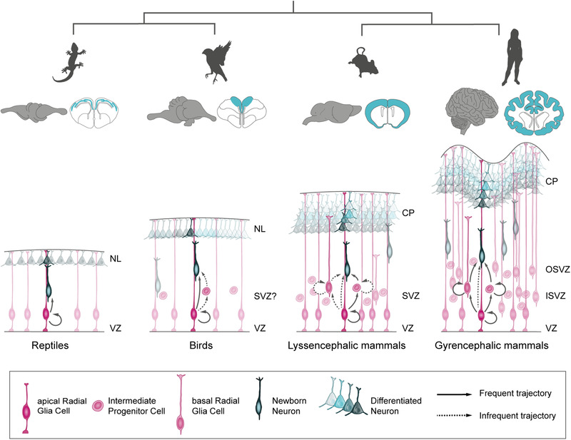
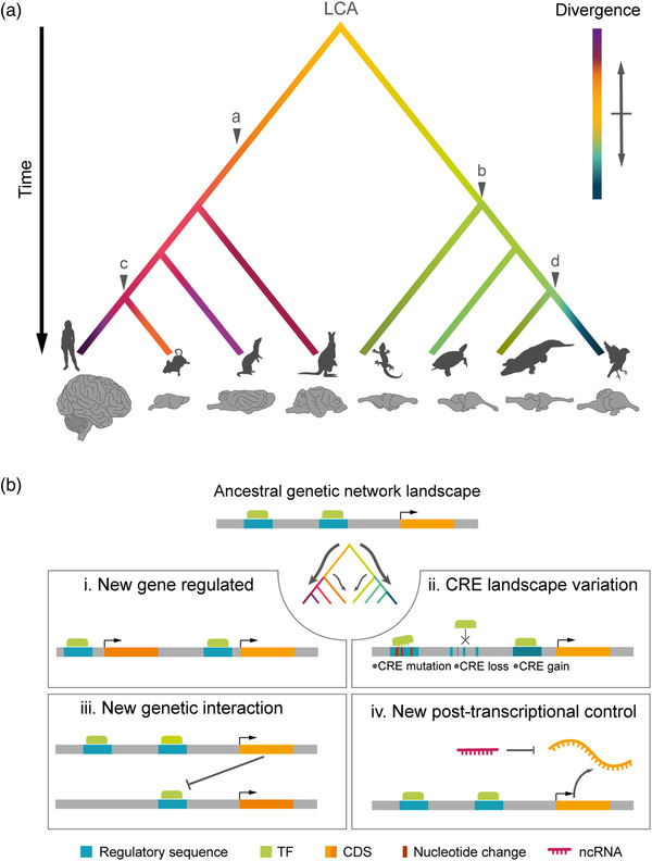
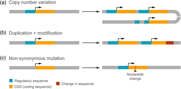
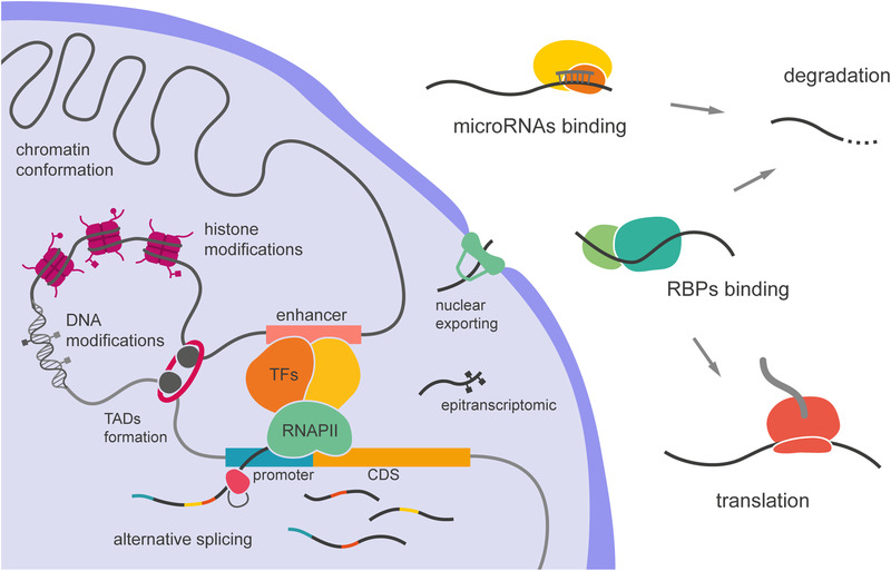
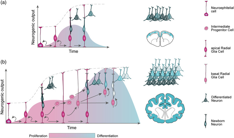
The size of the cerebral cortex increases dramatically across amniotes, from reptiles to great apes. This is primarily due to different numbers of neurons and glial cells produced during embryonic development. The evolutionary expansion of cortical neurogenesis was linked to changes in neural stem and progenitor cells, which acquired increased capacity of self-amplification and neuron production. Evolution works via changes in the genome, and recent studies have identified a small number of new genes that emerged in the recent human and primate lineages, promoting cortical progenitor proliferation and increased neurogenesis. However, most of the mammalian genome corresponds to noncoding DNA that contains gene-regulatory elements, and recent evidence precisely points at changes in expression levels of conserved genes as key in the evolution of cortical neurogenesis. Here, we provide an overview of basic cellular mechanisms involved in cortical neurogenesis across amniotes, and discuss recent progress on genetic mechanisms that may have changed during evolution, including gene expression regulation, leading to the expansion of the cerebral cortex.

Keywords: OSVZ; cerebral cortex; enhancer; ferret; intermediate progenitor; radial glia.


Conflict of interest statement

The authors declare no conflict of interest.

Figures

FIGURE 1
FIGURE 1
Regulation of gene expression levels across amniotes determines their predominant mode of neurogenesis, with consequences on the final size, folding, and cellular complexity of the cerebral cortex. CP, cortical plate; NL, neuronal layer; ISVZ, inner subventricular zone; OSVZ, outer subventricular zone; SVZ, subventricular zone; VZ, ventricular zone
FIGURE 2
FIGURE 2
The evolution of genomes paralleled the evolution of the cerebral cortex. (a) Schematic phylogenetic tree illustrating the relationships between amniote clades (from left to right: primates, rodents, carnivores, marsupials, lepidosaurs, testudines, crocodilians, and birds) and the accumulation of genomic changes on developmental programs leading to the diversification of brain size and structure. Evolutionary time and brain are not to scale. Phenotypic divergence from the last common ancestor (LCA) is color coded. Major changes in corticogenesis are indicated: (a) mammalian six‐layered neocortex, (b) reptile single‐layered cortex, (c) secondary loss of gyrencephaly in some mammalian clades, and (d) avian dorsal pallium. Animal silhouettes are from http://phylopic.org/. (B) Schemas of typical genomic changes occurring during evolution that alter gene regulatory networks within developmental programs. (i) Integration of new genes in the network, introducing new functions. (ii) Changes in cis‐regulatory elements (CRE) that control gene expression. (iii) Emergence of new genetic interactions between elements already existing in the network. (iv) New posttranscriptional control, such as the emergence of new noncoding RNAs (ncRNAs). CDS, coding sequence; TF, transcription factor
FIGURE 3
FIGURE 3
Mechanisms of genetic evolution. (a) Copy number variation caused by gene duplication, which may be complete or partial, and may go along with an original regulatory sequence. (b) Generation of a new gene by complete or partial duplication of an ancestral gene. The duplicated gene may be further subject to sequence modification. (c) Neofunctionalization of an existing gene by nonsynonymous mutation. Protein function may be altered by mutations modifying its gross structure or its functional properties, or even by nonsynonymous point mutations.
FIGURE 4
FIGURE 4
Layers of gene regulation. Pretranscriptional modalities include chromatin conformation, DNA and histone modifications, and the formation of topologically associated domains (TADs), which bring together different types of trans‐regulatory elements, affecting the binding of transcription factors (TFs). Posttranscriptional regulation involves alternative splicing, mRNA modification (epitranscriptomics), and binding to RNA binding proteins (RBPs), which control nuclear export and mRNA stability, including degradation by miRNA–RISC complex. CDS, coding sequence
FIGURE 5
FIGURE 5
Temporal progression of cortical neurogenesis. (a) Temporal progression of neurogenesis in the reptilian dorsal pallium. At the onset of neurogenesis, self‐amplifying neuroepithelial cells transit to apical radial glia cells, which then undergo long cell cycles to directly produce neurons, rapidly depleting the progenitor pool during the short neurogenic period. As a result, a low number and diversity of neurons are produced, arranged in a thin cortex. (b) Temporal progression of neurogenesis in the mammalian dorsal pallium. Apical radial glia cells self‐amplify prior to begin producing deep‐layer neurons (dark gray). A long neurogenic period allows for the unfolding of genetic programs that drive the emergence of basal progenitors, such as intermediate progenitor cells and basal radial glia cells. Basal progenitors amplify the output and diversity of neurons produced, especially those destined to upper layers, producing an expanded six‐layered neocortex. Developmental time is not to scale.

Similar articles

Cited by

References

    1. Acemel, R. D. , Maeso, I. , & Gómez‐Skarmeta, J. L. (2017). Topologically associated domains: A successful scaffold for the evolution of gene regulation in animals. Wiley Interdisciplinary Reviews: Developmental Biology, 6, 1–19. 10.1002/wdev.265 - DOI - PubMed
    1. Amadei, G. , Zander, M. A. , Yang, G. , Dumelie, J. G. , Vessey, J. P. , Lipshitz, H. D. , Smibert, C. A. , Kaplan, D. R. , & Miller, F. D. (2015). A smaug2‐based translational repression complex determines the balance between precursor maintenance versus differentiation during mammalian neurogenesis. Journal of Neuroscience, 35, 15666–15681. 10.1523/JNEUROSCI.2172-15.2015 - DOI - PMC - PubMed
    1. Antonacci, F. , Dennis, M. Y. , Huddleston, J. , Sudmant, P. H. , Steinberg, K. M. , Rosenfeld, J. A. , Miroballo, M. , Graves, T. A. , Vives, L. , Malig, M. , Denman, L. , Raja, A. , Stuart, A. , Tang, J. , Munson, B. , Shaffer, L. G. , Amemiya, C. T. , Wilson, R. K. , & Eichler, E. E. (2014). Palindromic GOLGA8 core duplicons promote chromosome 15q13.3 microdeletion and evolutionary instability. Nature Genetics, 46, 1293–1302. 10.1038/ng.3120 - DOI - PMC - PubMed
    1. Aprea, J. , & Calegari, F. (2015). Long non‐coding RNAs in corticogenesis: Deciphering the non‐coding code of the brain. EMBO Journal, 34, 2865–2884. 10.15252/embj.201592655 - DOI - PMC - PubMed
    1. Arcila, M. L. , Betizeau, M. , Cambronne, X. A. , Guzman, E. , Doerflinger, N. , Bouhallier, F. , Zhou, H. , Wu, B. , Rani, N. , Bassett, D. S. , Borello, U. , Huissoud, C. , Goodman, R. H. , Dehay, C. , & Kosik, K. S. (2014). Novel primate miRNAs coevolved with ancient target genes in germinal zone‐specific expression patterns. Neuron, 81, 1255–1262. 10.1016/j.neuron.2014.01.017 - DOI - PMC - PubMed

Publication types