. 2022 Jul;36(7):e22298.
doi: 10.1096/fj.201901024RR.

IL-10 promotes endothelial progenitor cell infiltration and wound healing via STAT3

Affiliations

IL-10 promotes endothelial progenitor cell infiltration and wound healing via STAT3

Walker D Short et al. FASEB J. 2022 Jul.

Abstract

Endothelial progenitor cells (EPCs) contribute to de novo angiogenesis, tissue regeneration, and remodeling. Interleukin 10 (IL-10), an anti-inflammatory cytokine that primarily signals via STAT3, has been shown to drive EPC recruitment to injured tissues. Our previous work demonstrated that overexpression of IL-10 in dermal wounds promotes regenerative tissue repair via STAT3-dependent regulation of fibroblast-specific hyaluronan synthesis. However, IL-10's role and specific mode of action on EPC recruitment, particularly in dermal wound healing and neovascularization in both normal and diabetic wounds, remain to be defined. Therefore, inducible skin-specific STAT3 knockdown mice were studied to determine IL-10's impact on EPCs, dermal wound neovascularization and healing, and whether it is STAT3-dependent. We show that IL-10 overexpression significantly elevated EPC counts in the granulating wound bed, which was associated with robust capillary lumen density and enhanced re-epithelialization of both control and diabetic (db/db) wounds at day 7. We noted increased VEGF and high C-X-C motif chemokine 12 (CXCL12) levels in wounds and a favorable CXCL12 gradient at day 3 that may support EPC mobilization and infiltration from bone marrow to wounds, an effect that was abrogated in STAT3 knockdown wounds. These findings were supported in vitro. IL-10 promoted VEGF and CXCL12 synthesis in primary murine dermal fibroblasts, with blunted VEGF expression upon blocking CXCL12 in the media by antibody binding. IL-10-conditioned fibroblast media also significantly promoted endothelial sprouting and network formation. In conclusion, these studies demonstrate that overexpression of IL-10 in dermal wounds recruits EPCs and leads to increased vascular structures and faster re-epithelialization.

Keywords: IL-10; VEGF; angiogenesis; diabetes; endothelial progenitor cells; wound healing.


Figures

FIGURE 1
FIGURE 1
IL‐10 overexpression enhanced wound closure in a STAT3‐dependent manner in vivo. (A–F) Hematoxylin and Eosin staining of wounds at day 3 post wounding. Wounds are marked in India ink; wound margins are indicated by arrowheads and the encroaching epithelial margins are indicated by arrows. (A–F) Original magnification: 4×, Scale Bar = 500 µm; As compared to PBS (A) or lentiviral GFP (B) control treatments, lentiviral IL‐10‐treated wounds show enhanced wound closure and robust granulation tissue formation (C) in STAT3Δ/Δ ctrl vehicle control mice. IL‐10's effects on wound healing were abrogated in STAT3−/− mice, as evidenced by significantly impaired wound morphology (D–F). (G and H) Quantitation of epithelial gap (G) and granulation tissue (H) amongst the different treatments based on image analysis show that while there is no significant difference in gap closure between PBS and lentiviral GFP wounds, lentiviral IL‐10 treated wounds exhibit increased gap closure and granulation tissue formation, whereas in STAT3−/− mice this effect is abrogated. Bar plots: mean ± SD, 2 sections/wound, n = 4 wounds from different mice/treatment group, ***p < .001 by ANOVA
FIGURE 2
FIGURE 2
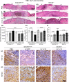
IL‐10 overexpression reduced inflammation and enhanced wound remodeling and regenerative phenotype in a STAT3‐dependent manner. (A–F) Hematoxylin and Eosin staining of wounds at day 7 post wounding. Wounds are marked in India ink; wound margins are indicated by arrowheads. (A–F) Original magnification: 4×, Scale Bar = 500 µm; (A–C) similar gap closure is observed between the three groups—PBS, lentiviral GFP and lentiviral IL‐10 treated wounds in STAT3Δ/Δ ctrl mice. However, IL‐10 treated cohort display thicker epidermis and robust granulation tissue as compared to the control treatments. (D–F) All treatment cohorts healed similarly in STAT3−/− mice and the effect of IL‐10 on the epidermis and granulation tissue deposition is not apparent. (F) Quantitation of granulation tissue between different treatment groups in STAT3Δ/Δ ctrl show a modest trend in IL‐10 treated mice, however, with no statistical significance between STAT3Δ/Δ ctrl and STAT3−/− with different treatments. (H) quantification of staining with CD45 show significantly lower CD45+ cells per high power field (HPF; 40×) in lentiviral IL‐10 wounds as compared to lentiviral GFP or PBS treatments in STAT3Δ/Δ ctrl mice, which is abrogated in STAT3−/− mice. (I) Elevated expression of Lamp2a positive cells was observed in lentiviral IL‐10 treated wounds as compared to the control treatments in STAT3Δ/Δ ctrl mice, which is abrogated in STAT3−/− mice. (J) Panels left to right show similar TGF‐β1 staining pattern in controls versus lentiviral IL‐10 treated wounds in STAT3Δ/Δ ctrl, mice, that remain similar in expression in STAT3−/− wounds. (K) A definite increase in TGF‐β3 expression is seen in lentiviral IL‐10 treated wounds as compared to control treated ones in STAT3Δ/Δ ctrl mice, however, this effect induced by IL‐10 waned in STAT3−/− wounds. Scale bar = 50 µm in (J and K); Bar plots: mean ± SD, 2 sections/wound, n = 4 wounds from different mice/treatment group, ***p < .001 by ANOVA
FIGURE 3
FIGURE 3
IL‐10 overexpression enhanced wound capillary lumen density and EPC infiltration in a STAT3‐dependent manner. (A–F) Capillary lumens are marked by MECA‐32 staining. (G–L) EPCs are identified by their characteristic morphology, large eccentric nuclei and CD133+/FLK‐1+ staining (CD133–green, FLK‐1–red, merged–yellow). Representative MECA‐32+ and CD133+/Flk‐1+ wound images for each treatment group at day 7 are shown here. Lentiviral IL‐10 treatment significantly enhanced wound vessel density (m) and EPC numbers (n) per high power field (HPF), compared to lentiviral GFP or PBS treatments in STAT3Δ/Δ ctrl mice, which was abrogated in STAT3−/− mice. CD133+/FLK‐1+ EPCs are indicated by arrow heads. Bar plots: mean ± SD. 2 sections/wound, n = 3–4 wounds from different mice/treatment group, ***p < .001 by ANOVA. Scale Bars = 50 µm and Original magnification: 40× in (A–F); Scale Bars = 20 µm and Original magnification: 80× in (G–L)
FIGURE 4
FIGURE 4
IL‐10 overexpression enhanced STAT3‐dependent EPC mobilization after cutaneous wounding. Peripheral blood was collected at baseline before wounding and again at day 3 post wounding from STAT3Δ/Δ ctrl and STAT3−/− mice that received lentiviral IL‐10, lentiviral GFP or PBS treatments. Cells were stained with 7AAD, CD34, CD133 and Flk‐1 for flow cytometry analysis. (A) Single cells were gated for 7AAD CD34+ populations; of which cells that were CD133+Flk‐1+ were quantified as EPCs (B). There is a clear difference in EPC levels in peripheral blood at baseline in uninjured mice (B) versus mice at day three post wounding (C). (D) Quantitative analysis show that following cutaneous wounding there is a significant increase in EPCs at day 3 post wounding as compared to uninjured control mice. Lentiviral IL‐10 treatment significantly increase circulating EPC levels as compared to lentiviral GFP or PBS treatments in STAT3Δ/Δ ctrl mice.IL‐10's effects were abrogated in STAT3−/− mice. Bar plots = mean ± SD of EPC numbers as analyzed by the FlowJo software, n = 3–4 wounds from different mice/treatment group, **p < .01 by ANOVA
FIGURE 5
FIGURE 5
IL‐10 regulates VEGF and CXCL12 gradients in wounds, serum, and bone marrow in a STAT3‐dependent manner. Wound tissue, peripheral blood, and bone marrow (BM) were collected at baseline before wounding and again at day 3 post wounding from STAT3Δ/Δ ctrl and STAT3−/− mice that received lentiviral IL‐10, lentiviral GFP, or PBS treatments. VEGF and CXCL12 levels were quantified with ELISA. The effect of different treatments on VEGF and CXCL12 levels within the wound tissue specimens, serum, and BM are respectively shown in (A–C) and (D–F). Bar plots = mean ± SD, n = 3–4 wounds from different animals/treatment group, *p < .05, **p < .01, **p < .001 by ANOVA
FIGURE 6
FIGURE 6
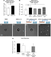
IL10 induced VEGF and CXCL12 production in primary murine dermal fibroblasts and increased cell sprouting and capillary‐like network formation in aortic ring assay. Primary murine dermal fibroblasts in culture were incubated +/− 200 ng/ml of IL‐10 for 48 h. Supernatants from the cultures were assayed for VEGF and CXCL12 expression by ELISA. (A) CXCL12 levels significantly increase in IL 10 treated fibroblasts as compared to untreated cells. (B) VEGF levels significantly increase in IL‐10 treated fibroblasts as compared to controls, however not when anti‐CXCL12 mAb is included in treatment. (C and D) Murine thoracic aortas are treated with either DMEM or DMEM supplemented with 200 ng/ml IL‐10 for 12 days. Aortic rings show an endothelial cell outgrowth when treated with DMEM (C), and the ones spiked with DMEM + IL10 respond but with a similar relative sprouting area (D). (E and F) The aortic ring assay was repeated with either conditioned media from primary murine dermal fibroblasts, or conditioned media from fibroblasts treated with IL‐10 from panel (A). The former produced no measurable endothelial outgrowth (E), whereas with the latter, a significant outgrowth with an increase in capillary‐like 2D network formation was observed. Relative sprouting area for treatment groups quantified in (G). White arrowheads indicate the capillary‐like networks. Bar plots: mean ± SD, experiments are conducted in triplicates with cells from 2 passages; *p < .05, by ANOVA. n = 3 aortic rings per treatment were studied and the experiment was repeated two times with aortas from different mice and conditioned media from different primary cell isolations
FIGURE 7
FIGURE 7
IL 10 enhanced wound re‐epithelialization and capillary lumen density in diabetic murine wound model. Dorsal wounds in db/db mice treated with either PBS, lentiviral GFP, or lentiviral IL‐10 were harvested at 7 days post injury. (A–C) Hematoxylin and Eosin staining reveals that lentiviral IL‐10 treated wounds re‐epithelialize faster as compared to the two control groups. Quantitation of epithelial gap (D) and granulation tissue is shown (E). (F–H) Representative images of MECA‐32 stained wounds section across the three treatment conditions—PBS, lentiviral GFP, or lentiviral IL‐10 respectively. Increased MECA‐32 stained‐capillary lumens are seen in lentiviral IL‐10 wounds, which is quantitatively significant (I). (J–M) Increase in the CD133+Flk1+ EPC levels in the wound beds is also observed in lentiviral IL‐10‐treated db/db wounds as compared to the control treatments. (N–O) qRT‐PCR on wound tissue homogenates collected at day 7 shows that VEGF (N) and (O) CXCL12 expression is significantly higher lentiviral IL‐10 treated cohort as compared to the PBS and lentiviral GFP controls. Scale bar = 500 µm in (A–C), 50 µm in (F–H) and 20 µm in (J–L); Bar plots: mean ± SD, 2 sections/wound, n = 4 wounds from different mice/treatment group, *p < .05 by ANOVA
FIGURE 8
FIGURE 8
IL‐10 can play a pivotal role in postnatal cutaneous wound healing and neovascularization by influencing EPC mobilization and recruitment via STAT3‐dependent increase in VEGF and CXCL12. IL10 overexpression, via a STAT3‐dependent mechanism, results in enhanced levels of VEGF and CXCL12, wound healing, and neovascularization associated with an increase in EPCs in the wound. We put forth a potential pathway that IL‐10 overexpression may induce wound fibroblasts to produce more VEGF and CXCL12, which creates a positive gradient for bone marrow derived EPC mobilization and homing to healing tissue. Image created using BioRender.com

References

    1. Economic costs of diabetes in the U.S. in 2017. Diabetes Care. 2018;41(5):917‐928. - PMC - PubMed
    1. Frykberg RG, Banks J. Challenges in the treatment of chronic wounds. Adv Wound Care. 2015;4(9):560‐582. - PMC - PubMed
    1. Casqueiro J, Casqueiro J, Alves C. Infections in patients with diabetes mellitus: a review of pathogenesis. Indian J Endocrinol Metab. 2012;16(Suppl 1):S27‐S36. - PMC - PubMed
    1. Balaji S, King A, Crombleholme TM, Keswani SG. The role of endothelial progenitor cells in postnatal vasculogenesis: implications for therapeutic neovascularization and wound healing. Adv Wound Care. 2013;2(6):283‐295. - PMC - PubMed
    1. Ribatti D, Vacca A, Nico B, Roncali L, Dammacco F. Postnatal vasculogenesis. Mech Dev. 2001;100(2):157‐163. - PubMed

Publication types

MeSH terms