. 2022 Jul 15;132(14):e158524.
doi: 10.1172/JCI158524.

Schwann cell nodal membrane disruption triggers bystander axonal degeneration in a Guillain-Barré syndrome mouse model

Affiliations

Schwann cell nodal membrane disruption triggers bystander axonal degeneration in a Guillain-Barré syndrome mouse model

Rhona McGonigal et al. J Clin Invest. .

Abstract

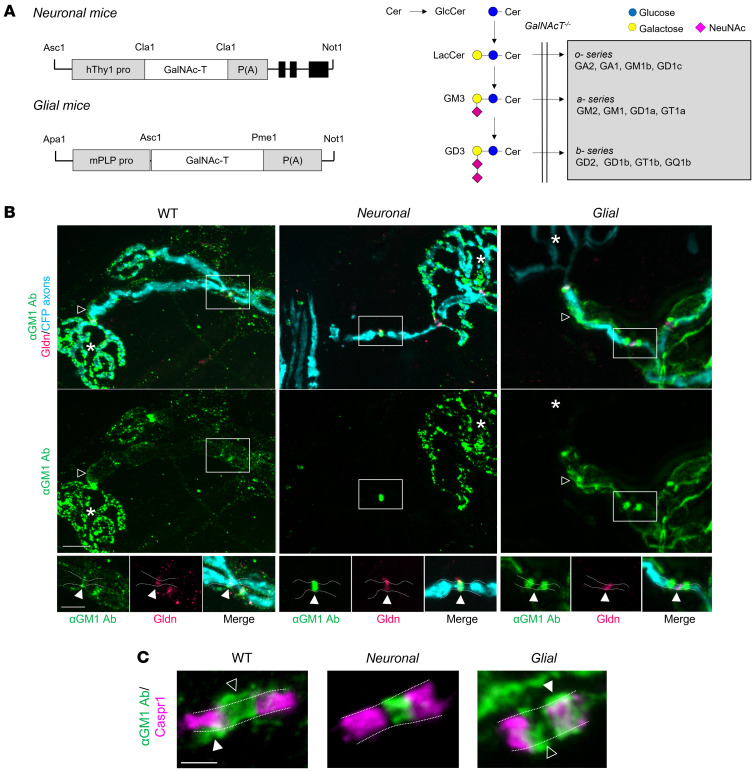
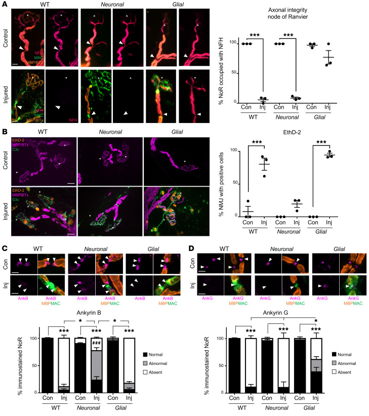
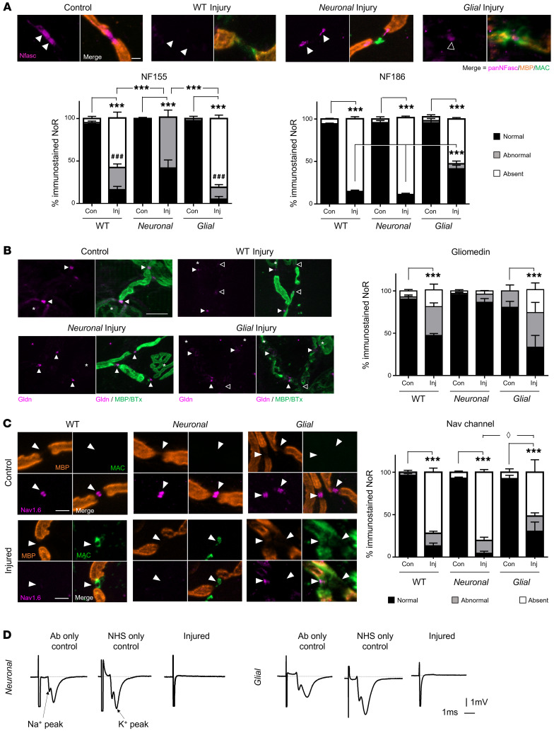
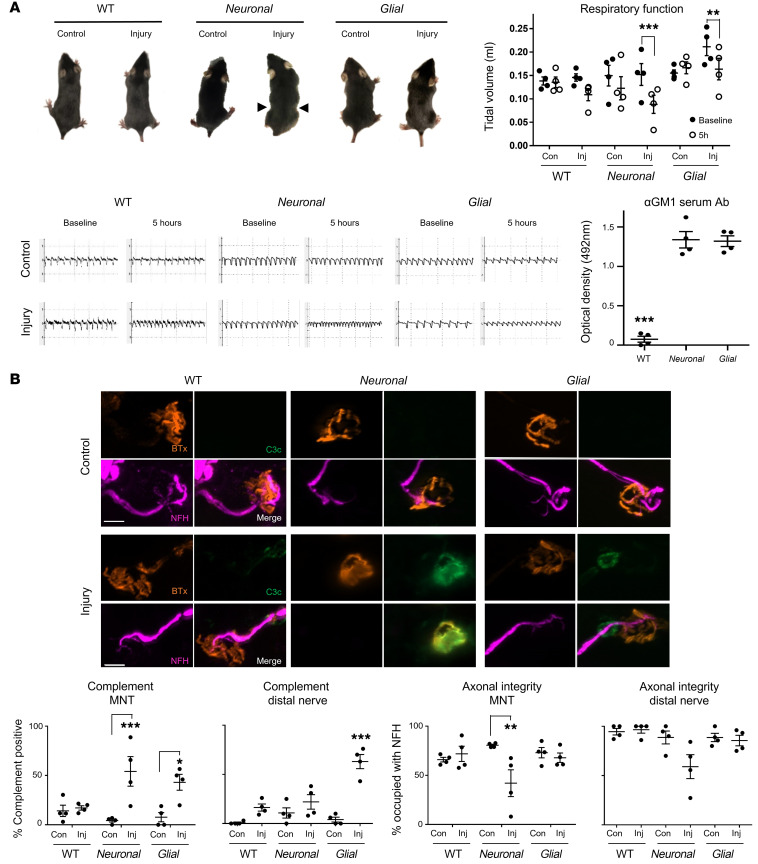
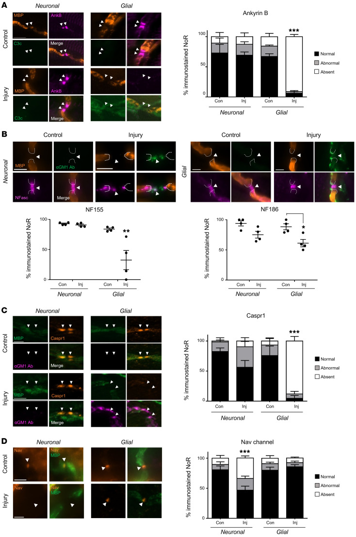
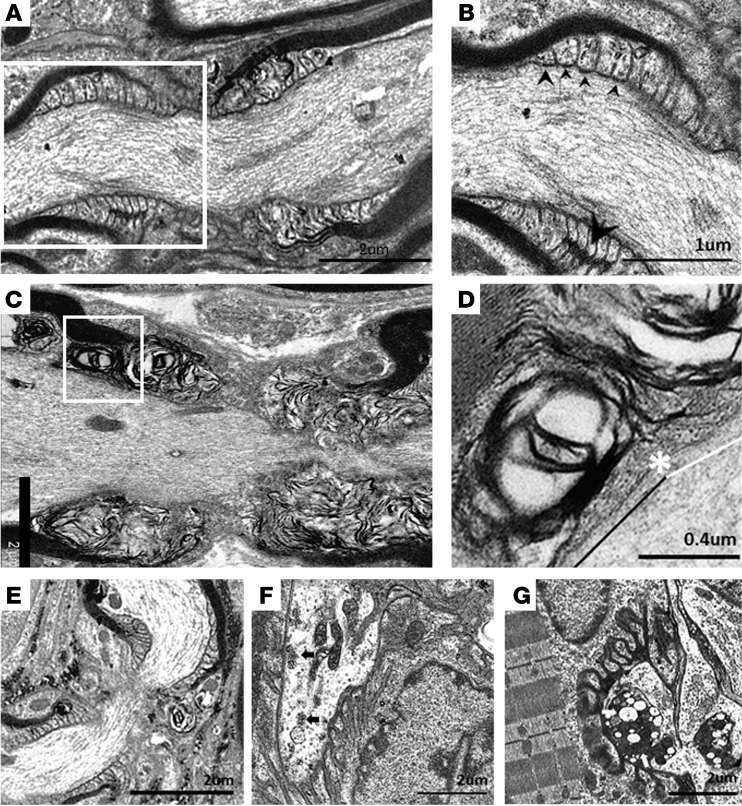
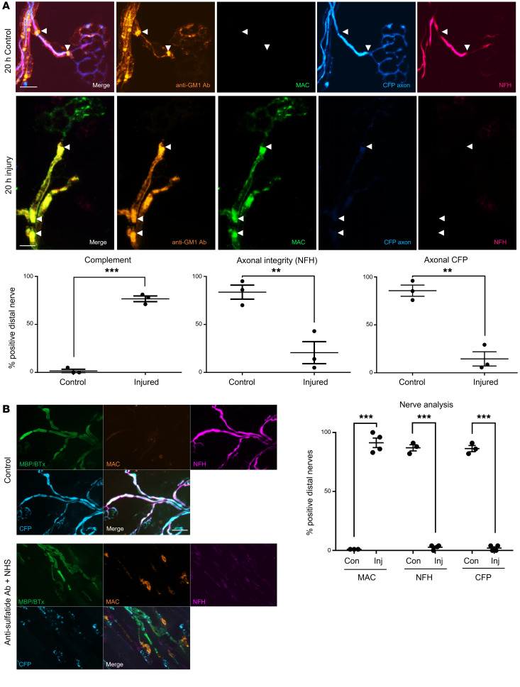
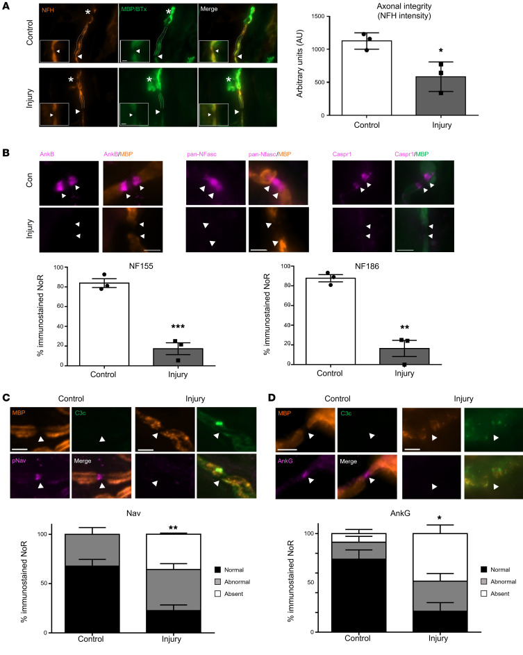
In Guillain-Barré syndrome (GBS), both axonal and demyelinating variants can be mediated by complement-fixing anti-GM1 ganglioside autoantibodies that target peripheral nerve axonal and Schwann cell (SC) membranes, respectively. Critically, the extent of axonal degeneration in both variants dictates long-term outcome. The differing pathomechanisms underlying direct axonal injury and the secondary bystander axonal degeneration following SC injury are unresolved. To investigate this, we generated glycosyltransferase-disrupted transgenic mice that express GM1 ganglioside either exclusively in neurons [GalNAcT-/--Tg(neuronal)] or glia [GalNAcT-/--Tg(glial)], thereby allowing anti-GM1 antibodies to solely target GM1 in either axonal or SC membranes, respectively. Myelinated-axon integrity in distal motor nerves was studied in transgenic mice exposed to anti-GM1 antibody and complement in ex vivo and in vivo injury paradigms. Axonal targeting induced catastrophic acute axonal disruption, as expected. When mice with GM1 in SC membranes were targeted, acute disruption of perisynaptic glia and SC membranes at nodes of Ranvier (NoRs) occurred. Following glial injury, axonal disruption at NoRs also developed subacutely, progressing to secondary axonal degeneration. These models differentiate the distinctly different axonopathic pathways under axonal and glial membrane targeting conditions, and provide insights into primary and secondary axonal injury, currently a major unsolved area in GBS research.

Keywords: Autoimmunity; Demyelinating disorders; Neuroscience.


Conflict of interest statement

Conflict of interest: The authors have declared that no conflict of interest exists.

Figures

Figure 1
Figure 1. Anti–GM1 ganglioside antibody binding in transgenic mice with selective neuronal or glial complex ganglioside expression.
(A) Constructs used to direct GalNAc-T expression in neurons (human Thy1.2 promoter) or glia (mouse Plp promoter) of GalNAc-T–/–-Tg(neuronal) (Neuronal) and GalNAc-T–/–-Tg(glial) (Glial) mice, respectively. Ganglioside biosynthesis pathway indicates the reexpression of complex ganglioside expression (gray box) following construct insertion on a GalNAc-T–/– background (20). (B) Using a single anti-GM1 antibody (Ab, green), differential binding was observed at the distal motor nerves from triangularis sterni nerve–muscle preparations among genotypes. Open arrowheads indicate internodal Schwann cell (SC) abaxonal membrane anti-GM1 Ab deposition on WT and Glial nerves (absent along Neuronal nerves). Gliomedin (Gldn) immunostaining identifies the nodal gap. Boxed areas are enlarged underneath and represent differential anti-GM1 Ab binding at nodes of Ranvier (NoRs) among genotypes in relation to gliomedin (closed arrowheads). Dashed lines delineate the border of the axonal membrane determined by cytoplasmic CFP–positive axons. Scale bars: 10 μm (top panels) and 5 μm (lower panels). Asterisks indicate motor nerve terminals. (C) Caspr1 immunostaining (magenta) indicates the paranodes. Dashed lines delineate the border of the axonal membrane and arrowheads indicate anti-GM1 Ab binding beyond this membrane, suggesting binding on the glial membranes of the SC microvilli (open arrowheads) or paranodal loops (closed arrowheads) at WT and Glial NoRs. Scale bar: 2 μm.
Figure 2
Figure 2. Distal motor nerve integrity following selective targeting and acute injury of neural membranes ex vivo.
Triangularis sterni nerve–muscle preparations from WT, Neuronal, and Glial mice were treated ex vivo with anti-GM1 Ab and a source of complement (injury, Inj) or anti-GM1 Ab alone (control, Con). (A) Loss of axonal integrity due to injury at the motor nerve terminal (MNT, identified by α-bungarotoxin, BTx, orange, asterisk) and node of Ranvier (NoR, orange, arrowheads) was monitored by presence of neurofilament H immunostaining (NFH, magenta). Membrane attack complex (MAC) complement pore deposition (green) was present in all injured preparations compared with control. (B) Ethidium homodimer–positive (EthD-2–positive, orange) cells overlying MNT (magenta, asterisk) were compared among treatment groups. Representative images show the presence of complement deposition (green) in all injured tissue. (C and D) The sites where ankyrin B (AnkB) or AnkG immunostaining should be located are indicated by arrowheads. The presence of normal (black bars, statistical comparisons indicated with asterisks) or abnormal (gray bars) AnkB and AnkG immunostaining was compared to associated controls for each genotype. A lengthened gap between AnkB domains is shown in a representative image from injured Neuronal tissue. Weakened, uneven AnkG staining in injured Glial tissue is shown in the representative image. Scale bars: 20 μm (A), 50 μm (B), and 5 μm (C and D). Results are represented as the mean ± SEM. n = 3/genotype/treatment: 10–46 NoRs/mouse (median = 21, NFH); 11–29 neuromuscular junctions (NMJs)/mouse (median = 18, EthD-2); 10–26 NoRs/mouse (median = 23, AnkG); and 12–31 NoRs/mouse (median = 21, AnkB) were analyzed. *P < 0.05; ***P < 0.001; ###P < 0.001 (for abnormal AnkB and AnkG immunostaining in C) compared with control by 2-way ANOVA with Tukey’s post hoc test.
Figure 3
Figure 3. Differential disruption to the node of Ranvier when neuronal and glial membranes are injured selectively ex vivo.
Triangularis sterni nerve–muscle preparations from WT, Neuronal, and Glial mice were treated ex vivo with anti-GM1 Ab and a source of complement (injury, Inj) or anti-GM1 Ab alone (control, Con). Disruption to nodal protein (magenta) organization at the node of Ranvier (NoR) due to injury was assessed; the site of expected staining is indicated by arrowheads for each marker. Representative images demonstrate normal nodal protein localization in all control tissue and absent or abnormal staining in injury groups, which coincides with nodal complement deposition (A and C, green). (A) A pan-neurofascin (Nfasc) Ab was used to assess paranodal NF155 (closed arrowheads) and nodal NF186 (open arrowhead). (B) SC microvilli marker gliomedin (Gldn) immunostaining at NoRs was assessed compared to controls. Asterisks indicate motor nerve terminals. (C) Changes to normal (black bars) Nav1.6 labeling were observed in injured tissue from all genotypes compared with associated controls. Diamond defines statistical comparisons of absent immunostaining (white bars). (D) Perineural recordings from distal motor nerves were performed on tissue from Neuronal and Glial mice treated with anti-GM1 Ab only, a source of complement (normal human serum, NHS) only, or a combination of Ab and NHS (injured). Representative recordings from 1 mouse per treatment demonstrate that normal Na+ and K+ waveforms were lost when the tissue was injured. Scale bar: 5 μm. Results are represented as the mean ± SEM. n = 3/genotype/treatment: 13–36 NoRs/mouse (median = 24, pNFasc); 15–33 NoRs/mouse (median = 19, gliomedin); and 11–30 NoRs/mouse (median = 23, Nav1.6) were analyzed. *P < 0.05, **P < 0.01, ***P < 0.001 (for comparisons between normal immunostaining); ###P < 0.001 (for abnormal NF155 immunostaining in Neuronal injury group compared to WT or Glial imjury in A) compared with control by 2-way ANOVA with Tukey’s post hoc test.
Figure 4
Figure 4. Distal motor nerve axonal integrity remains intact following selective glial membrane targeting in vivo.
WT, Neuronal, and Glial mice were dosed i.p. with 50 mg/kg anti-GM1 Ab followed 16 hours later with 30 μL/g normal human serum (NHS) (injury, Inj) or NHS only (control, Con). Respiratory function was monitored and diaphragm distal nerves assessed by immunoanalysis 5 hours after NHS delivery. (A) Injured Neuronal mice displayed the most severe respiratory phenotype: a pinched, wasp-like abdomen (arrowheads) and significantly reduced tidal volume (TV) measured using whole-body plethysmography (EMMS). Injured Glial mice also had significantly reduced TV compared with baseline. Representative respiratory flow charts for each treatment group show reduced TV and an increase in respiratory rate. Serum analysis indicates that circulating anti-GM1 Ab could be detected in Neuronal and Glial but not WT mice. Results are represented as the mean ± SEM, n = 4/genotype/treatment. (B) Complement deposition and axonal integrity (neurofilament H [NFH] occupancy) were compared at the diaphragm motor nerve terminals (MNTs) and along distal nerves. Representative images illustrate complement deposits (green) overlying the MNT, identified by bungarotoxin (BTx, orange), in injured Neuronal mice, and on the distal nerve in injured WT and Glial mice. Scale bar: 10 μm. Results are represented as the mean ± SEM. n = 4/genotype/treatment: 68–133 MNTs/mouse (median = 103) and 7–30 NoRs/mouse (median = 15) were analyzed. *P < 0.05, **P < 0.01, ***P < 0.001 by repeated measures 2-way ANOVA with Bonferroni post-hoc tests (A) or 2-way ANOVA with Tukey post-hoc tests (B).
Figure 5
Figure 5. Disruption of paranodal proteins following glial membrane targeting in vivo.
Neuronal and Glial mice were dosed i.p. with 50 mg/kg anti-GM1 Ab followed 16 hours later with 30 μL/g normal human serum (NHS) (injury, Inj) or NHS only (control, Con). The site of expected nodal protein immunostaining is indicated by arrowheads. (A) The presence of normal ankyrin B (AnkB) immunostaining at the distal paranode (black bars) was significantly reduced in injured Glial mice compared with all treatment groups in the presence of complement (green). (B) A pan-neurofascin (Nfasc) Ab was used to assess glial NF155 and axonal NF186 (magenta). Representative images show loss of NF155 staining at paranodal regions, indicated by dashed lines, and the preservation of NF186 when NoRs are decorated with anti-GM1 Ab (green) in Glial mice. (C) Normal Caspr1 (orange) immunostaining at the distal paranodes was significantly reduced in injured Glial mice compared with all other treatment groups. (D) There was a reduction in distal NoRs with normal Nav channel (orange) staining in injured Neuronal mice. Scale bar: 5 μm. Results are represented as the mean ± SEM. n = 4/genotype/treatment: 5–46 NoRs/mouse (median = 21, AnkB); 7–53 NoRs/mouse (median = 25, NFasc); 5–15 NoRs/mouse (median = 11, Caspr1); and 11–27 NoRs/mouse (median = 16, Nav) were analyzed. *P < 0.05, **P < 0.01, ***P < 0.001 for comparisons with the other treatment groups (A, C, and D) or compared with control (B) by 2-way ANOVA with Tukey’s post hoc test.
Figure 6
Figure 6. Ultrastructural evaluation of diaphragms from in vivo injury models.
Neuronal and Glial mice were dosed i.p. with 50 mg/kg anti-GM1 Ab followed 16 hours later with 30 μL/g normal human serum (NHS) (injury) or NHS only (control). (A) A normal paranode from Glial control tissue. N.B. This image is also representative of the Neuronal control NoR (not shown). (B) Higher magnification of boxed region from A shows tight junctions (large arrowhead) between the paranodal loops, and transverse bands (TBs, small arrowheads) at the axo-glial junction between the axon and paranodal loops. (C) Injured Glial NoRs show severely disrupted paranodal loop organization compared with control. (D) Magnification of boxed area from C, shows TBs are present between the paranodal loops and axon at the juxtaparanodal-proximal paranode (above black line); however, they are absent at the node-proximal border (above white line, right of asterisk). (E) Injured Neuronal NoRs show no architectural disruption. (F) Neuronal control motor nerve terminal (MNT) displays normal architecture and contains synaptic vesicles (black arrows). (G) Disturbance to the injured Neuronal MNT includes an absence of neurofilament, synaptic vesicles, and the formation of dense or vacuolated mitochondria (white arrows). Results are representative of analysis from 8–10 NoRs per mouse (n = 3/genotype/treatment).
Figure 7
Figure 7. Extended ex vivo injury selectively targeting glial membranes results in secondary axonal degeneration.
Triangularis sterni nerve–muscle preparations from Glial (A) and WT (B) mice were treated ex vivo with anti-GM1 Ab or anti-sulfatide Ab, respectively, and a source of complement (injury, Inj) or Ab alone (control, Con) for 20 hours. (A) Anti-GM1 Ab (orange) and complement (green) deposition along the distal motor nerve was strongly enriched at the paranodes (arrowheads) in injured compared with control tissue. Loss of axonal integrity along the distal nerve was monitored by presence of neurofilament H immunostaining (NFH, magenta) and cytosolic CFP (blue). (B) The experiment was repeated in WT mice using an anti-sulfatide Ab; the results reflect those reported in A. Scale bars: 10 μm (A) and 20 μm (B). Results are represented as the mean ± SEM. n = 3/treatment: 25–54 NoRs/mouse (median = 39) were analyzed. **P < 0.01, ***P < 0.001 compared with control by 1-tailed Student’s t test.
Figure 8
Figure 8. Extended in vivo injury selectively targeting glial membrane results in secondary axonal degeneration.
Glial mice were dosed i.p. with 50 mg/kg anti-GM1 Ab followed 16 hours later with 30 μL/g normal human serum (NHS) (injury, Inj) or NHS only (control, Con). The experiment was terminated 24 hours after NHS delivery. The site of expected nodal protein immunostaining is indicated by arrowheads. (A) At this time point there was loss of neurofilament H staining (NFH, orange) at the motor nerve terminal (MNT, asterisk) and the staining intensity was significantly reduced at the first distal node of Ranvier (NoR). (B) Normal ankyrin B (AnkB), NF155, NF186, and Caspr1 (magenta) immunostaining was assessed at distal paranodes after injury compared to control. (C) There was a further reduction in distal NoRs with normal voltage-gated sodium (Nav) channel staining (magenta) in injured Glial mice compared with control at this extended time point. (D) Additionally, the Nav channel–tethering protein AnkG was notably absent. Scale bar: 5 μm. Results are represented as the mean ± SEM. n = 3/genotype/treatment: 5–15 NoRs/mouse (median = 11, NFH intensity); 4–25 NoRs/mouse (median = 18, panNFasc); 9–23 NoRs/mouse (median = 12, Nav); and 10–28 NoRs/mouse (median = 18, AnkG) were analyzed. *P < 0.05, **P < 0.01, ***P < 0.001 by 2-tailed Student’s t test (A and B) or 2-way ANOVA with Tukey’s post hoc test (C).

Similar articles

Cited by

References

    1. Willison HJ, et al. Guillain-Barre syndrome. Lancet. 2016;388(10045):717–727. doi: 10.1016/S0140-6736(16)00339-1. - DOI - PubMed
    1. Ho TW, et al. Motor nerve terminal degeneration provides a potential mechanism for rapid recovery in acute motor axonal neuropathy after Campylobacter infection. Neurology. 1997;48(3):717–724. doi: 10.1212/WNL.48.3.717. - DOI - PubMed
    1. Uncini AK, et al. Nodo-paranodopathy: beyond the demyelinating and axonal classification in anti-ganglioside antibody-mediated neuropathies. Clin Neurophysiol. 2013;124(10):1928–1934. doi: 10.1016/j.clinph.2013.03.025. - DOI - PubMed
    1. Feasby TE, et al. An acute axonal form of Guillain-Barré polyneuropathy. Brain. 1986;109(pt 6):1115–1126. - PubMed
    1. Altmann P, et al. Increased serum neurofilament light chain concentration indicates poor outcome in Guillain-Barré syndrome. J Neuroinflammation. 2020;17(1):86. doi: 10.1186/s12974-020-01737-0. - DOI - PMC - PubMed

Publication types