Review
. 2022 Jun 20;51(12):5214-5236.
doi: 10.1039/d2cs00193d.

PROTACs: past, present and future

Affiliations
Review

PROTACs: past, present and future

Ke Li et al. Chem Soc Rev. .

Abstract

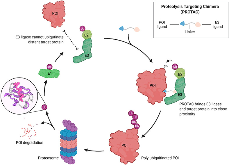
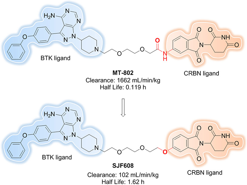
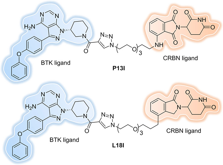
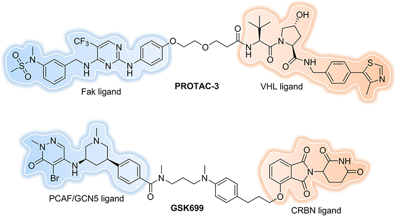
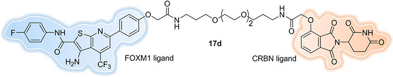
Proteolysis-targeting chimeras (PROTACs) are heterobifunctional molecules consisting of one ligand that binds to a protein of interest (POI) and another that can recruit an E3 ubiquitin ligase. The chemically-induced proximity between the POI and E3 ligase results in ubiquitination and subsequent degradation of the POI by the ubiquitin-proteasome system (UPS). The event-driven mechanism of action (MOA) of PROTACs offers several advantages compared to traditional occupancy-driven small molecule inhibitors, such as a catalytic nature, reduced dosing and dosing frequency, a more potent and longer-lasting effect, an added layer of selectivity to reduce potential toxicity, efficacy in the face of drug-resistance mechanisms, targeting nonenzymatic functions, and expanded target space. Here, we highlight important milestones and briefly discuss lessons learned about targeted protein degradation (TPD) in recent years and conjecture on the efforts still needed to expand the toolbox for PROTAC discovery to ultimately provide promising therapeutics.


Conflict of interest statement

Conflicts of Interest

C. M. C. is a shareholder and consultant to Halda Therapeutics and Siduma Therapeutics. He is also a shareholder in Arvinas Inc.

Figures

Figure 1.
Figure 1.
PROTACs hijack the UPS to induce targeted protein degradation. The figure was created with BioRender.com.
Figure 2.
Figure 2.
Early small-molecule PROTACs.
Figure 3.
Figure 3.
Representative IMiDs and VHL ligands.
Figure 4.
Figure 4.
More recent small-molecule PROTACs.
Figure 5.
Figure 5.
Representative ternary complex structures of POI-PROTAC-E3 ligase. The figure was created with ChimeraX.
Figure 6.
Figure 6.
Linker length can define target selectivity.
Figure 7.
Figure 7.
Linker orientation can define target selectivity.
Figure 8.
Figure 8.
Linker rigidity can influence cooperativity.
Figure 9.
Figure 9.
Linker composition can improve the PK properties.
Figure 10.
Figure 10.
PROTACs derived from ibrutinib show both WT and C481S mutated BTK degradation.
Figure 11.
Figure 11.
Fak and PCAF/GCN5 PROTACs.
Figure 12.
Figure 12.
FOXM1 PROTAC.
Figure 13.
Figure 13.
Oligonucleotide-based PROTACs Targeting TFs. (A) TRAFTAC recruits E3 ligase complex through dCas9-HT7 in the presence of haloPROTAC. (B) TF-PROTAC formed in-situ via a copper-free strain-promoted azide–alkyne cycloaddition (SPAAC) reaction to recruit E3 ligase complex. (C) O’PROTAC or oligoTRAFTAC with E3 ligand and oligonucleotide directly attached. The figure was created with BioRender.com.
Figure 14.
Figure 14.
MIF and SF3B1 PROTACs.
Figure 15.
Figure 15.
Trivalent PROTACs.
Figure 16.
Figure 16.
Folate-Caged PROTAC.
Figure 17.
Figure 17.
Hypoxia-activated PROTAC.
Figure 18.
Figure 18.
ERα PROTAC with ligand discovered from a DEL.
Figure 19.
Figure 19.
L3MBTL3-recruiting PROTACs.
Figure 20.
Figure 20.
PROTAC with pM BRD4 degradation potency.
Figure 21.
Figure 21.
PROTAC displays folded conformations in nonpolar solvent.
Figure 22.
Figure 22.
Potent PROTAC degraders can be obtained using an E3 ligase ligand with moderate binding affinity.
Figure 23.
Figure 23.
Targeting virus-related proteins/RNA.

Similar articles

Cited by

References

    1. Hopkins AL and Groom CR, Nat. Rev. Drug Discov, 2002, 1, 727–730. - PubMed
    1. Nandi D, Tahiliani P, Kumar A and Chandu D, J. Biosci, 2006, 31, 137–155. - PubMed
    1. Kumar SV, Bugg CE and Cook WJ, J. Mol. Biol, 1987, 194, 531–544. - PubMed
    1. Livneh I, Cohen-Kaplan V, Cohen-Rosenzweig C, Avni N and Ciechanover A, Cell Res., 2016, 26, 869–885. - PMC - PubMed
    1. Sakamoto KM, Kim KB, Kumagai A, Mercurio F, Crews CM and Deshaies RJ, Proc. Natl. Acad. Sci. U.S.A, 2001, 98, 8554–8559. - PMC - PubMed

MeSH terms

Substances