Mechanical nociceptive assessment of the equine hoof after navicular bursa anesthetic infiltration validated by bursography
- PMID: 35671268
- PMCID: PMC9173607
- DOI: 10.1371/journal.pone.0269532
Mechanical nociceptive assessment of the equine hoof after navicular bursa anesthetic infiltration validated by bursography
Abstract
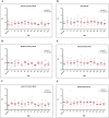
The analgesic specificity of navicular bursa (NB) anesthetic infiltration is still questionable. The study aimed to determine the mechanical nociceptive threshold of non-specific analgesia in the dorsal lamellar stratum, as well as in the sole, coronary band, and heel bulbs of the hoof, after navicular bursa anesthetic infiltration. Six healthy horses with no clinical or radiographic changes of the digits and no communication between the NB and the distal interphalangeal joint, were used. After random selection, the NB of one of the forelimbs was infiltrated with 2% lidocaine and the contralateral one with lactated ringer's solution. Contrast was added to confirm radiographic infiltration. The mechanical nociceptive threshold was determined using a portable pressure dynamometer, before and at various times after the infiltration, in 10 points of the hoof. The effects of time and treatment were verified by ANOVA (P<0.05). There was no statistical difference in the values of the mechanical nociceptive threshold (P>0.05) in all regions evaluated. However, in one of the six hooves that receives lidocaine, complete absence of response to the painful stimulus (maximum force of 6 Kg over an area of 38.46 mm2, for a maximum of 4 seconds) was observed in the dorsal lamellae between 30 and 60 min after infiltration. In conclusion, lidocaine infiltration of NB did not promote significant increases in the nociceptive threshold of the sole, coronary band, bulbs of the heel and dorsal lamellae clinically healthy horses. However, the occurrence of analgesia in one of the six hooves subjected to NB anesthesia indicates that the technique may not be fully specific in few horses.
Conflict of interest statement
The authors have declared that no competing interests exist.
Figures


References
-
- Schumacher J, Taylor DR, Schramme MC, Schumacher J. Localization of Pain in the Equine Foot Emphasizing the Physical Examination and Analgesic Techniques. AAEP Proceedings. 2012; 58: 138–156.
-
- Schumacher J, Shramme MC, Schumacher J, Degraves J. Diagnostic analgesia of the equine digit. Equine Veterinary Education. 2013; 25: 408–421.
-
- Schumacher J, Schramme M. Diagnostic and Regional Surgical Anesthesia of the limbs and axial skeleton. In: Equine Surgery. 4a Ed. Elsevier, St. Louis. 2018; 1220–1227.
-
- Adams OR. Lameness in Horses. 3a Ed. Philadelphia, PA, USA, 1974. pp. 91–118.
Publication types
MeSH terms
Substances
LinkOut - more resources
Full Text Sources

