. 2022 Jun 7;39(6):msac123.
doi: 10.1093/molbev/msac123. Online ahead of print.

Recurrent Plant-Specific Duplications of KNL2 and Its Conserved Function as a Kinetochore Assembly Factor

Affiliations

Recurrent Plant-Specific Duplications of KNL2 and Its Conserved Function as a Kinetochore Assembly Factor

Sheng Zuo et al. Mol Biol Evol. .

Abstract

KINETOCHORE NULL2 (KNL2) plays key role in the recognition of centromeres and new CENH3 deposition. To gain insight into the origin and diversification of the KNL2 gene, we reconstructed its evolutionary history in the plant kingdom. Our results indicate that the KNL2 gene in plants underwent three independent ancient duplications in ferns, grasses and eudicots. Additionally, we demonstrated that previously unclassified KNL2 genes could be divided into two clades αKNL2 and βKNL2 in eudicots and γKNL2 and δKNL2 in grasses, respectively. KNL2s of all clades encode the conserved SANTA domain, but only the αKNL2 and γKNL2 groups additionally encode the CENPC-k motif. In the more numerous eudicot sequences, signatures of positive selection were found in both αKNL2 and βKNL2 clades, suggesting recent or ongoing adaptation. The confirmed centromeric localization of βKNL2 and mutant analysis suggests that it participates in loading of new CENH3, similarly to αKNL2. A high rate of seed abortion was found in heterozygous βKNL2 plants and the germinated homozygous mutants did not develop beyond the seedling stage. Taken together, our study provides a new understanding of the evolutionary diversification of the plant kinetochore assembly gene KNL2, and suggests that the plant-specific duplicated KNL2 genes are involved in centromere and/or kinetochore assembly for preserving genome stability.

Keywords: CENH3; KNL2; adaptive evolution; centromere; endopolyploidy; gene duplication; kinetochore.


Figures

Fig. 1.
Fig. 1.
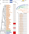
Identification of the KNL2 gene homologs across major plant lineages. (A) Protein structure of KNL2 in Arabidopsis. SANTA domain and CENPC-k motif are indicated by differently colored boxes. (B) The number of KNL2 homologs in 90 representative plant species. The phylogenetic tree is adopted from the NCBI common tree. The blue-, green-, and orange-colored species names indicate alga, bryophytes, and vascular plants, respectively. The red filled boxes mean that we could not retrieved KNL2 from these species. (C) Phylogenetic relationships of the analyzed species were adapted from Banks et al. (2011). (D) The number of KNL2 homologs identified in analyzed crucifer (Brassicaceae) genomes.
Fig. 2.
Fig. 2.
Evolutionary relationship of KNL2 homologs in land plants. Maximum likelihood phylogenetic analysis was performed using IQ-tree with a protein alignment of KNL2 homologs in land plants. The KNL2 genes cluster into two branches in three plant clades—heterosporous water ferns (Salviniaceae), eudicots, and grasses (Poaceae)—indicating ancient gene duplications (arrows). The KNL2 in eudicots and grasses can be classified into two major groups (αKNL2 and βKNL2, and γKNL2 and δKNL2, respectively). Bootstrap values obtained after 1,000 ultrafast bootstrap replicates (bb) are shown in the tree. The scale bar indicates the number of substitutions per site. The tree is arbitrarily rooted between bryophytes and tracheophytes.
Fig. 3.
Fig. 3.
Alignments of SANTA domain and CENPC-k motif in KNL2 homologs presented in LOGO format. (A) Variation map of the SANTA domain in the KNL2 homologs. The WebLogo program (http://weblogo.berkeley.edu/logo.cgi) was used to present SANTA domain alignments. The upper panel aligns SANTA domains of all KNL2 homologs from Brassicales, whereas the middle and bottom panels represent SANTA domain alignments of αKNL2 and βKNL2 homologs, respectively. The conserved N-terminal and C-terminal hydrophobic motifs are marked by blue and orange bars, respectively. Putative Aurora kinase phosphorylation consensus sites are underlined with red bars. (B) Alignment of CENPC-k motif of KNL2 homologs from land plants.
Fig. 4.
Fig. 4.
Evolutionary pressures on the KNL2 paralogs. (A) Summary of tests for positive selection performed on KNL2 paralogs from Brassicaceae species. Statistically significant tests (P < 0.05) are indicated with asterisks. (B) A schematic of a representative KNL2 protein, showing sites evolving under positive selection identified by Bayes empirical Bayes analysis (posterior probability > 0.95).
Fig. 5.
Fig. 5.
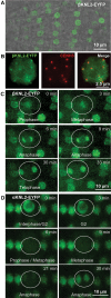
Subcellular localization of βKNL2 in Arabidopsis. (A) Live imaging of root tip cells of Arabidopsis transformed with the βKNL2-EYFP and αKNL2-EYFP fusion constructs. Fluorescent signals showed distinct centromeric and diffused nucleoplasmic distribution. (B) Nucleus isolated from seedlings of the βKNL2-EYFP transformants after immunostaining with anti-GFP (left panel) and anti-CENH3 (middle panel) antibodies. Merge of both immunosignals (right panel). (C) Live imaging of root tip cells of Arabidopsis transformed with the βKNL2-EYFP fusion construct. (D) Live imaging of root tip cells of Arabidopsis transformed with the αKNL2-EYFP fusion construct. Cell undergoing mitosis is encircled.
Fig. 6.
Fig. 6.
The CENH3, CENP-C, and KNL2 gene expression profiles in Arabidopsis. Column charts showing different expression levels of the CENH3, CENP-C, and KNL2 genes in tissues enriched for dividing cells. The relative fragments per kilobase of exon per million mapped fragments (RPKM) values of CENH3, CENP-C, and KNL2 were normalized to the reference gene MON1 (At2g28390) in RNA-seq data sets. The corresponding gene id numbers are: CENH3 (At1g01370), CENP-C (At1g15660), αKNL2 (At5g02520), and βKNL2 (At1g58210).
Fig. 7.
Fig. 7.
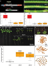
Identification and primary analysis of βknl2 mutant. (A) Schematic representation of the T-DNA insertion position in the genomic fragment and protein with the position of the SANTA domain. (B) Representative siliques with red arrowheads showing abnormal whitish glossy-seed phenotype from heterozygous βknl2-1 and βknl2-2 plants. (C,D) Boxplots depicting the number of abnormal seeds per silique data from the reciprocal crossing of WT and heterozygous βknl2-1 and βknl2-2 (***P ≤ 0.001). (E) Two weeks old in vitro germinated seedlings from Col-0, βknl2-1, and βknl2-2 heterozygous (+/−) and homozygous mutants (−/−). (F) βknl2 homozygous (−/−) and heterozygous (+/−) mutants on soil, homozygous mutants turning yellow in the red circle. (G–I) Representative dry seeds of Col-0, βknl2-1, and βknl2-2. Red arrowheads indicate the abnormal seeds. (J) Boxplot depicting the significant increase of abnormal dry seeds per silique of heterozygous βknl2-1 and βknl2-2 compared with WT as control.
Fig. 8.
Fig. 8.
Analysis of single siliques for seeds germination and presence of abnormal seedlings. (A) Two-week-old in vitro germinated seeds collected from single siliques of WT as control and heterozygous self-pollinated βknl2-1 and βknl2-2 plants. βknl2 homozygous seedlings are indicated by red circles. Bars: 1 cm. (B) Boxplot depicting the significant decrease of germination percentage per silique of heterozygous βknl2-1 and βknl2-2 compared with WT as control (*P ≤ 0.05, ***P ≤ 0.001). (C) Boxplot depicting the significant increase of abnormal seedlings (red color circled seedlings in (A) germinated from single silique seeds of heterozygous βknl2-1 and βknl2-2 compared with WT as control (**P ≤ 0.01), ***P ≤ 0.001). (D) RT-PCR amplification of βKNL2 from βknl2-1 and βknl2-2 homozygous null mutants and WT as the positive control with βKNL2 (EMB1674) gene-specific primers and EF1B primers as housekeeping gene.
Fig. 9.
Fig. 9.
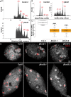
Reduced CENH3 levels in βknl2 null mutants leading to endoreduplication. (A) Representative ploidy analysis histogram of normal (green) seeds of heterozygous βknl2 mutants and WT as control (upper panel) and white abnormal seeds from βknl2 heterozygous mutants (lower panel). (B) Representative ploidy analysis histogram of WT seedlings as control (left panel) and abnormal seedlings of βknl2 null mutants (right panel). (C) Boxplot showing a significant decrease in the number of centromeric CENH3 signals in βknl2-1 and βknl2-2 compared with WT as a control (***P ≤ 0.001). (D) Super-resolution microscopy images showing nuclei of WT and βknl2 null mutants immune-stained with anti-CENH3 antibodies in meristematic cells (top) and differentiated cells (bottom).

Similar articles

Cited by

References

    1. Ahmadli U, Kalidass M, Khaitova LC, Fuchs J, Cuacos M, Demidov D, Zuo S, Pecinkova J, Mascher M, Heckmann S, et al. 2022a. High temperature increases centromere-mediated genome elimination frequency in Arabidopsis deficient in cenH3 or its assembly factor KNL2. BioRxive. - PMC - PubMed
    1. Ahmadli U, Sandmann M, Fuchs J, Lermontova I. 2022b. Immunolabeling of nuclei/chromosomes in Arabidopsis thaliana. In: Caillaud MC, editor. Plant cell division. Methods in molecular biology, vol. 2382. New York: (NY: ): Humana. p. 19–28. - PubMed
    1. Bailey TL, Boden M, Buske FA, Frith M, Grant CE, Clementi L, Ren JY, Li WW, Noble WS. 2009. MEME SUITE: tools for motif discovery and searching. Nucleic Acids Res. 37(2):W202–W208. - PMC - PubMed
    1. Bailey TL, Gribskov M. 1998. Combining evidence using p-values: application to sequence homology searches. Bioinformatics. 14(1):48–54. - PubMed
    1. Banks JA, Nishiyama T, Hasebe M, Bowman JL, Gribskov M, dePamphilis C, Albert VA, Aono N, Aoyama T, Ambrose BA, et al. 2011. The Selaginella genome identifies genetic changes associated with the evolution of vascular plants. Science. 332(6032):960–963. - PMC - PubMed