. 2022 Jun 14;119(24):e2201862119.
doi: 10.1073/pnas.2201862119. Epub 2022 Jun 7.

Hepatitis E virus infects brain microvascular endothelial cells, crosses the blood-brain barrier, and invades the central nervous system

Affiliations

Hepatitis E virus infects brain microvascular endothelial cells, crosses the blood-brain barrier, and invades the central nervous system

Debin Tian et al. Proc Natl Acad Sci U S A. .

Abstract

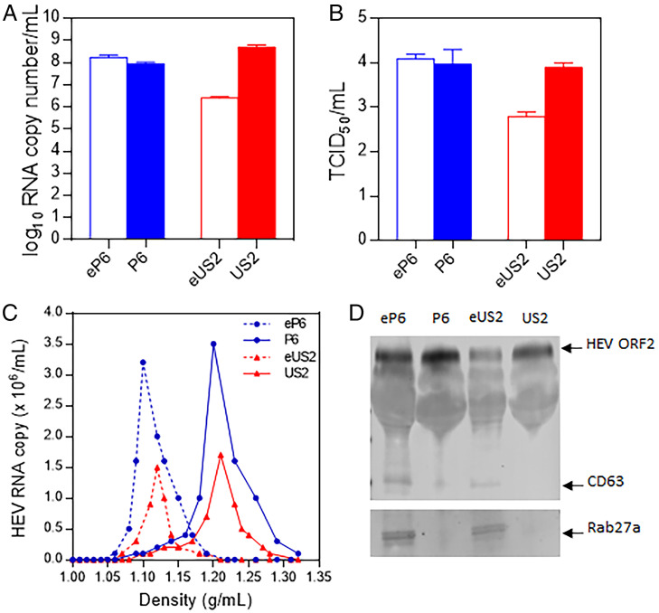
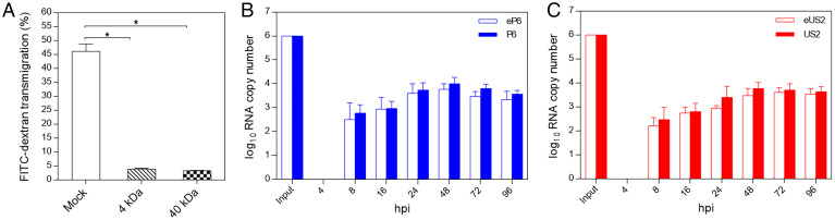
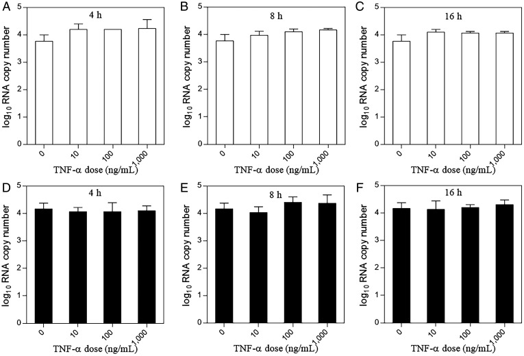
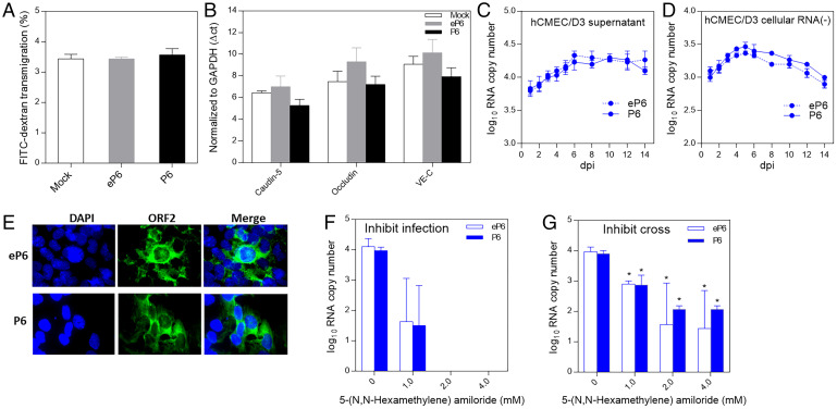
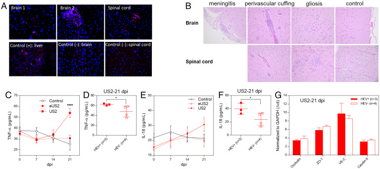
Hepatitis E virus (HEV) is an important but understudied zoonotic virus causing both acute and chronic viral hepatitis. A proportion of HEV-infected individuals also developed neurological diseases such as Guillain-Barré syndrome, neuralgic amyotrophy, encephalitis, and myelitis, although the mechanism remains unknown. In this study, by using an in vitro blood-brain barrier (BBB) model, we first investigated whether HEV can cross the BBB and whether the quasi-enveloped HEV virions are more permissible to the BBB than the nonenveloped virions. We found that both quasi-enveloped and nonenveloped HEVs can similarly cross the BBB and that addition of proinflammatory cytokine tumor necrosis factor alpha (TNF-α) has no significant effect on the ability of HEV to cross the BBB in vitro. To explore the possible mechanism of HEV entry across the BBB, we tested the susceptibility of human brain microvascular endothelial cells lining the BBB to HEV infection and showed that brain microvascular endothelial cells support productive HEV infection. To further confirm the in vitro observation, we conducted an experimental HEV infection study in pigs and showed that both quasi-enveloped and nonenveloped HEVs invade the central nervous system (CNS) in pigs, as HEV RNA was detected in the brain and spinal cord of infected pigs. The HEV-infected pigs with detectable viral RNA in CNS tissues had histological lesions in brain and spinal cord and significantly higher levels of proinflammatory cytokines TNF-α and interleukin 18 than the HEV-infected pigs without detectable viral RNA in CNS tissues. The findings suggest a potential mechanism of HEV-associated neuroinvasion.

Keywords: blood–brain barrier (BBB); brain microvascular endothelial cells; central nervous system (CNS); hepatitis E virus (HEV); neurological disorder.


Conflict of interest statement

The authors declare no competing interest.

Figures

Fig. 1.
Fig. 1.
Generation of membrane-associated quasi-enveloped and nonenveloped HEV virions from two different strains of genotype 3 HEV. In vitro-transcribed full-length genomic RNA from the genotype 3 HEV (strain Kernow-C1/P6) infectious clone was transfected into Huh-7 S10-3 liver cells. The culture supernatants of the transfected cells were collected and concentrated via ultracentrifugation as the virus stock of the quasi-enveloped eP6 virus. The eP6 virus was treated by detergents to produce the nonenveloped P6 virus stock. The 10% fecal suspension from a pig experimentally infected with HEV (strain US2) was purified via centrifugation and filtration to produce the nonenveloped US2 virus stock. The US2 virus was then used to infect HepG2 liver cells to generate the quasi-enveloped eUS2 virus stock. The virus stocks produced in this study were quantified by qRT-PCR to determine the genomic RNA copy numbers (A) and the infectious virus titer (TCID50 per milliliter) in Huh-7 S10-3 cells (B). (C) Sucrose density gradient fractionation of the virus stocks produced in this study. (D) The virus stocks were analyzed via Western blot analyses using antibodies against HEV ORF2 protein and exosome markers (CD63 and Rab27a).
Fig. 2.
Fig. 2.
Establishment of in vitro BBB culture model and demonstration of the ability of quasi-enveloped and nonenveloped HEV virions to cross the BBB. (A) The human brain microvascular endothelial cells (hCMEC/D3) were cultured in collagen-coated 0.4-μm PTFE membrane transwell inserts with growth medium for 2 d and then replaced with astrocyte-conditioned media for TJ formation and cultured for 3 d. The FITC-dextran (4 kDa and 40 kDa) were used to test the permeability of the established BBB. Bare PTFE membrane transwell inserts under the same condition were used as the mock control. Each assay was performed in triplicates. *P < 0.05, one way ANOVA for statistics analysis. (B) Genomic RNA copies (1.0 × 10⁶) of eP6 virus or P6 virus were added in the luminal space of the BBB transwell inserts. The culture medium in the abluminal space of the BBB were collected at different time points for quantification of HEV RNAs by qRT-PCR. Each assay was performed in triplicates. (C) The quasi-enveloped eUS2 virus and the nonenveloped US2 virus were analyzed using procedures similar to those described in B.
Fig. 3.
Fig. 3.
HEV virions cross the in vitro BBB in a TNF-α–independent manner. The in vitro BBB model was established by growing human brain microvascular endothelial cells (hCMEC/D3) in collagen-coated 0.4-μm PTFE membrane transwell inserts as described in Materials and Methods. Prior to HEV infection, the cells were incubated with an increasing dose of TNF-α (0 to 1,000 ng/mL) for different times (4, 8, or 16 h). The cells were washed once with PBS, followed by inoculation with 1.0 × 10⁶ genomic RNA copies of the quasi-enveloped eP6 virus (AC) or with the nonenveloped P6 virus (DF) in the luminal space of the BBB transwell inserts. The medium in the abluminal space of the BBB was collected at 48 hpi for quantification of HEV RNAs. Each assay was performed in triplicates. One-way ANOVA statistics analysis was used.
Fig. 4.
Fig. 4.
HEV crosses the in vitro BBB probably via direct infection of brain microvascular endothelial cells. (A) Genomic RNA copies (1.0 × 10⁶) of quasi-enveloped eP6 virus or nonenveloped P6 virus or medium only as control were inoculated onto the luminal space of the BBB. At 48 h later, the permeability of the barrier was tested by a permeability assay using 40-kDa FITC-dextran. (B) The hCMEC/D3 cells were inoculated with 1.0 × 10⁶ genomic RNA copies of eP6 virus or P6 virus or medium only as control. At 48 h later, cellular RNAs were isolated and the mRNA levels of TJ proteins were quantified by qRT-PCR. The CT values were normalized to GAPDH. The hCMEC/D3 cells in a 12-well plate were inoculated with eP6 or P6 (1.0 × 10⁶) for 2 h. After washing twice, the cells were cultured with medium for 14 d. The culture supernatants (C) and cell monolayers (D) were collected at indicated time points and subjected to quantification of HEV RNA (C) and negative-strand HEV RNA (D) by qRT-PCR. (E) The hCMEC/D3 cells inoculated with the eP6 virus or P6 virus were stained by IFA using anti-HEV ORF2 antibody at 3 dpi. (F) Confluent hCMEC/D3 cells were preinoculated with different concentrations of 5-(N,N-hexamethylene) amiloride for 30 min. After washing, the cells were inoculated with 1.0 × 10⁶ genomic RNA copies of eP6 or P6 for 2 h and after washing incubated for 48 h. The amounts of HEV RNA in the culture supernatants were quantified by qRT-PCR. (G) The in vitro BBB culture inserts were treated with 5-(N,N-hexamethylene) amiloride, and then HEV virions were added as described in F. The amounts of virus crossed into the abluminal space were quantified by qRT-PCR. *P < 0.05, one-way ANOVA.
Fig. 5.
Fig. 5.
HEV invades the CNS tissues in HEV-infected pigs. Four-week-old HEV-negative SPF pigs were intravenously inoculated with the quasi-enveloped eUS2 virus or nonenveloped US2 or medium as control. Samples of brain and spinal cord were collected at 21 dpi. (A) The formalin-fixed tissues of brains and spinal cords were paraffin-embedded and in situ-hybridized with a fluorescent-labeled (red) HEV-specific probe (representative pictures showing the detection of HEV RNAs in CNS tissues by FISH are shown). (B) The tissues of brains and spinal cords were H&E-stained and paraffin-embedded. The histological lesions were evaluated, in a blind fashion, by a board-certified veterinary pathologist (T. L.). Representative histopathology pictures are shown. Weekly sera were collected from each infected pig and the serum levels of TNF-α (C) and IL-18 (E) were determined. Comparison of the serum levels of TNF-α (D) and IL-18 (F) between HEV-infected pigs with detectable HEV RNAs in brain tissues (n = 3) and infected pigs with no detectable HEV RNA in brain tissues (n = 4) at 21 dpi. (G) Total RNAs were isolated from the brain tissues of the US2-infected pigs at 21 dpi. The mRNA levels of the TJ proteins in brain tissues (occludin, ZO-1, VE-C, and caudin-5) were quantified by qRT-PCR. *P < 0.05, ****P < 0.0001, one-way ANOVA.

Similar articles

Cited by

References

    1. Wang B., Meng X. J., Hepatitis E virus: Host tropism and zoonotic infection. Curr. Opin. Microbiol. 59, 8–15 (2021). - PMC - PubMed
    1. Nimgaonkar I., Ding Q., Schwartz R. E., Ploss A., Hepatitis E virus: Advances and challenges. Nat. Rev. Gastroenterol. Hepatol. 15, 96–110 (2018). - PMC - PubMed
    1. Rein D. B., Stevens G. A., Theaker J., Wittenborn J. S., Wiersma S. T., The global burden of hepatitis E virus genotypes 1 and 2 in 2005. Hepatology 55, 988–997 (2012). - PubMed
    1. Geng Y., et al. , High seroprevalence of hepatitis E virus in rabbit slaughterhouse workers. Transbound. Emerg. Dis. 66, 1085–1089 (2019). - PubMed
    1. Nan Y., Wu C., Zhao Q., Zhou E. M., Zoonotic Hepatitis E., Zoonotic hepatitis E virus: An ignored risk for public health. Front. Microbiol. 8, 2396 (2017). - PMC - PubMed

Publication types

LinkOut - more resources