Review
. 2022 Sep 29;9(1):213-238.
doi: 10.1146/annurev-virology-100120-012345. Epub 2022 Jun 7.

The Role of Viral RNA Degrading Factors in Shutoff of Host Gene Expression

Affiliations
Review

The Role of Viral RNA Degrading Factors in Shutoff of Host Gene Expression

Léa Gaucherand et al. Annu Rev Virol. .

Abstract

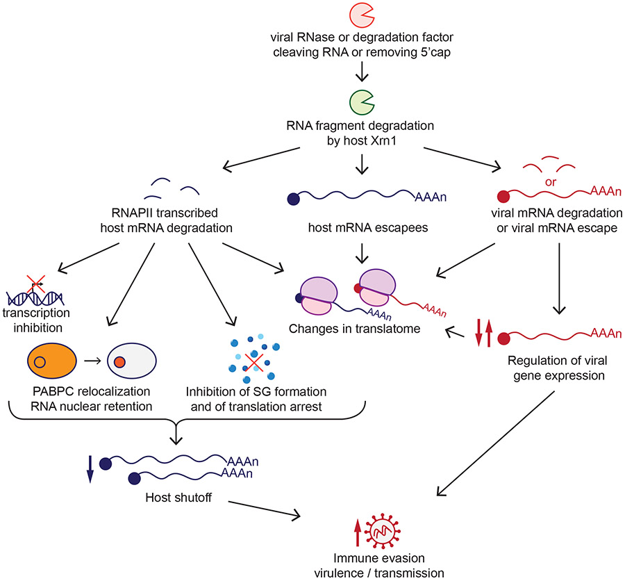
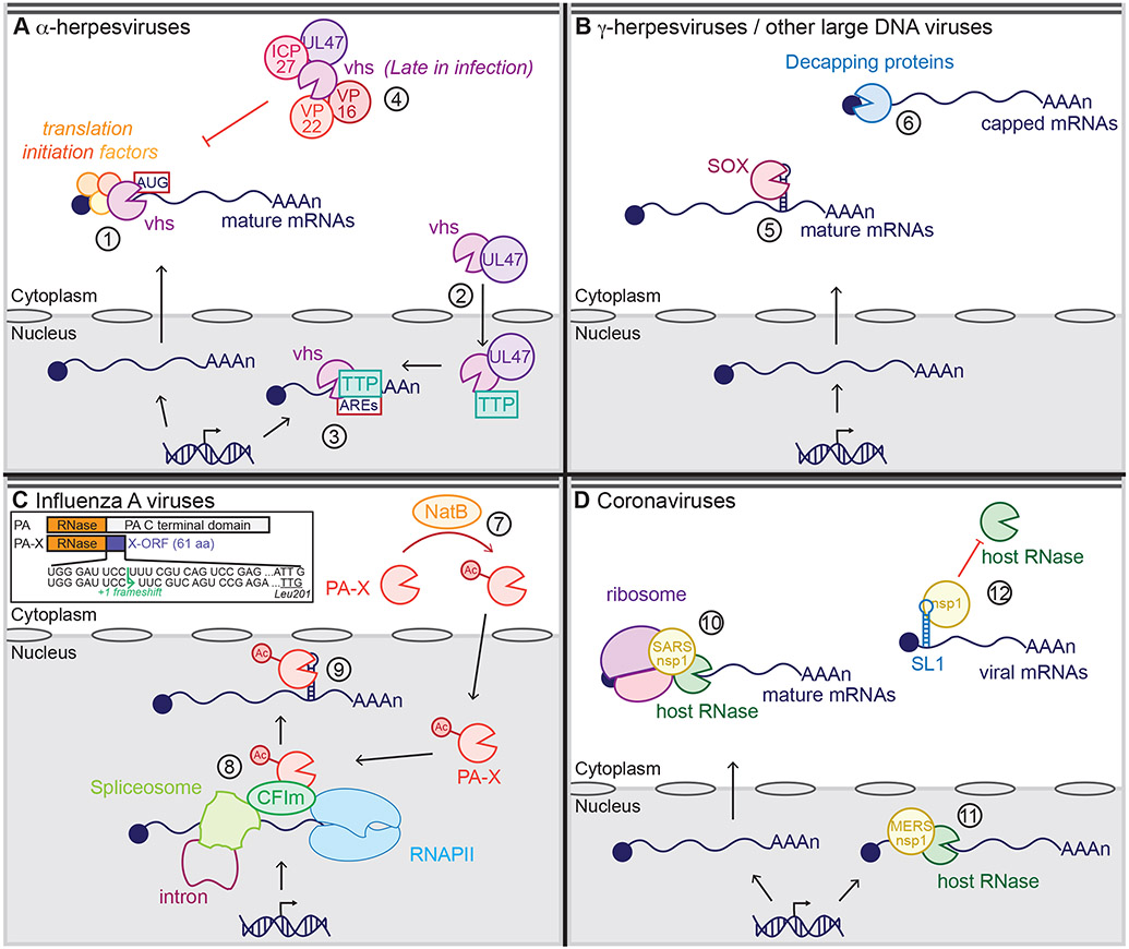
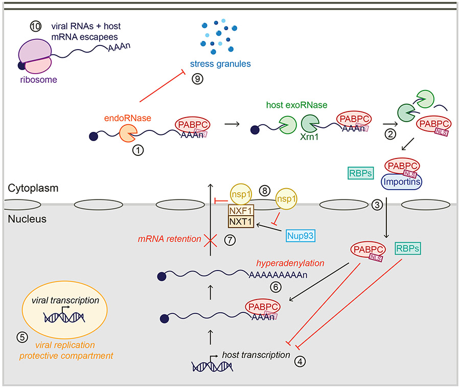
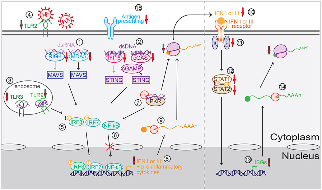
Many viruses induce shutoff of host gene expression (host shutoff) as a strategy to take over cellular machinery and evade host immunity. Without host shutoff activity, these viruses generally replicate poorly in vivo, attesting to the importance of this antiviral strategy. In this review, we discuss one particularly advantageous way for viruses to induce host shutoff: triggering widespread host messenger RNA (mRNA) decay. Viruses can trigger increased mRNA destruction either directly, by encoding RNA cleaving or decapping enzymes, or indirectly, by activating cellular RNA degradation pathways. We review what is known about the mechanism of action of several viral RNA degradation factors. We then discuss the consequences of widespread RNA degradation on host gene expression and on the mechanisms of immune evasion, highlighting open questions. Answering these questions is critical to understanding how viral RNA degradation factors regulate host gene expression and how this process helps viruses evade host responses and replicate.

Keywords: RNA decay; decapping enzymes; host shutoff; ribonuclease; virus.


Figures

Figure 1
Figure 1
Role of viral RNA degradation factors in host shutoff and the viral infectious cycle. Viral ribonuclease (RNase) or RNA degradation factors degrade host RNAs transcribed by RNA polymerase II (RNAPII). RNA degradation can then lead to host transcription inhibition, nuclear localization of the poly(A)-binding protein (PABPC) and nuclear retention of RNAs, inhibition of stress granule (SG) formation, and changes in the RNAs that are translated by the cell (translatome). The escape of some host messenger RNAs (mRNAs) from degradation and the presence of viral mRNAs also contribute to changes to the translatome. Viral mRNAs in some cases escape degradation, while in other viruses the degradation of viral mRNAs is integrated into the viral replication cycle as a mechanism of viral gene regulation. Overall, all these processes allow the virus to evade host immunity, affecting its virulence and transmission.
Figure 2
Figure 2
Current models of the mechanisms of action of viral RNA degradation factors. (a) α-Herpesviruses and virion host shutoff (vhs): vhs binds translation initiation factors in the cytoplasm to access its target RNAs, then cleaves capped RNAs or RNAs containing an internal ribosome entry site (IRES) close to the translation start site (#1). The viral protein pUL47 also shuttles vhs to the nucleus (#2), where it binds the host protein tristetraprolin (TTP) to cleave short-lived messenger RNAs (mRNAs) containing AU-rich elements (AREs) (#3). Late in infection, vhs activity is dampened through interactions with the virion proteins (VPs) VP16, VP22, pUL47, and infected cell protein (ICP) 27 (#4). (b) γ-Herpesviruses and SOX; large DNA viruses and decapping proteins: SOX recognizes and cleaves a specific sequence and structure within cytoplasmic mRNAs associated with translation complexes (#5). The decapping proteins African swine fever virus (ASFV)-DP and L357 bind to the RNA to locate its 5′ cap, while D9 and D10 bind to both the RNA and the 5′ cap to locate the cap and cleave it (#6). (c) Influenza A viruses and polymerase acidic (PA)-X: The cellular N-terminal acetylase (Nat) complex NatB protein N-terminally acetylates PA-X (#7), leading to an active PA-X, which accumulates in the nucleus, where it binds the cleavage factor Im (CFIm) complex and RNA processing factors to access its target spliced RNAs transcribed by RNA polymerase II (RNAPII) (#8). PA-X then recognizes and cleaves a specific sequence and structure (#9). The inset shows the mechanism of PA-X production via ribosomal frameshifting during translation of the PA mRNA. PA-X thus has the same N terminal ribonuclease (RNase) domain as PA, but a unique C terminal domain termed the X-ORF. Panel c inset adapted from Reference (47). (d) Coronaviruses and nonstructural protein 1 (nsp1): Severe acute respiratory syndrome coronavirus (SARS-CoV) and SARS-CoV-2 nsp1 bind to the 40S ribosome subunit to target capped or IRES-containing mRNAs that are actively translated (#10), inducing RNA degradation by an unknown host RNase in the cytoplasm. Middle East respiratory syndrome coronavirus (MERS-CoV) nsp1 also induces RNA degradation, but within the nucleus and without binding to the ribosome (#11). SARS and MERS-CoV nsp1 bind to stem loop 1 (SL1) within the 5′ untranslated region (UTR) of viral mRNAs, leading to viral mRNA protection from degradation (#12).
Figure 3
Figure 3
Consequences of widespread RNA degradation by viral host shutoff factors on cellular and viral RNA metabolism. Poly(A)-binding protein (PABPC) usually binds RNAs in the cytoplasm. However, upon endonucleolytic cleavage of RNAs by viral ribonucleases RNases or the host RNase activated by nonstructural protein 1nsp1 (endoRNase) (#1) and subsequent RNA fragment degradation by Xrn1 and other host exoribonucleases (exoRNase) (#2), PABPC is released from the RNA, exposing its nuclear localization signal (NLS). PABPC thus interacts with nuclear import proteins and is shuttled to the nucleus (#3), along with other RNA-binding proteins (RBPs) that are also no longer RNA bound. In the nucleus, these proteins inhibit host transcription (#4), while viral transcription continues, likely in protective compartments (#5). PABPC accumulation in the nucleus leads to hyperadenylation (#6) and nuclear mRNA retention (#7). nsp1 also induces nuclear retention of RNAs independently of PABPC by preventing localization of Nup93 to the nuclear pore and interfering with the function of the NXF1-NXT1 complex (#8). In parallel, viral RNases prevent formation of stress granules (SGs) (#9), allowing translation of the viral mRNAs and host mRNAs that escape host shutoff (#10).
Figure 4
Figure 4
Mechanisms of immune evasion by viral RNA degradation factors. Viral RNA degradation factors can block viral sensing by downregulating the levels of the double-stranded RNA (dsRNA) sensors retinoic acid-inducible gene-I (RIG-I) and melanoma differentiation-associated gene 5 (MDA5) (#1) that signal through mitochondrial antiviral-signaling protein (MAVS), the double-stranded DNA (dsDNA) sensors interferon gamma-inducible protein 16 (IFI16) and cyclic GMP-AMP (cGAMP) synthase (cGAS) (#2) that signal through stimulator of interferon genes (STING), the endosomal RNA and DNA sensors Toll-like receptor (TLR) TLR3 and TLR9 (#3), and the surface receptor TLR2 (#4). These sensors signal to activate and translocate transcription factors nuclear factor-kappa B (NF-κB), interferon regulatory factor (IRF) 3 and IRF7 to the nucleus, although phosphorylation (represented by the yellow P) of IRF3 (#5) and translocation of NF-κB can also be blocked by some RNA degradation factors (#6). RNA degradation also dampens viral sensing through the RNA sensor protein kinase R (PKR) (#7), which activates NF-κB and inhibits translation. As a result of these signaling changes, transcription of messenger RNAs (mRNAs) for interferons (IFNs) and other proinflammatory cytokines is decreased (#8). Also, their mRNAs are degraded (#9), leading to less cytokine secretion (#10). Secreted type I and III IFNs are normally sensed by the IFN I and III receptors, respectively, which activate tyrosine kinase 2 (TYK2) and Janus kinase (JAK). Viral RNA degradation factors also decrease signaling downstream of the IFN receptors by downregulating the levels of TYK2 (#11) and signal transducer and activator of transcription (STAT) 1 and STAT2 proteins (#12), orinhibiting phosphorylation of STAT1 (#12). This leads to a decrease in IFN-stimulated gene (ISG) transcription (#13), in addition to direct degradation of ISG mRNAs (#14), which ultimately reduce ISG translation. Viral RNA degradation factors can also contribute to evasion of the adaptive immune response by downregulating antigen-presenting molecules (#15).

Similar articles

Cited by

References

    1. Minskaia E, Hertzig T, Gorbalenya AE, Campanacci V, Cambillau C, et al. 2006. Discovery of an RNA virus 3’->5’ exoribonuclease that is critically involved in coronavirus RNA synthesis. Proc Natl Acad Sci U S A. 103(13):5108–13 - PMC - PubMed
    1. Hackbart M, Deng X, Baker SC. 2020. Coronavirus endoribonuclease targets viral polyuridine sequences to evade activating host sensors. Proc Natl Acad Sci US A. 117(14):8094–8103 - PMC - PubMed
    1. Decroly E, Ferron F, Lescar J, Canard B. 2012. Conventional and unconventional mechanisms for capping viral mRNA. Nat Rev Microbiol. 10(1):51–65 - PMC - PubMed
    1. Gaglia MM, Covarrubias S, Wong W, Glaunsinger BA. 2012. A Common Strategy for Host RNA Degradation by Divergent Viruses. Journal of Virology. 86(17):9527–30 - PMC - PubMed
    1. Khaperskyy DA, Schmaling S, Larkins-Ford J, McCormick C, Gaglia MM. 2016. Selective Degradation of Host RNA Polymerase II Transcripts by Influenza A Virus PA-X Host Shutoff Protein. PLoS Pathog. 12(2):e1005427. - PMC - PubMed

Publication types

LinkOut - more resources