Review
. 2022 Jul;298(7):102105.
doi: 10.1016/j.jbc.2022.102105. Epub 2022 Jun 6.

3D cryo-EM imaging of bacterial flagella: Novel structural and mechanistic insights into cell motility

Affiliations
Review

3D cryo-EM imaging of bacterial flagella: Novel structural and mechanistic insights into cell motility

Sonia Mondino et al. J Biol Chem. 2022 Jul.

Abstract

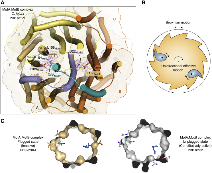
Bacterial flagella are nanomachines that enable cells to move at high speeds. Comprising 25 and more different types of proteins, the flagellum is a large supramolecular assembly organized into three widely conserved substructures: a basal body including the rotary motor, a connecting hook, and a long filament. The whole flagellum from Escherichia coli weighs ∼20 MDa, without considering its filament portion, which is by itself a ∼1.6 GDa structure arranged as a multimer of ∼30,000 flagellin protomers. Breakthroughs regarding flagellar structure and function have been achieved in the last few years, mainly because of the revolutionary improvements in 3D cryo-EM methods. This review discusses novel structures and mechanistic insights derived from such high-resolution studies, advancing our understanding of each one of the three major flagellar segments. The rotation mechanism of the motor has been unveiled with unprecedented detail, showing a two-cogwheel machine propelled by a Brownian ratchet device. In addition, by imaging the flagellin-like protomers that make up the hook in its native bent configuration, their unexpected conformational plasticity challenges the paradigm of a two-state conformational rearrangement mechanism for flagellin-fold proteins. Finally, imaging of the filaments of periplasmic flagella, which endow Spirochete bacteria with their singular motility style, uncovered a strikingly asymmetric protein sheath that coats the flagellin core, challenging the view of filaments as simple homopolymeric structures that work as freely whirling whips. Further research will shed more light on the functional details of this amazing nanomachine, but our current understanding has definitely come a long way.

Keywords: Spirochetes; allosteric regulation; cell motility; molecular motor; protein self-assembly; protein–protein interaction; proton motive force; proton transport; structural biology.


Conflict of interest statement

Conflict of interest The authors declare that they have no conflicts of interest with the contents of this article.

Figures

Figure 1
Figure 1
Molecular architecture of the bacterial flagellum. Cartoon representation of a complete bacterial flagellum, juxtaposing high-resolution experimental structures of subassemblies, with proper interconnecting geometries. All the components are drawn to scale. The left panel shows solid molecular surfaces for most of the appendage. The right panel is identical, except that outermost solvent-exposed protein complexes are rendered semitransparent, uncovering the inner composition of the protein export apparatus and rod. There are no structures of assembled hook-associated proteins FlgL and FlgK (only structures of individual protomers); they are thus represented by two schematic rings at the hook–filament junction. There is no deposited PDB for the FliG–FliM–FliN C ring assembly (only structures of protomers or subregion complexes), corresponding to the fitted model within the available cryo-ET reconstruction volumes (41), hence a schematic illustration is shown (adapted from Ref. (41)). PDB IDs used to build this drawing: 6SIH (FliD filament–capping complex from Campylobacter jejuni); 1UCU (FliC flagellin filament from Salmonella enterica); 6K3I (FlgE hook from S. enterica); 7CGO (FlgH L ring; FlgI P ring; FliF MS ring; the rod components FlgG, FlgF, FlgC, FlgB, and FliE; the export apparatus components FliP, FliQ, and FliR; all from S. enterica; note that the transmembrane and C ring–connecting regions of the MS ring were not well resolved in the original cryo-EM maps (110), hence depicted as a semitransparent red surface); 6S3L (FlhB, from the complex with FliPQR from Vibrio mimicus); 7AMY (FlhA ring, cytoplasmic region, from Vibrioparahaemolyticus; the FlhA transmembrane region is represented as a semitransparent violet volume, since it could not be solved (111)); 5B0O (part of the ATPase complex corresponding to the FliH–FliI complex from S. enterica; ATP-synthase 6OQR was used as template to generate the FliH–FliI hexameric assembly; FliH N-terminal extension arms are shown semitransparent going up toward the C ring, they were modeled with AlphaFold2 with high reliability); 3AJW (FliJ subunit of the ATPase complex from S. enterica); 6YKM (transmembrane portion of the MotA–MotB complex from Campylobacter jejuni); and 2ZVY (MotB PG-binding OmpA-like domain from S. enterica). ET, electron tomography; IM, inner membrane; OM, outer membrane; PDB, Protein Data Bank; PG, peptidoglycan.
Figure 2
Figure 2
The flagellar motor acts as a Brownian ratchet.A, cartoon depicting the cryo-EM structure of the stator MotA–MotB complex from Campylobacter jejuni in the “plugged” inactive state (PDB ID: 6YKM) (36). Toward the center, the MotB dimer is colored in blue, with different tones distinguishing the two monomers. The proton-transporter aspartate (D22) is labeled on each monomer and drawn as sticks. The pentamer of MotA chains, labeled from A to E, surrounds MotB. MotA monomers are distinguished in different tones of yellow-to-orange, and transparent surfaces with silhouettes also emphasize each MotA monomer’s position. MotA residues in contact with D22 are shown as sticks, together with their van der Waals surfaces displayed as dots. Note the distinct asymmetric environments that surround the proton transporter D22 in both MotB monomers. B, schematic drawing revisiting the mechanism of ratchet and pawl. Because of force applied to the pawl(s), they tend to remain close to the ratchet, whereas Brownian motion is induced in the entire system because of environmental thermal energy. The mechanism thus transduces the geometric asymmetry of the ratchet’s structure, into unidirectional rotation motion. C, simplified views of the MotA–MotB complex comparing two functional states, inactive (left) and active (right). The “plugged” inactive state is identical to A but simplified for clarity: MotB proteins are not shown except for the proton-carrier aspartates, and MotA is shown mostly as a molecular surface, with D22-contacting residues in stick representation. To the right, the constitutively active state—that is promoting proton transfer—was obtained by the authors using an “unplugged” N-term-truncated MotB mutant (PDB ID: 6YKP). Asymmetric features of MotA’s surface (“ratchet”) interfere with MotB’s aspartates (“pawls”) such that random motion of MotA only results in effective rotation in one direction. PDB, Protein Data Bank.
Figure 3
Figure 3
Basal bodies of Borrelia burgdorferi (endoflagellum) and Salmonella enterica (exoflagellum). Schematic illustrations highlighting the positions of different components of the basal body, drawn to scale for comparative purposes, based on currently available in situ cryo-electron tomographic data. Note the presence of the collar: a large, 79 nm wide, protein complex unique to Spirochetal endoflagella, with identified protein subunits indicated and additional components that remain to be localized. The rod is shorter in endoflagellates (17 nm in B. burgdorferi versus 25 nm in S. enterica), but the motor has a larger radius (54 nm versus 40 nm), key to generate the high torques observed in spirochetal endoflagella. IM, inner membrane; OM, outer membrane; PG, peptidoglycan.
Figure 4
Figure 4
FlgE protomers in the flagellar hook adopt a continuous array of conformations, one for each protofilament.A, 3D structure of the native hook from Salmonella enterica (PDB ID: 6K3I), viewed from two orthogonal perspectives. Each of the 11 protofilaments are distinguished with different colors, and the natural curvature of the assembly allows to identify outermost (convex surface) and innermost (concave surface) lines. FlgE protomers are most separated among them along the former and most tightly packed along the latter line. B, each protofilament exhibits a distinct conformation, contradicting a simpler two-state conformational rearrangement switch for flagellin-like protomers. To the left of the panel, one FlgE protomer from each of the 11 protofilaments is highlighted in strong colors, and to the bottom, they are superposed, showing a substantial and continuous rearrangement among them. By changing the perspective to the right, one protofilament is chosen (marked with an orange line through its trajectory), zooming in to confirm that all protomers within the protofilament are nearly identical in conformation. Note that the conformational rearrangement of FlgE protomers is highly cooperative (consolidating each protofilament) and happens fast and synchronously as the hook rotates. PDB, Protein Data Bank.
Figure 5
Figure 5
FlgE protomers in Spirochetes are covalently crosslinked in the assembled hook.A, autocatalytic cross-linking reaction at the D1–D2′ domain interface, generating a lysinoalanine covalent adduct. The reaction occurs between D1 residue Lys165 and Cys178 on the D2′ domain of an adjacent FlgE subunit in the assembly. The reaction involves three distinct biochemical steps: oligomerization of FlgE subunits via D0 interactions, β-elimination of the Cys178 thiol with the release of hydrogen sulfide as a byproduct, and aza-Michael addition of Lys165 from an adjacent FlgE monomer to yield lysinoalanine. B, structure of the resulting crosslinked FlgED1D2:D2 dimer (PDB ID: 6NDX). Note the lysinoalanine residue (blue dashed box) located at the interface between the D2 domain of one FlgE protomer (green) and the beginning of domain D1 of a neighboring protomer (gray), near the linker segment between domains D1 and D2. PDB, Protein Data Bank.
Figure 6
Figure 6
The flagellar filament.A, the protomers of the hook (FlgE), the filament capping complex (FliD), and the filament core itself (FliC flagellin) are shown as cartoons, with domains highlighted in different colors. Note that the three share a similar 3D architecture and domain organization (D0 to D3 as indicated), defining a flagellin-like fold. B, evolutionary constraints have selected filaments with increasing and variable complexity. A flagellin core comprising the two all-helical domains D0 and D1 is always present. Leftmost panel, a bare D0–D1 core composes the native filaments of, for example, Firmicutes including Bacillus subtilis (the filament from the related Firmicute Kurthia sp. is shown; PDB ID: 6T17). Center panel, in Enterobacteria, the flagellin protomers have two extra domains, D2 and D3, decorating the core and protruding as spikes from it, such as in Salmonella enterica (PDB ID: 1UCU, applying the reported helical symmetry to generate the ensemble). Rightmost panel, a case of extreme complexity is observed in Spirochetes, where a D0–D1 flagellin core is sheathed asymmetrically by several different protein species on either side of the curved appendage, illustrated by Leptospira biflexa (PDB ID: 6PWB; the sheath protein FlaA, and likely additional unannotated proteins, is not localized with certainty, yet preliminary data support their indicated position). PDB, Protein Data Bank.

References

    1. Turner L., Ryu William S., Berg Howard C. Real-time imaging of fluorescent flagellar filaments. J. Bacteriol. 2000;182:2793–2801. - PMC - PubMed
    1. Magariyama Y., Sugiyama S., Kudo S. Bacterial swimming speed and rotation rate of bundled flagella. FEMS Microbiol. Lett. 2001;199:125–129. - PubMed
    1. Magariyama Y., Sugiyama S., Muramoto K., Kawagishi I., Imae Y., Kudo S. Simultaneous measurement of bacterial flagellar rotation rate and swimming speed. Biophys. J. 1995;69:2154–2162. - PMC - PubMed
    1. Bente K., Mohammadinejad S., Charsooghi M.A., Bachmann F., Codutti A., Lefèvre C.T., et al. High-speed motility originates from cooperatively pushing and pulling flagella bundles in bilophotrichous bacteria. Elife. 2020;9 - PMC - PubMed
    1. Berg H.C. The rotary motor of bacterial flagella. Annu. Rev. Biochem. 2003;72:19–54. - PubMed

MeSH terms

LinkOut - more resources