. 2022 Aug:148:323-335.
doi: 10.1016/j.actbio.2022.05.051. Epub 2022 Jun 6.

Mesh deformation: A mechanism underlying polypropylene prolapse mesh complications in vivo

Affiliations

Mesh deformation: A mechanism underlying polypropylene prolapse mesh complications in vivo

Katrina M Knight et al. Acta Biomater. 2022 Aug.

Abstract

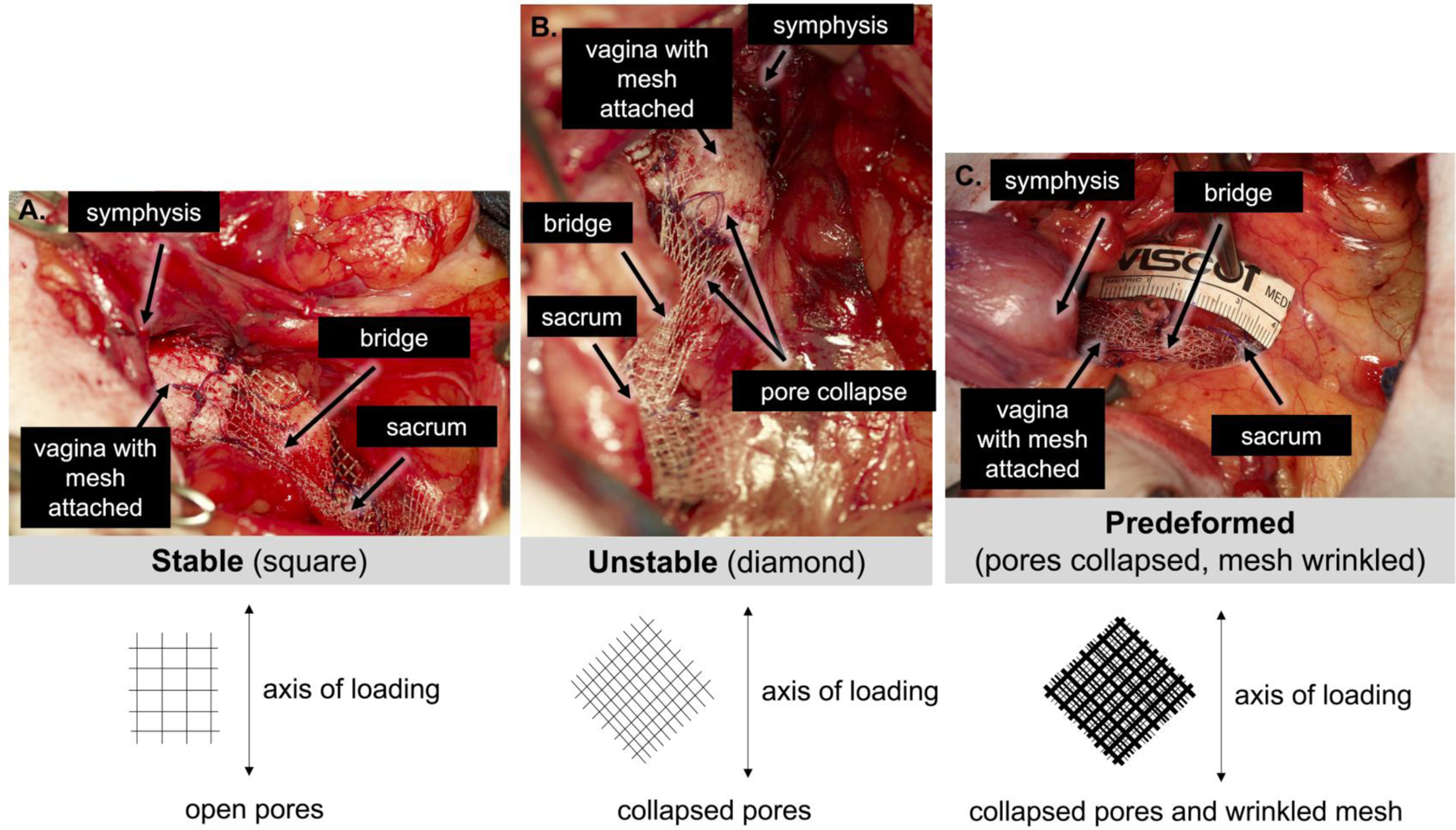
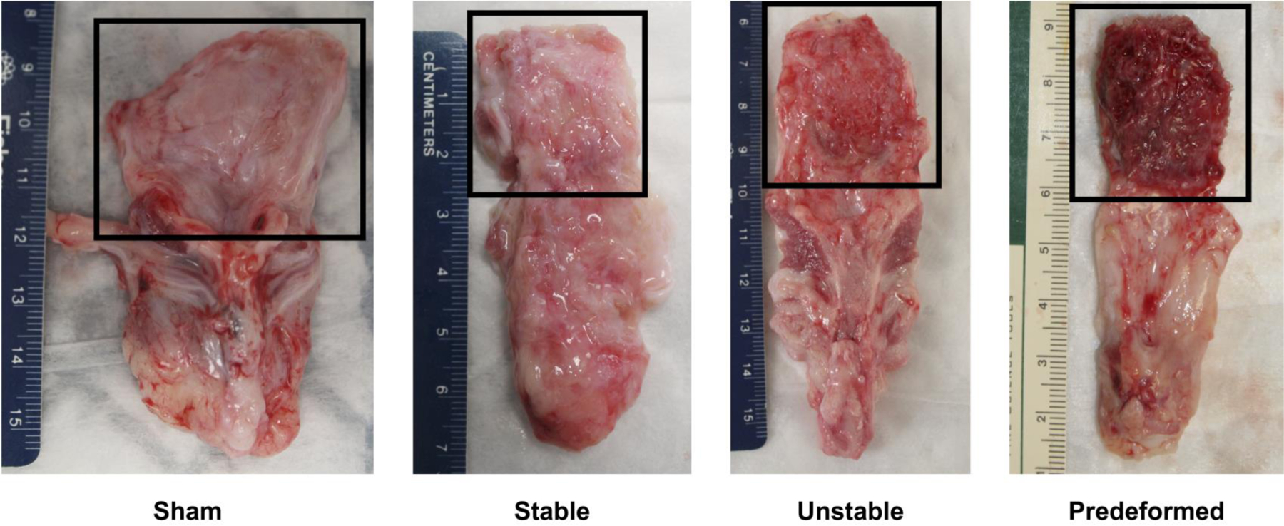
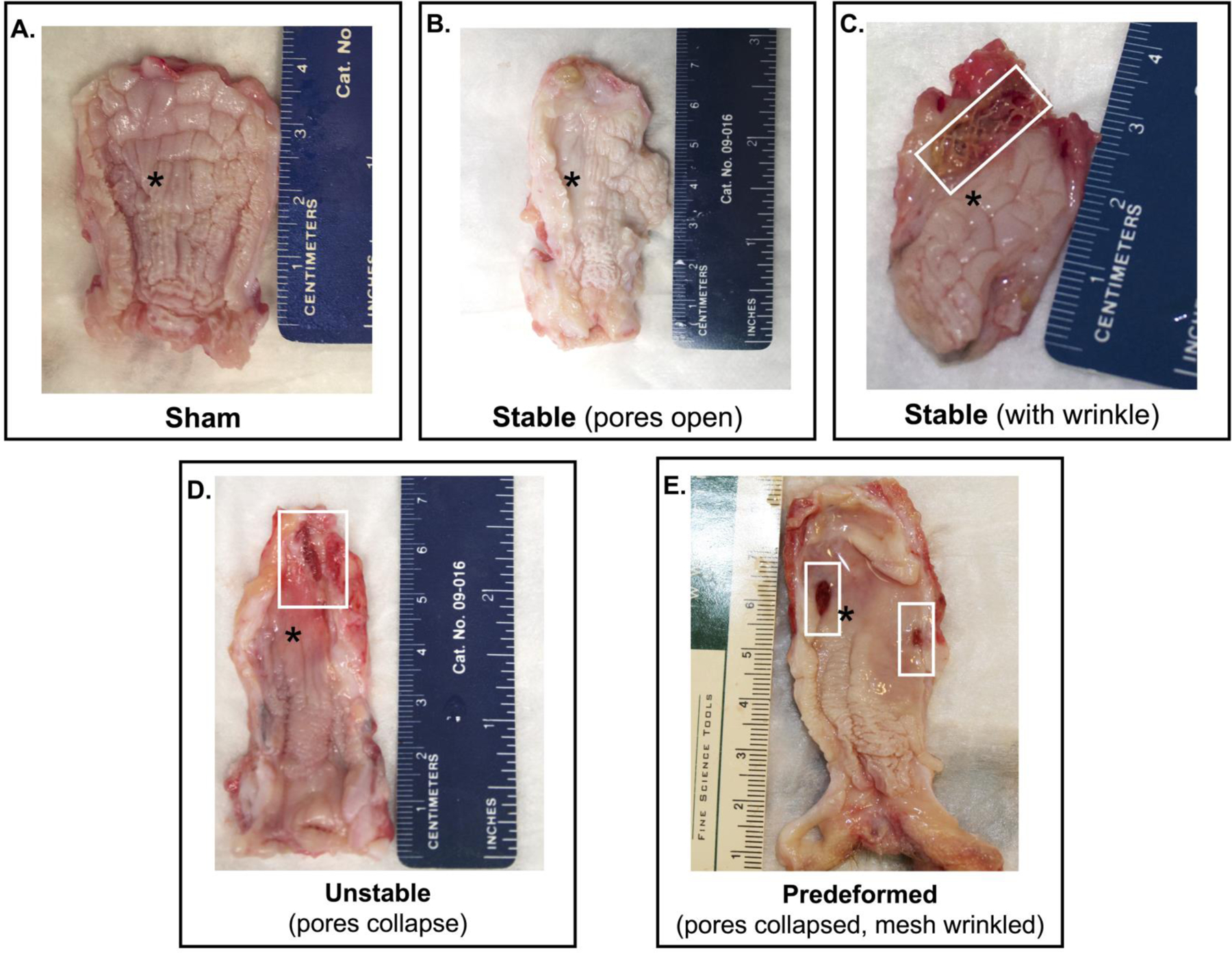
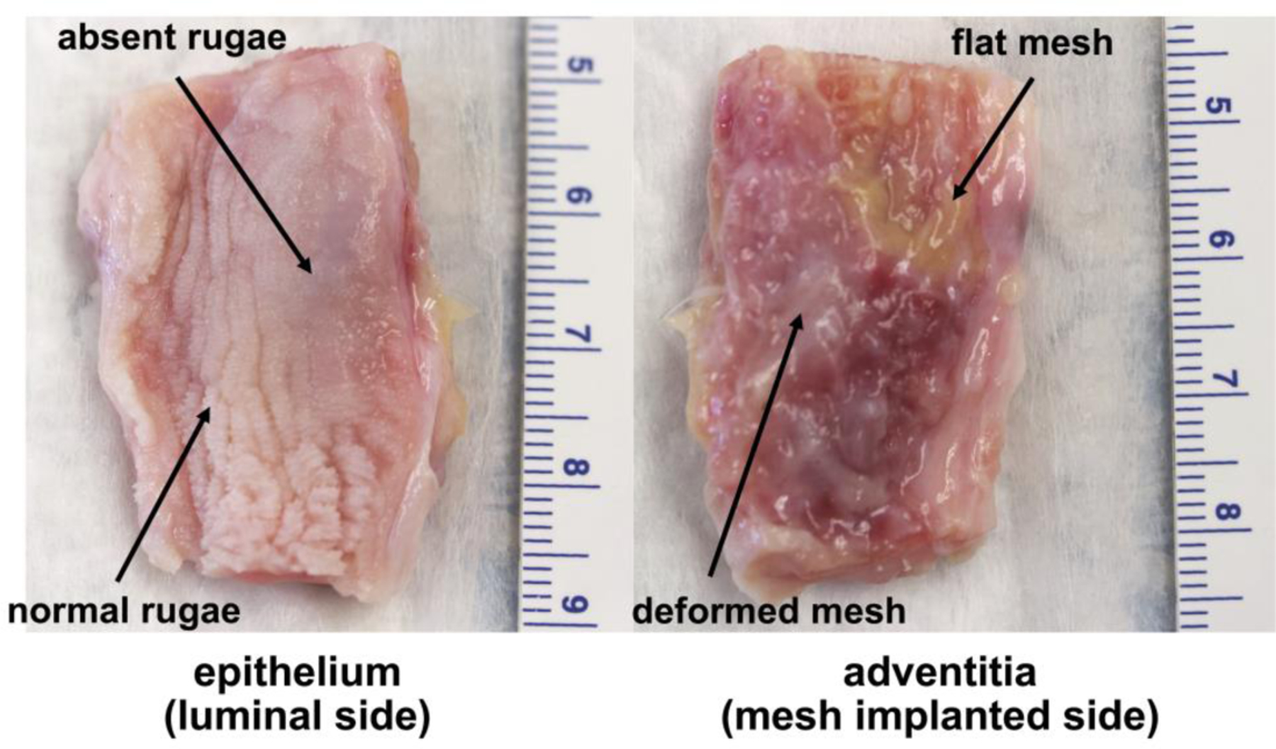
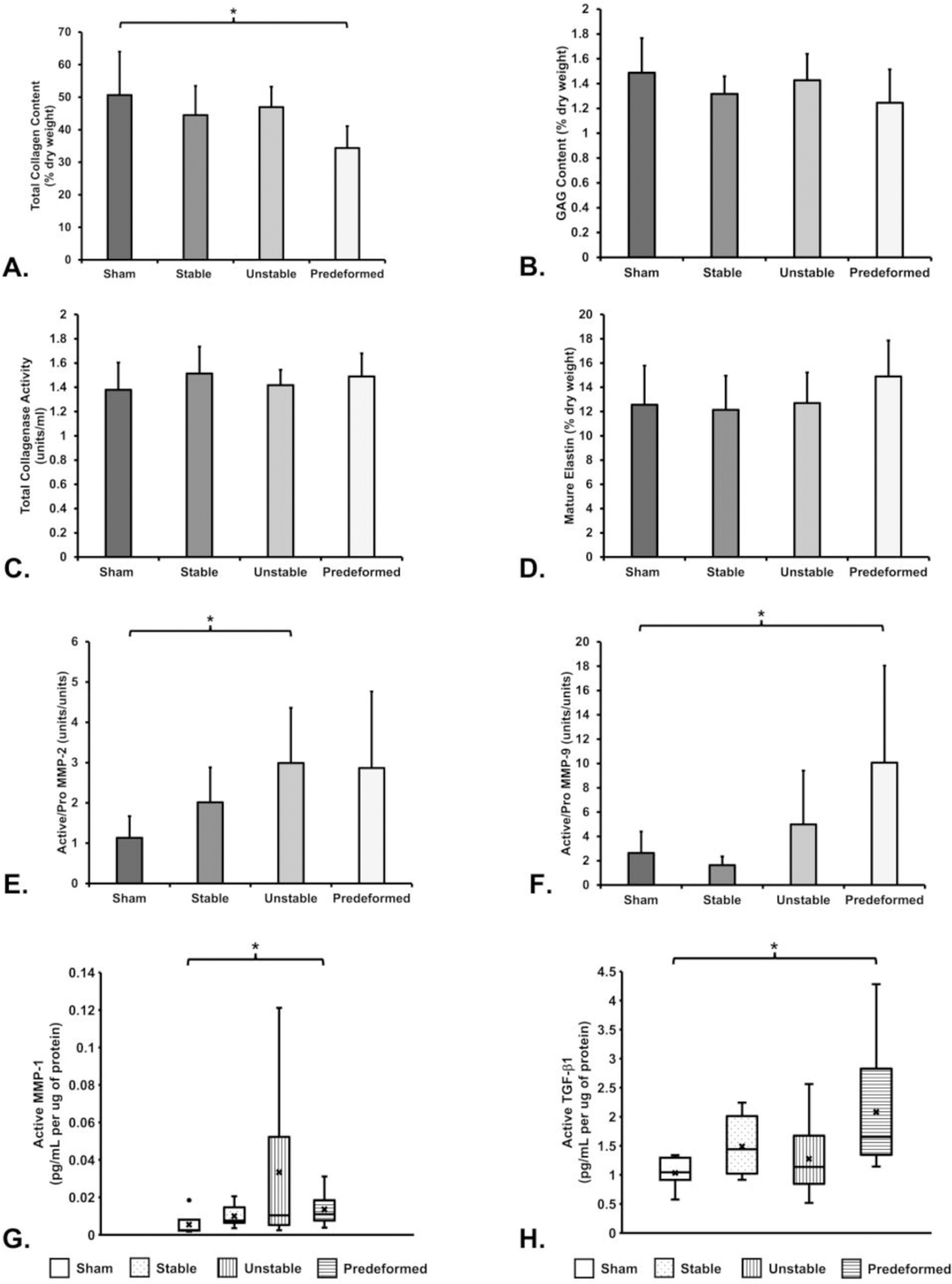
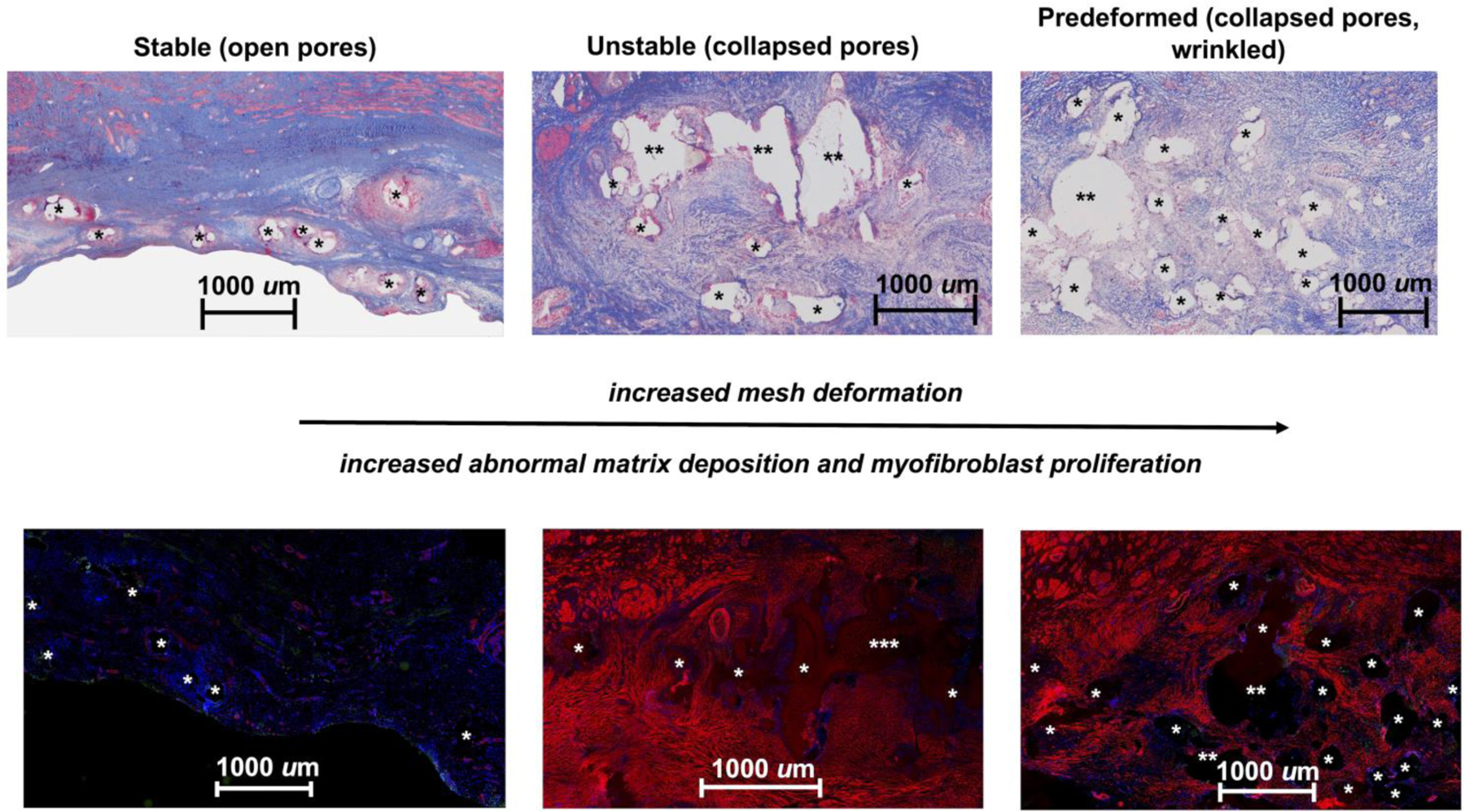
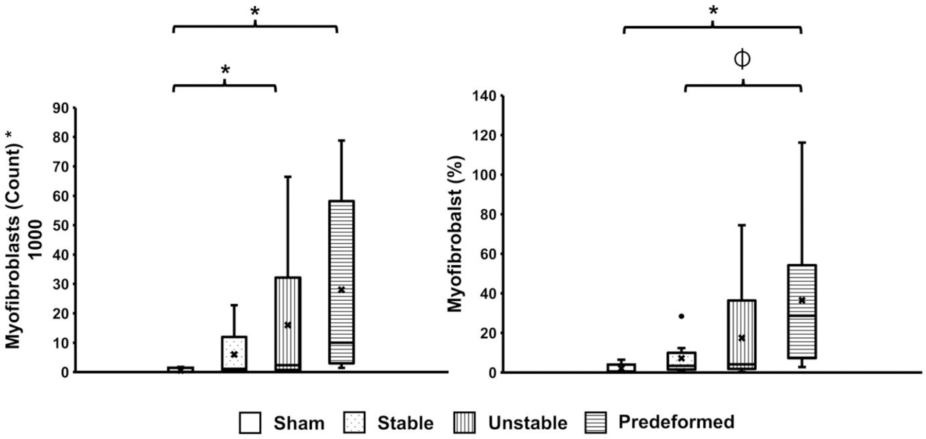
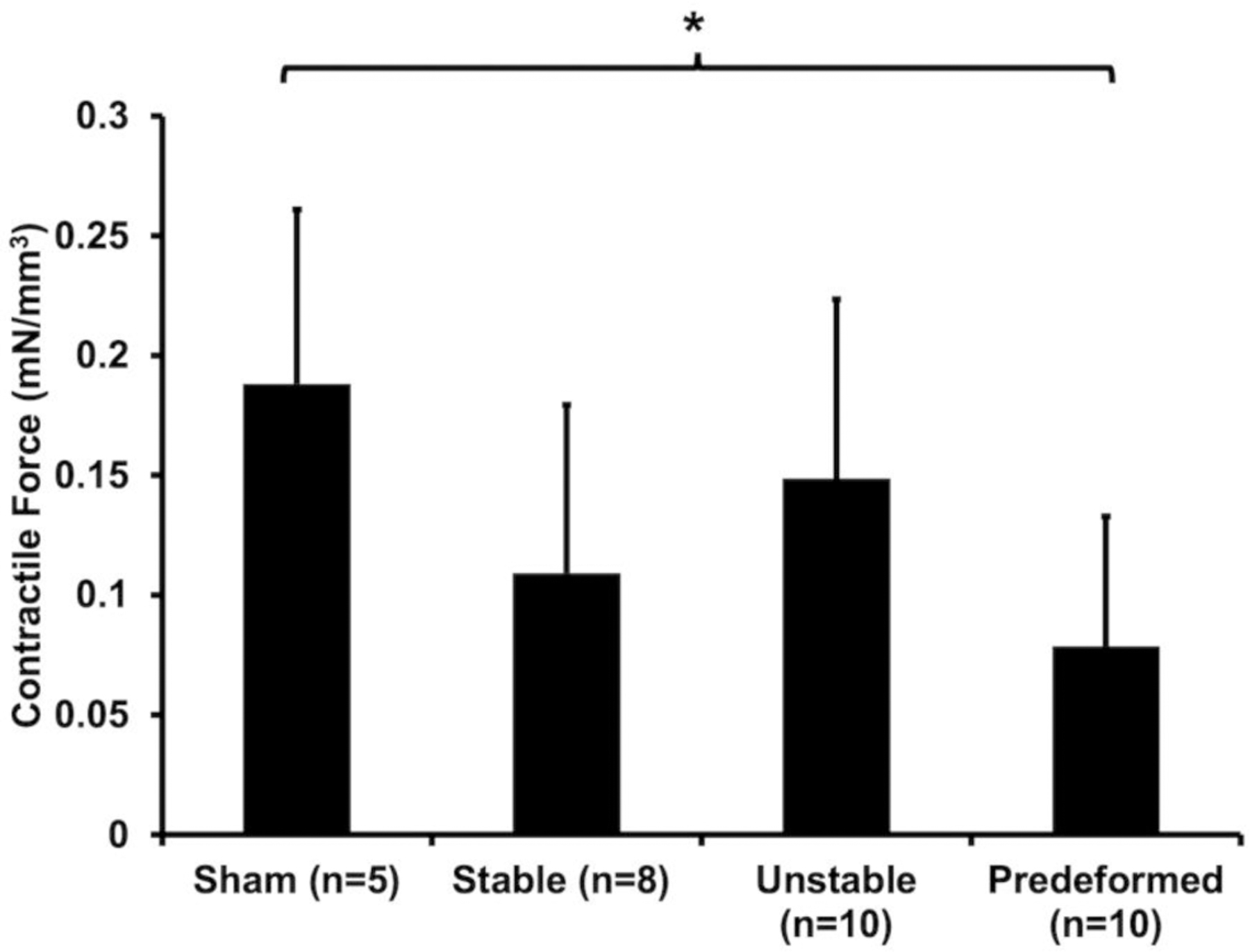
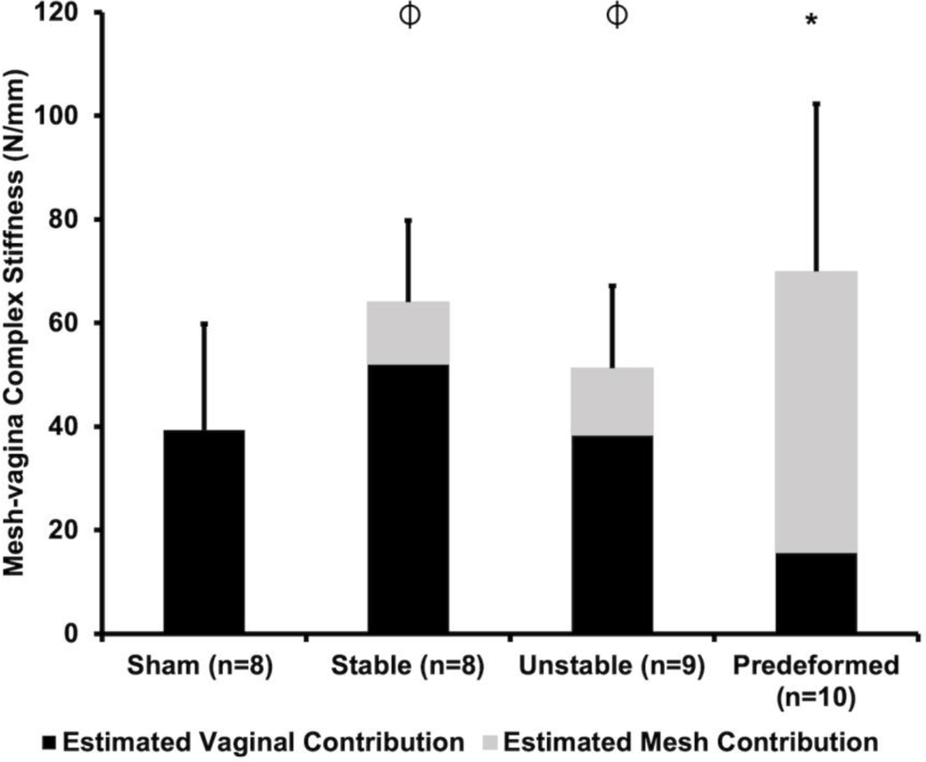
Polypropylene meshes used in pelvic organ prolapse (POP) repair are hampered by complications. Most POP meshes are highly unstable after tensioning ex vivo, as evidenced by marked deformations (pore collapse and wrinkling) that result in altered structural properties and material burden. By intentionally introducing collapsed pores and wrinkles into a mesh that normally has open pores and remains relatively flat after implantation, we reproduce mesh complications in vivo. To do this, meshes were implanted onto the vagina of rhesus macaques in nondeformed (flat) vs deformed (pore collapse +/- wrinkles) configurations and placed on tension. Twelve weeks later, animals with deformed meshes had two complications, (1) mesh exposure through the vaginal epithelium, and (2) myofibroblast proliferation with fibrosis - a mechanism of pain. The overarching response to deformed mesh was vaginal thinning associated with accelerated apoptosis, reduced collagen content, increased proteolysis, deterioration of mechanical integrity, and loss of contractile function consistent with stress shielding - a precursor to mesh exposure. Regional differences were observed, however, with some areas demonstrating myofibroblast proliferation and matrix deposition. Variable mechanical cues imposed by deformed meshes likely induce these two disparate responses. Utilizing meshes associated with uniform stresses on the vagina by remaining flat with open pores after tensioning is critical to improving outcomes. STATEMENT OF SIGNIFICANCE: Pain and exposure are the two most reported complications associated with the use of polypropylene mesh in urogynecologic procedures. Most meshes have unstable geometries as evidenced by pore collapse and wrinkling after tensioning ex vivo, recapitulating what is observed in meshes excised from women with complications in vivo. We demonstrate that collapsed pores and wrinkling result in two distinct responses (1) mesh exposure associated with tissue degradation and atrophy and (2) myofibroblast proliferation and matrix deposition consistent with fibrosis, a tissue response associated with pain. In conclusion, mesh deformation leads to areas of tissue degradation and myofibroblast proliferation, the likely mechanisms of mesh exposure and pain, respectively. These data corroborate that mesh implantation in a flat configuration with open pores is a critical factor for reducing complications in mesh-augmented surgeries.

Keywords: Biomechanical properties; Extracellular matrix; Stress shielding; Synthetic mesh complications; Vagina.


Conflict of interest statement

Declaration of Competing Interest The authors declare that they have no known competing financial interests or personal relationships that could have appeared to influence the work reported in this paper. We would like to draw the attention of the Editor to the following facts which may be considered as potential declaration of interests: Pamela A. Moalli reports equipment, drugs, or supplies was provided by Coloplast Corp. Katrina M. Knight reports financial support was provided by Magee-Womens Research Institute & Foundation. Pamela A. Moalli reports a relationship with Hologic Inc that includes: board membership. However, there has been no significant financial support for this work that could have influenced its outcome.

Figures

Figure 1:
Figure 1:. In vivo live images of Restorelle implanted in 3 different geometries
1) square pore, a Stable configuration (A), in which the pores stay open on the vagina with loading; 2) diamond pores, an Unstable configuration (B), in which the pores collapse, primarily in the mesh bridge to the sacrum and at the vaginal apex; 3) Predeformed, the most unstable configuration, achieved by allowing the pores to collapse and the mesh to wrinkle prior to implantation (C). Bottom schematics depict the orientation of the pores on the vagina with respect to the loading direction. Note: due to the 2D nature of the image, wrinkling of the mesh is not depicted in Predeformed schematic. Position of the pubic symphysis, vagina, mesh, mesh bridge to the sacrum, and sacrum are shown.
Figure 2:
Figure 2:. Mesh-vagina complex explants.
Exemplar mesh-vagina complex explants depicting the adventitial side of the vagina following sham surgery (no mesh) and the implantation of Stable, Unstable, and Predeformed. The grafted region is highlighted within the box. The mesh remained relatively flat with tissue incorporated within the mesh pores for the Stable and Unstable configurations whereas the mesh was wrinkled with poor quality tissue into the mesh for the Predeformed configuration.
Figure 3:
Figure 3:. Mesh deformations reproduce mesh complications.
The luminal or epithelial side of the vagina at 12 weeks after surgery demonstrates that 1) deformations successfully reproduced mesh exposures, and 2) the degree of disruption of normal vaginal structure increased with increased deformations. Mesh exposures were most prevalent in the Unstable and Predeformed groups. Proof of this principle is shown in C, a specimen in which a wrinkle was incidentally introduced due to loss of a fixation suture. In the area of the wrinkle, a mesh exposure is observed. Mesh exposures were associated with a loss of rugae (*) or vaginal “smoothing,” which was most pronounced in the Predeformed configuration relative to Sham.
Figure 4:
Figure 4:. Profound morphological changes correspond to areas of mesh deformation.
Images of the vaginal epithelium and adventitia of the same mesh-vagina complex from the Predeformed group 12 weeks after surgery. An area where the mesh remained flat after loading is adjacent to an area of pore collapse and wrinkling. In the area where the mesh is flat, the vaginal rugae are normal but are noticeably absent in the areas where the mesh is deformed (right image). Dense fibrotic encapsulation (within the adventitia) is observed in the area of deformed mesh.
Figure 5:
Figure 5:. Deformed mesh results in vaginal thinning consistent with stress shielding.
Lateral view of a Predeformed mesh-vagina complex demonstrating dramatic vaginal thinning over an area of deformed mesh (left). Representative Masson’s trichrome images of A) Sham, B) Stable, C) Unstable, and D) Predeformed configurations, demonstrating progressively increased crowding of mesh fibers with pore collapse in the Unstable configuration and pore collapse and wrinkling in the Predeformed group. Also apparent is marked thinning of the vagina, especially the smooth muscle layer (*), particularly in the Predeformed group, in which a high mesh burden, minimal tissue incorporation, and excessive matrix deposition (evidenced by faint pink staining within the adventitia layer) between the mesh pores (arrow) was observed (D).
Figure 6:
Figure 6:. Biochemical analysis of the structural components of the vagina.
Significant differences from Sham indicated by (*).
Figure 7:
Figure 7:. Abnormal matrix deposition and myofibroblast proliferation is increased with increasing mesh deformation.
Thin sections (7 µm) labeled with α-smooth muscle actin (red), apoptotic cells (green), and DAPI (blue) reveal normal matrix deposition (top left) and a very limited number of myofibroblasts (bottom left) in the open-pore Stable configuration. However, abnormal matrix deposition, as depicted by the faint pink staining surrounding mesh fibers (top middle and right images), which corresponds to myofibroblast, is observed in the Unstable (bottom middle) and Predeformed (bottom right) configurations. The small amount of red staining in the Stable group corresponds to the smooth muscle in blood vessels. Mesh fibers are delineated by asterisks (*), with more than one asterisk (** or ***) indicating more than one mesh fiber. The widely spaced fibers of the Stable group, corresponding to open pores, contrasts with the fiber crowding due to mesh deformation in the remaining two groups.
Figure 8:
Figure 8:. Number and percentage of myofibroblasts in the adventitia.
The percentage of myofibroblasts was obtained by normalizing the total number of myofibroblasts by the total number of cells. A significant difference in the percentage of myofibroblasts from Sham is indicated by *, and a significant difference in the percentage of myofibroblasts compared to Predeformed is indicated by ⏀.
Figure 9:
Figure 9:. The contractile force, or force per volume (mN/mm3) of the vagina following stimulation with 120 mM KCl.
A significant difference from Sham is indicated by *.
Figure 10:
Figure 10:. The stiffness (N/mm) of the mesh-vagina complexes (MVCs) obtained via ball-burst testing.
The estimated contributions of the vagina (black) and mesh (grey) to the overall MVC stiffness are delineated in black and grey, respectively. A significant difference in MVC stiffness from Sham is indicated by * and a significant difference in the estimated vaginal contribution compared to Predeformed is indicated by ⏀.

References

    1. Samuelsson EC, Victor A, Tibblin G, Svardsudd KF, Signs of genital prolapse in a Swedish population of women 20 to 59 years of age and possible related factors, American Journal of Obstetrics and Gynecology 180(2 I) (1999) 299–305. - PubMed
    1. Wu JM, Matthews CA, Conover MM, Pate V, Jonsson Funk M, Lifetime risk of stress urinary incontinence or pelvic organ prolapse surgery, Obstetrics and Gynecology 123(6) (2014) 1201–1206. - PMC - PubMed
    1. Barber MD, Brubaker L, Burgio KL, Richter HE, Nygaard I, Weidner AC, Menefee SA, Lukacz ES, Norton P, Schaffer J, Nguyen JN, Borello-France D, Goode PS, Jakus-Waldman S, Spino C, Warren LK, Gantz MG, Meikle SF, Comparison of 2 transvaginal surgical approaches and perioperative behavioral therapy for apical vaginal prolapse: The OPTIMAL randomized trial, JAMA - Journal of the American Medical Association 311(10) (2014) 1023–1034. - PMC - PubMed
    1. Jelovsek JE, Barber MD, Norton P, Brubaker L, Gantz M, Richter HE, Weidner A, Menefee S, Schaffer J, Pugh N, Meikle S, Effect of uterosacral ligament suspension vs sacrospinous ligament fixation with or without perioperative behavioral therapy for pelvic organ vaginal prolapse on surgical outcomes and prolapse symptoms at 5 years in the OPTIMAL randomized clinical trial, JAMA - Journal of the American Medical Association 319(15) (2018) 1554–1565. - PMC - PubMed
    1. U.S.F.a.D. Administration, Surgical Mesh for Treatment of Women with Pelvic Organ Prolapse and Stress Urinary Incontinence: FDA Executive Summary, 2011.

Publication types